IN STI TU TO D E CIÊ N CIA S BIO M ÉD ICA S A BE L SA LA ZA R
Marta Laranjeiro Pinto
Colorectal tumor microenvironment: unravelling the interplay
between macrophages and the extracellular matrix
Colorectal tumor microenvironment:
unravelling the interplay between macrophages and the extracellular matrix
Marta Laranjeiro Pinto
Colorectal tumor microenvironment:
unravelling the interplay between
macrophages and the extracellular matrix
Marta Laranjeiro Pinto
D
2018D
.IC
B
A
S
2018
i
MARTA LARANJEIRO PINTO
COLORECTAL TUMOR MICROENVIRONMENT: UNRAVELLING THE
INTERPLAY BETWEEN MACROPHAGES AND THE
EXTRACELLULAR MATRIX
Tese de Candidatura ao grau de Doutor em Ciências Biomédicas, submetida ao Instituto de Ciências Biomédicas Abel Salazar da Universidade do Porto.
Orientadora – Doutora Maria José Cardoso Oliveira Categoria – Investigadora Auxiliar, Professora Afiliada Afiliação – i3S-Instituto de Investigação e Inovação em Saúde/INEB- Instituto de Engenharia Biomédica
Departamento de Patologia da Faculdade de Medicina da Universidade do Porto
Coorientadora – Doutora Maria de Fátima Machado Henriques Carneiro
Categoria – Professora catedrática
Afiliação – i3S-Instituto de Investigação e Inovação em Saúde/Ipatimup - Instituto de Patologia e Imunologia Molecular da Universidade do Porto
Departamento de Patologia da Faculdade de Medicina da Universidade do Porto
Coorientador – Doutor Mário Adolfo Monteiro Rocha Barbosa
Categoria – Professor catedrático
Afiliação – i3S-Instituto de Investigação e Inovação em Saúde/INEB- Instituto de Engenharia Biomédica
Departamento de Biologia Molecular, ICBAS - Instituto de Ciências Biomédicas Abel Salazar, Universidade do Porto
This work was financially supported by the projects PTDC-SAU-ONC/112511/2009, NORTE-01-0145-FEDER-000012, POCI-01-0145-FEDER-016390, through support by Norte Portugal Regional Operational Programme (NORTE 2020), under the PORTUGAL 2020 Partnership Agreement, through the European Regional Development Fund (ERDF) and also by the Prize L’Óreal for Women Science (Foundation L'Óreal/FCT/UNESCO). Marta Pinto received a PhD fellowship (SFRH/BD/81103/2011) by FCT-Portuguese Foundation for Science and Technology and a fellowship from Liga Portuguesa Contra o Cancro - Núcleo Regional Do Norte.
iii
The research described in this thesis was conducted at:
- i3S, Instituto de Investigação e Inovação em Saúde da Universidade do Porto - INEB, Instituto de Engenharia Biomédica da Universidade do Porto, Porto, Portugal
v Pinto ML, Rios E, Silva AC, Neves SC, Caires HR, Pinto AT, Carvalho FA, Durães C, Cardoso AP, Santos NC, Barrias CC, Nascimento DS, Pinto-do-Ó P, Barbosa, MA, Carneiro F, Oliveira MJ. Decellularized human colorectal cancer matrices polarize
macrophages towards an anti-inflammatory phenotype promoting cancer cell invasion via CCL18. Biomaterials. 2017 Apr;124:211-224.
Pinto ML, Rios E, Durães C, Ribeiro R, Machado JC, Mantovani A, Barbosa, MA, Carneiro F, Oliveira MJ. Profiling macrophage populations in human colorectal cancer. To be submitted.
vii
Agradecimentos ... ix
Sumário ... xiii
Abstract ... xv
Abbreviations ... xv
Thesis aims ... xix
CHAPTER 1 –
General Introduction... 1
1. Hallmarks of cancer ... 3 2. Tumor microenvironment ... 3 2.1 Tumor cells ... 3 2.2 Fibroblasts ... 6 2.4 Adipocytes ... 8 2.5.1 Macrophages ...15 2.5.1.1 Origin ...15
2.5.1.2 Functions and classification ...16
2.5.1.3 Tumor-associated macrophages ...19
2.6 Extracellular matrix ...27
2.6.1 Composition ...28
2.6.2 ECM in homeostasis and disease ...32
2.6.2.1 ECM and cancer ...33
2.6.3 ECM and macrophages ...38
CHAPTER 2 -
Profiling macrophage populations in human colorectal cancer...
41CHAPTER 3 -
Decellularized human colorectal cancer matrices polarize macrophages towards an anti-inflammatory phenotype promoting cancer cell invasion via CCL18 ...63CHAPTER 4 -
CCL18 stimulates gastric and colorectal cancer cell invasion: dissecting the underlying molecular mechanisms...
95CHAPTER 5 –
General Discussion and Future Perspectives...
117ix Após todos estes anos de trabalho é um verdadeiro desafio escrever estes agradecimentos sem verter umas lágrimas. Foram muitas as pessoas que, direta ou indiretamente, contribuíram para que eu conseguisse chegar a esta fase e espero não deixar ninguém de fora.
Primeiro que tudo quero agradecer à minha orientadora, Maria, pela oportunidade. Agradeço-te por todos os ensinamentos e por Agradeço-teres visto desde muito cedo algo especial em mim. Considero que foi um longo percurso de aprendizagem para ambas e que valeu a pena. De seguida queria agradecer à minha coorientadora, Professora Fátima Carneiro por toda a ajuda, apoio e disponibilidade constantes. Não me esqueço que aceitou embarcar nesta aventura sem me conhecer e por isso espero ter estado à altura. É uma verdadeira honra ter podido trabalhar de perto consigo e queria que soubesse que acho que é um exemplo e uma verdadeira inspiração.
Queria igualmente agradecer ao meu coorientador, Professor Mário Barbosa, pelo voto de confiança. Obrigada pela sua visão inovadora e por continuar a estimular a descoberta de algo que faça efetivamente a diferença.
Elisabete, já te disse várias vezes e não me canso de o repetir: foste o meu anjinho da guarda e nunca te conseguirei retribuir toda a ajuda! Foste arrastada para este processo sem o pedires e acabaste por te tornar numa das peças mais importantes. Ficarei para sempre grata pelo facto de, mesmo com mil e uma coisas para fazeres, arranjares sempre um tempinho para me aturares com um sorriso na cara.
Ao meu grupo: obrigada do fundo do coração! Patrícia, Ana, Flávia e Ângela, ao longo destes anos partilhámos alegrias e tristezas e vocês foram, sem dúvida, uma peça chave para levar isto a bom porto. Patrícia e Ana, obrigada por todos os momentos de descontração sempre indispensáveis! Ana, agradeço-te ainda pela amizade e por teres tido sempre uma palavra de apoio nas fases difíceis. Flávia, chegaste de mansinho mas rapidamente de impuseste! Obrigada por todas as discussões científicas e, mais importante, pela preocupação constante e disponibilidade total para me ouvires. Um obrigado muito especial à Ângela. Sinto que te devo muito daquilo que sou hoje enquanto investigadora. Ensinaste-me imenso e foste sempre um exemplo de trabalho e rigor.
A todos os coautores, agradeço todo o contributo científico que permitiu o desenvolvimento de todo este trabalho. Sem vocês não o teria conseguido. Um obrigado especial à Ana Silva por toda a ajuda na descelularização, à Sara pelas análises com o reómetro e ao Hugo pelas análises morfológicas. Hugo, ainda uma palavra especial pela tua amizade e apoio.
gratidão por me terem recebido da melhor forma possível e por me terem feito crescer tanto a nível pessoal como profissional. Um agradecimento especial à Céu por ter apostado em mim e por me ter aberto as portas da ciência. Ao grupo da Helicobacter pylori, nomeadamente Ana Costa e Rui, obrigada por toda a paciência e ajuda. Um beijinho muito especial à Renata, uma pessoa que me acompanhou praticamente desde o início! Passámos por muito o que só tornou a nossa amizade mais forte. Obrigada por estares presente em todos os momentos. Xu, gradualmente fomo-nos aproximando e é com grande alegria que vejo em ti uma verdadeira amiga com quem posso contar. Cecília, apesar de sermos muito diferentes, quero que saibas que tens um lugarzinho muito especial no meu coração. Para além de toda a tua ajuda preciosa em todas as questões de estatística, quero agradecer-te por teres sido alguém em quem me pude apoiar. Nair, diz que não costumas receber muito bem as pessoas mas desde início senti o contrário! Obrigada pelo apoio, pelos jantares e pela comunhão futebolística! Joaninha, Soraia, Hugo Pinheiro, Joana Carvalho, André Vieira, Patrícia Oliveira, Babi, Ana Margarida, Marta Teixeira, Marta Correia, Sérgia, Gonçalo e António Carlos, obrigada por todos os momentos que recordo com saudades! Um agradecimento ainda especial ao Nuno Mendes pela disponibilidade constante!
Não podia igualmente de deixar uma palavra ao Professor Sobrinho Simões e à Raquel Seruca. São duas verdadeiras forças da Natureza que lutam por aquilo em que acreditam com a mesma energia de sempre! Espero conseguir começar uma nova etapa com igual força e entusiasmo.
Sr. Mário, Sr. Oliveira, Sr. Mendes, Zezinha e Cátia, obrigada por, mesmo estando nos bastidores, terem conseguido manter a máquina em andamento sempre com um sorriso e uma palavra de carinho.
Queria ainda agradecer a todas as pessoas do INEB que me ajudaram a chegar aqui. Marta Oliveira, queria deixar-te uma palavra de sincera gratidão. Partilhámos a bancada mas, mais importante, partilhámos conversas e confidências que levo no coração.
À minha família: Mãe e Pai, o que sou devo-o a vocês. Mãe, amo-te pela mulher que foste, Pai, amo-te pelo homem que és. Foram sempre um exemplo de bondade, generosidade, dedicação e trabalho, valores estes que espero conseguir aplicar na minha vida! Maninho, quero que saibas que és um verdadeiro pilar. Olho para ti com tremenda admiração e sinto uma enorme felicidade pela relação que temos. Obrigada por tudo o que ensinaste e por me desafiares constantemente. Queria ainda agradecer ao meu sobrinho André que, mesmo sem
xi Rita e Zé, não podia deixar de vos deixar uma palavra. Conheço-vos quase desde que me conheço a mim e quero que saibam que são essenciais na minha vida. Mais ou menos distantes, sei que posso contar com a vossa amizade sempre e quero agradecer-vos por isso. Uma palavra final para o Nuno: obrigada por me apoiares incondicionalmente e por me aceitares como sou. Ao longo deste percurso sofreste comigo e puxaste por mim quando era preciso! Obrigada ainda por, mesmo sem teres vontade ou interesse, me deixares falar de descelularização, macrófagos ou do espetáculo que é a microscopia eletrónica. Acredito que fazes de mim melhor pessoa e espero que possamos continuar de mãos dadas nas próximas etapas que se avizinham.
xiii Os tumores não são apenas células com a capacidade de proliferar de forma descontrolada e que, após adquirirem determinadas características, irão ter a capacidade de migrar, invadir os tecidos adjacentes e eventualmente metastizar para nódulos linfáticos adjacentes e órgãos distantes, culminando numa doença maligna. A verdade é que o processo tumorigénico é extremamente complexo e envolve vários elementos, celulares e não celulares, presentes no microambiente tumoral.
De entre destes, os macrófagos representam as células imunes predominantes e estão ativamente envolvidos no desenvolvimento e progressão tumorais. Vários estudos epidemiológicos têm, na generalidade, descrito uma correlação entre uma elevada infiltração de macrófagos associados ao tumor (TAMs) e pior prognóstico. Múltiplos trabalhos, realizados tanto in vitro como in vivo, demostraram a capacidade dos macrófagos em promoverem a proliferação, sobrevivência, migração e invasão das células tumorais. Os macrófagos são, na verdade, células extremamente plásticas com capacidade de adotarem diferentes perfis, de acordo com os estímulos externos. Hoje em dia, é aceite que existe um contínuo de perfis de diferenciação entre duas populações extremas: os macrófagos pro-inflamatórios ou M1, ativados classicamente, e os macrófagos anti-inflamatórios ou M2, ativados alternativamente. Para além das várias células presentes no estroma tumoral, existe um componente não-celular muito importante denominado matriz extranão-celular (ECM). Trata-se de uma rede de macromoléculas de elevada complexidade que aprisiona vários elementos envolvidos na sinalização celular, incluindo fatores de crescimento e citocinas/quimiocinas. Assim sendo a ECM, além de providenciar suporte às células e tecidos, modela também vias de sinalização, desempenhando múltiplas funções como a regulação da diferenciação e migração celulares. Os tecidos tumorais apresentam uma ECM com composição, organização e propriedades biomecânicas anómalas, mas com um papel fulcral no desenvolvimento e progressão tumorais. Apesar do reconhecido papel pro-tumoral dos TAMS noutros tumores, existe ainda algum debate sobre o seu papel no cancro colo-retal. Por conseguinte, o objetivo geral deste
trabalho foi de clarificar a importância dos macrófagos, e de definir o perfil de distribuição de subpopulações macrofágicas de distintos perfis inflamatórios em tumores de doentes com cancro colo-retal. Simultaneamente, tendo em conta a capacidade da ECM em modelar o comportamento celular, procurámos compreender o papel da ECM tumoral colo-rectal na polarização macrofágica e quais as possíveis consequências para a invasão celular mediada pelos macrófagos. O derradeiro objetivo foi a identificação de novos alvos moleculares para o desenvolvimento de estratégias terapêuticas mais eficientes dirigidas à modelação do microambiente tumoral.
subpopulações, identificadas pelo CD80, expresso por macrófagos pro-inflamatórios, e o CD163, expresso por macrófagos do tipo anti-inflamatório. Neste estudo, incluímos ainda a mucosa normal adjacente ao tumor, proveniente do mesmo paciente, que foi usada como termo de comparação. Esta estratégia permitiu demonstrar que há uma completa inversão na proporção de macrófagos do tipo pro- e anti-inflamatório entre os tecidos normais e tumorais, maioritariamente devido a um desaparecimento quase completo de células positivas para o CD80 nos tecidos neoplásicos. Em tumores colo-rectais do estadio III, uma infiltração mais elevada de macrófagos e uma redução do rácio CD80/CD163 revelaram estar associado a menor sobrevida. Por outro lado, a expressão de CD80 apresentou-se como tendo um efeito protetor na prevenção da recorrência ou recidiva loco-regional.
Após descrever a alteração do perfil macrofágico em tumores colo-rectais, procurou-se clarificar qual o papel da ECM na polarização dos macrófagos. Por forma a obter ECM que recapitulasse com exatidão o tecido nativo, fragmentos humanos obtidos a partir de ressecções cirúrgicas de pacientes com CRC foram descelularizados e caracterizados a nível estrutural, bioquímico e biomecânico. Posteriormente, estas matrizes foram repopuladas com monócitos humanos. Após diferenciação, revelou-se que, nas matrizes derivadas de tumores, os macrófagos adotavam um perfil mais do tipo anti-inflamatório, com uma produção mais elevada de TGF-β, IL-10 e CCL18 e uma expressão reduzida de TNF e CCR7. Por outro lado, estes macrófagos condicionados pelas matrizes tumorais induziram a invasão de linhas celulares colo-rectais, um processo mediado pelo CCL18. Finalmente, mostrou-se que o CCL18 ativa uma cascata de sinalização que envolve a fosforilação da FAK, EGFR, Akt, Src, ERK e p38, e induz uma transição epitélio-mesenquimal parcial.
Como conclusão, esta tese providenciou novos conhecimentos sobre o papel dos macrófagos no carcinoma colo-retal. Os nossos resultados realçam o papel do microambiente tumoral, mais especificamente da ECM, na modelação da polarização macrofágica num fenótipo do tipo anti-inflamatório. Por outro lado, perante o papel protetor das células CD80+ na prevenção da recorrência/recidiva loco-regional combinado com o efeito pro-invasivo do CCL18 proveniente dos macrófagos, este trabalho reforça a relevância de modelar os TAMs pelas novas estratégias terapêuticas, nomeadamente a imunoterapia.
xv Tumors are not simply a group of cells proliferating uncontrollably which, upon acquiring specific features, will be able to migrate, invade the adjacent tissues and eventually metastasize to regional lymph nodes adistant organs, culminating in malignant disease. The reality is that the tumorigenic process is much more complex and involves several players, cellular and non-cellular, present within the tumor microenvironment.
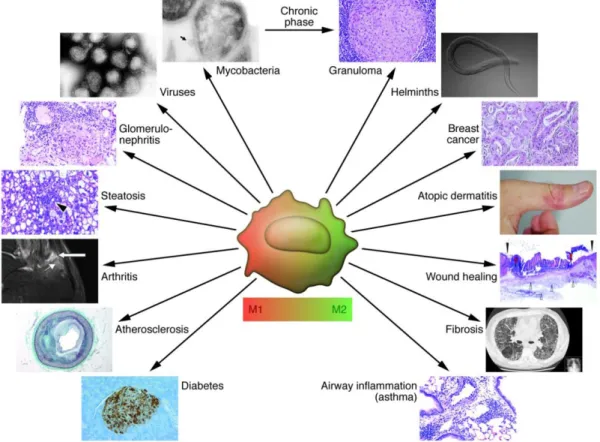
Among these, macrophages represent the predominant immune cells and are actively implicated in tumor development and progression. Epidemiological studies have described an association between increased tumor-associated macrophages (TAMs) infiltration and worst prognosis. In addition, extensive in vitro and in vivo work reported the capacity of macrophages to promote tumor cell proliferation, survival, migration and invasion. Macrophages are extremely plastic cells and can adopt different profiles according to the external environment and, nowadays, it is accepted that there is a continuum of polarization status between two extreme populations: the pro-inflammatory, M1 or classically-activated macrophages, and the anti-inflammatory, M2 or alternatively-activated macrophages.
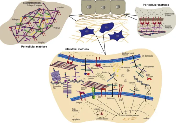
Aside from stromal cells, there is also an important non-cellular component within the tumor microenvironment designated as the extracellular matrix (ECM). The ECM is a complex network of macromolecules with several arrested signaling molecules, namely growth factors and cytokines/chemokines, which, besides providing support to cells and tissues, also performs other functions, such as regulating cell differentiation and migration. Cancer tissues are known for having an abnormal ECM, specifically in composition, organization and biomechanical characteristics, and it is recognized that this aberrant ECM is also involved in cancer development and progression. Despite the known pro-tumoral role of macrophages in cancer, there is still great debate on their role in colorectal cancer (CRC). Therefore, the
overall aim of this thesis was to clarify the relevance of TAMs in CRC. Simultaneously, given the ECM role in shaping cell behavior, we sought to unravel the role of colorectal tumor ECM on the modulation of macrophage polarization and the implications on macrophage-mediated cancer cell invasion. The ultimate goal was to identify novel targets for the development of more efficient therapeutic strategies focusing on the modulation of the tumor microenvironment.
In order to profile TAMs in CRC, we quantitatively assessed macrophages in a series of well-characterized CRC cases, including the analysis of different subpopulations, identified by CD80, expressed by pro-inflammatory macrophages, and CD163, expressed by their anti-inflammatory counterparts. In the present study, we have also included the tumor adjacent
macrophages between normal and tumor tissues, mainly due to an almost complete disappearance of CD80+ cells in neoplastic tissues. In stage III tumors, higher macrophage infiltration and decreased CD80/CD163 ratio associated with decreased survival. Moreover, CD80 expression provided a protective role in preventing relapse and locoregional recurrence.
After describing the alterations on macrophage profile within colorectal tumors, the role of the tumor ECM on macrophage polarization was also assessed. To obtain an ECM which accurately resembled the native tissue, human fragments originated from CRC patients’ surgical resections were decellularized and subsequently characterized regarding their biochemical, structural and biomechanical properties. These matrices were then repopulated with human monocytes derived from healthy blood donors. Their characterization revealed that, in tumor-derived ECM, macrophages adopted a more anti-inflammatory profile, with increased production of TGF-β, IL-10 and CCL18 and decreased expression of TNF and CCR7. Moreover, tumor ECM-educated macrophages stimulated CRC cell invasion, a process mediated by CCL18. Finally, the chemokine CCL18 was shown to activate a signaling cascade involving FAK, EGFR, Akt, Src, ERK and p38, and to induce a partial epithelial to mesenchymal transition.
Taken together, this thesis provided new insights regarding macrophages in CRC. Our findings highlight the role of the tumor microenvironment, specifically the ECM, on the modulation of macrophage polarization towards an anti-inflammatory phenotype. Additionally, given the protective role of CD80+ cells and the pro-invasive role of macrophage-derived CCL18, this PhD work further supports the relevance of targeting macrophages by new therapeutic approaches, namely immunotherapy.
xv 3D - three dimensional
A
ACF - aberrant crypt foci AM - alveolar macrophages ANM - adjacent normal mucosa
APC - adenomatous polyposis coli
APCs - antigen presenting cells ANG2 - angiopoietin-2
B
bFGF - basic FGF
BMDC - bone marrow-derived cells
C
CAFs – cancer-associated fibroblasts CAM-RR - cell-adhesion-mediated-radio-resistance
CAR - chimeric antigen receptors CCL - chemokine (C-C motif) ligand CCR2 - C-C chemokine receptor type 2 CIMP - CpG island methylator phenotype CIN - chromosomal instability
CNS - central nervous system CRC - colorectal cancer
CSF-1/M-CSF - Colony stimulating factor 1/Macrophage colony-stimulating factor CSFR1 - colony stimulating factor receptor 1 CTCs - circulating tumor cells
CTLA-4 - cytotoxic T-lymphocyte protein 4 CXCL - chemokine (C-X-C motif) ligand CD – cluster of differentiation
D
DCs - dendritic cells
E
ECM - extracellular matrix EGF - epidermal growth factor
EGFR - epidermal growth factor receptor EMT – epithelial-mesenchymal transition
F
FACITs - fibril-associated collagens with interrupted triple helices
FAK - focal adhesion kinase
FDA - Food and Drug Administration DFS - disease free survival
FGF - fibroblast growth factor FOXP3 - forkhead boxP3
G
GAGs - glycosaminoglycans GPR30/GPER1 - G protein-coupled estrogen receptor 1H
HA - hyaluronic acid/hyaluronanHB EGF - heparin-binding EGF-like growth factor
HCC - hepatocellular carcinoma HGF – hepatocyte growth factor HGFR/c-Met - HGF receptor
HSC - hematopoietic stem cells
I
IF - tumor invasive front IFN - interferon
IGF - insulin growth factor IL - interleukin
IRA % - percentage of the immunoreactive area
IT - intratumoral region
J
JAK - Janus kinase
L
LPS – lipopolysaccharide LOL - lysyl oxidase LOXL - LOX-like LXs - lipotoxins
M
MACITs - membrane-associated collagens with interrupted triple helices
MCP1 - monocyte chemoattractant protein-1 MDSCs - myeloid-derived suppressor cells MHC - major histocompatibility complex miRNA - microRNAs
MMPs - matrix metalloproteinases MMR - mismatch repair
MPS - mononuclear phagocyte system MSCs - mesenchymal stem cells (MSCs) MSI - microsatellite instability
producing collagens
N
NF-κB - nuclear factor-κB NK - natural killer cells NO - nitric oxide
NOS2/iNOS - NO synthase 2/inducible NO synthase
O
OPN - osteopontin
P
PAMPs - pathogen-associated molecular patterns
PAR1 - protease-activated receptors 1 PBMCs - peripheral blood mononuclear cells
PDAC - pancreatic ductal adenocarcinoma PD1 - programmed cell death protein 1 PDGF - platelet-derived growth factor PD-L1 - programmed death-ligand 1 PEMs - polyelectrolyte multilayers PGA - chitosan/poly γ-glutamic acid PGE2 - prostaglandin E2
PGF - placental growth factor PGs - proteoglycans
PITPNM3 - Membrane-associated phosphatidylinositol transfer protein 3 PPRs - pattern recognition receptors Pyk2 - proline-rich tyrosine kinase 2 PyMT - polyoma middle T
xvii RGD - Arg-Gly-Asp
S
SDF-1 - stromal cell-derived factor-1 SEM - Scanning electron microscopy SLRP - small leucine-rich proteoglycans STAT3 - signal transducer and activator of transcription 3
STI1 - stress inducible protein 1
T
TAMs - Tumor-associated macrophages TEMs - TIE2-expressing monocytes TF - tissue factor
TGF - Transforming growth factor Th1 - T helper type I
Th2 -T helper type II
TILs - tumor-infiltrating lymphocytes
TLR - Toll-like receptors
TMEM - tumor microenvironment of metastasis
TNC - tenascin-C
TNF - tumor necrosis factor TNM - tumor/node/metastasis Tregs - regulatory T cells
U
UICC - Union for International Cancer Control
uPA - urokinase-type plasminogen activator
V
VEGF - vascular endothelial growth factor VCAM-1 - vascular cell adhesion molecule-1
xix In many malignancies, including melanoma, breast or ovarian cancer, the presence of high levels of tumor associated macrophages (TAMs) correlates with more aggressive disease and worst prognosis. TAMs are actively involved in tumor progression, since they may promote tumor growth, survival, angiogenesis, cancer invasion and metastasis. Nevertheless, in colorectal cancer (CRC), the data regarding macrophage’s clinicopathological importance is contradictory, with some studies describing an association between macrophage infiltration and decreased survival and others reporting the exactly the opposite. Therefore, in this PhD thesis, we sought to clarify the significance of TAMs within CRC. Subsequently, the focus of our research was centered on the role of the extracellular matrix (ECM), present at the tumor microenvironment, on the modulation of macrophage inflammatory profile but also on identifying the molecular mechanisms through which macrophages enhance cancer cell invasion.
Accordingly, the subsequent specific objectives were established:
1. Characterize the macrophage populations, specifically the inflammatory subtypes, present in CRC tissues, and explore possible associations with prognosis and clinical outcome
Many studies have addressed the relevance of macrophage infiltration in CRC but, probably due to disparities in the selected markers and in the methodologies used across studies, the available results are conflicting. For this reason, we performed a quantitative characterization of macrophages, including different subpopulations, across CRC tissues. Our approach consisted on profiling macrophage subtypes in 150 CRC cases (dated from 2007-2012) from Centro Hospitalar São João Tumor Bank. Importantly, all tumor fragments analyzed also contained the adjacent normal mucosa. Consecutive sections were stained with specific antibodies for CD68, a macrophage lineage marker, CD80, a co-stimulatory molecule expressed by pro-inflammatory macrophages, and CD163, a scavenger receptor expressed by anti-inflammatory macrophages. Following immunohistochemistry, tissue slides were digitalized and the immunoreactive area for each marker was quantified, using Fiji software, in three different regions: normal adjacent mucosa, intratumoral region and tumor invasive front. For each region, 10 distinct areas were randomly selected for each marker quantification and analysis. The data was crossed with patient’s clinicopathologic information in an attempt to unravel the clinical relevance of the distinct macrophage
xx
2. Recreate an organotypic 3D culture system that resembles the colorectal tumor ECM
Having shown, in the previous chapter, that there is an alteration in macrophage profile within colorectal tumors, we sought to unravel whether tumor ECM, being the most prevalent element at the tumor microenvironment, had any role in this differential macrophage polarization. Due to the biochemical and biomechanical complexity of the human ECM, difficult to recreate by bioengineered scaffolds, we decided to take advantage of human colorectal samples and use decellularization as a method to obtain reliable ECM that accurately resembles the native tissue. Accordingly, using fragments obtained from CRC patients’ surgical resections, we optimized a decellularization protocol able to remove DNA and cell debris from both normal and tumor colorectal tissues. Furthermore, to ensure that decellularized tissues preserved native tissues’ characteristics, their composition, architecture, and biomechanical properties were monitored through immunohistochemistry, scanning electron microscopy and rheometer analysis, respectively. These results are included in Chapter 3.
3. Dissect the role of tumor extracellular matrix on human macrophage polarization
Knowing that macrophages adjust their phenotype according to microenvironmental factors, and that the tumor ECM displays modifications in its biochemical and physical properties, we assessed the effect of such altered ECM on macrophage polarization. Therefore, decellularized normal and tumor matrices, described in the previous objective, were repopulated with human monocytes derived from healthy blood donors. After 14 days of differentiation within such matrices, macrophages were characterized in terms of their morphology, RNA expression for specific cell surface receptors and pro- and anti-inflammatory cytokine/chemokine production. Furthermore, the effect of such ECM-educated macrophages on CRC cell invasion was also evaluated. These results are included in Chapter 3.
4. Unravel the effects of CCL18 on colorectal and gastric cancer cell invasion
Our group has previously reported that macrophages, particularly the anti-inflammatory, stimulate gastric and CRC cell invasion through the secretion of factors, as EGF or MMPs. In Chapter 3, we report that macrophages differentiated in tumor- derived matrices secrete high levels of CCL18, an anti-inflammatory and immunosuppressive chemokine. Moreover,
xxi CCL18 mechanism of action which was leading to this increased pro-invasive capacity. To address this question, CRC cells were stimulated with CCL18 and the activated signaling pathways were evaluated. Additionally, the effect of CCL18 on the expression of some epithelial-mesenchymal transition (EMT) related genes was assessed. Due to previous work from our group regarding the effect of macrophages on gastric cancer cell migration and invasion, a gastric cancer cell line was also included in this work. These results are included in Chapter 4.
1
CHAPTER 1
3
1. Hallmarks of cancer
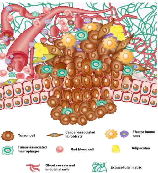
Cancer was known for a long time as a genetic disease. Alterations in genes, either by somatic or germinal mutations, resulted in uncontrolled cell growth, escape to apoptosis, enhanced migration or ability to migrate across basement membranes, which ultimately could lead to metastatic disease. The central role of the neoplastic cell was well illustrated in the review from 2000 by Hanahan and Weinberg, describing the capabilities that cells had to acquire during the carcinogenic process, which they coined as the six hallmarks of cancer [1]. These included the self-sufficiency in growth signals, limitless replicative potential, insensitivity to growth-inhibitory signals, evasion of apoptosis, sustained angiogenesis and tissue invasion and metastasis. At the time, it was already recognized that, albeit the unquestionable fundamental role of the cancer cell, a tumor was much more complex than initially though. In fact, neoplastic disease develops in a complex microenvironment also composed by normal non-malignant stromal cells, namely fibroblasts, endothelial cells, adipocytes and immune cells, from both the innate and adaptive immune system (Figure 1.1). All these cells intercommunicate and influence each other’s behavior through direct interaction or by exchanging a myriad of soluble factors, including growth factors, cytokines or matrix metalloproteinases (MMPs) [2]. Moreover, supporting the cellular components, there is also a complex network of macromolecules called the extracellular matrix (ECM). Once largely neglected, nowadays the ECM is recognized to be extremely dynamic and to have a major impact in cell behavior, in both health and disease [3, 4]. This evolution in cancer biology knowledge culminated in a 2011 revised version of the hallmarks of cancer, according to which tumors are in fact considered intricate organs with multiple components with active roles in the carcinogenic process [5].
2. Tumor microenvironment
2.1 . Tumor cells
Carcinomas are malignant tumors that originate from epithelial cells. Accumulation of mutations in specific genes confers a growth advantage to cells enabling them with the capacity to resist the control mechanisms which would cause their elimination [6]. In fact, errors during DNA synthesis are not a rare event, but there exist specific mechanisms, namely the mismatch repair (MMR) system, which assure the correction of the majority of these mistakes before resulting in fixed mutations [7]. Those cells in which the mutations are not efficiently corrected are generally directed to cell death, through induction of apoptosis or autophagy. It was proposed that cancer cells arise from an evolutionary process, where mutations and a process of natural selection act together [8]. Epigenetic alterations, namely disruptions in DNA methylation and histone modifications, add another layer of complexity to this process and, in
4
Figure 1.1. Schematic representation of the primary tumor microenvironment. Tumor cells, derived from
normal epithelial cells, are surrounded by a complex microenvironment formed by stromal cells, namely fibroblasts, immune cells, such as macrophages and lymphocytes, adipocytes and endothelial cells, and by a complex non-cellular component that is the extranon-cellular matrix. Adapted from Berindan-Neagoe I, Clin Can Res, 2014, with
permission from AACR [9].
fact, cannot be dissociated from mutations [10]. The combination of all these alterations represents an advantage to the cells, providing them the capacity to migrate and invade adjacent basement membranes. Once tumor cells reach a blood vessel, they will enter the bloodstream, a process named intravasation, which will allow their transport to a secondary site. There, they will leave the blood vessel, or extravasate, and colonize the new organ forming a metastasis [11].
In the specific case of colorectal cancer (CRC), the pathogenesis of the disease is fairly well established. Nowadays it is accepted that only 20% of CRC have familial origin, some being associated with hereditary nonpolyposis colorectal cancer (HNPCC) or familial adenomatous polyposis (FAP), while others remain without known mechanism. The other 80% are sporadic, being generally divided in three groups: about 85% present chromosomal instability (CIN), with great losses or gains of chromosomal material, others are characterized by epigenetic instability presenting the CpG island methylator phenotype (CIMP) pathway while others
5 display accumulation of numerous mutations throughout the genome, mainly caused by inactivation of MMR genes, resulting in a phenotype known as microsatellite instability (MSI), or [12].
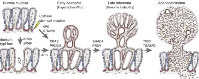
Sporadic CRC development follows a step-wise progression of mutations in oncogenes and tumor suppressor genes that translate into the classical adenoma-carcinoma sequence (Figure
1.2). The earliest genetic change is, most frequently, the mutation and/or loss of the adenomatous polyposis coli (APC) gene that mediates the transition of single preneoplastic
cells to aberrant crypt foci (ACF). The exact sequence of acquired genetic changes, accumulated subsequently to inactivation of APC is variable. K-ras mutations are found in about 50% of CRC and are thought to be relatively early events which correlate, in terms of histology, with the transition from early to intermediate adenomas. Disruption of the transforming growth factor (TGF)-βIIR/mothers against DPP homolog (SMAD)-2-4 pathway and mutations in MMR genes [e.g. MutL-homolog (hMLH) 1 and MutS-homolog (hMSH) 2] have also been identified as key factors in the development and progression of CRC, while p53 mutations are believed to mark the transition from adenoma to carcinoma [13]. Once tumor cells accumulate these mutations they will start to invade the underlying tissue and, contrary to what happens in most tumors, will most likely form distant metastasis rapidly without the common latency period. One plausible explanation is that, after cells acquire the mutations needed to invade, very few, if any, genetic alterations are required to be able to colonize other organs, namely the liver [14].
Figure 1.2. Step-wise progression in sporadic colorectal cancer. Upon initial mutations in the epithelial stem
cells, most frequently in the adenomatous polyposis coli (APC) gene, there will be accumulation of mutations in both oncogenes and tumor-suppressor genes, resulting in sequential development of pre-malignant lesions, culminating in an invasive adenocarcinoma. BRAF, B-Raf proto-oncogene, serine/threonine kinase; KRAS, Kirsten rat sarcoma viral oncogene homolog; CTNNB1, catenin beta-1; PIK3CA, phosphatidylinositol-4,5-bisphosphate 3-kinase catalytic subunit alpha; SMAD4, Mothers against DPP homolog family member 4; PTEN, phosphatase and tensin homolog; TGFBR2, transforming growth factor beta receptor 2; TP53, tumor protein p53. From Strubberg
6
2.2 . Fibroblasts
Fibroblasts are the major ECM producers, being particularly relevant during wound healing, embryonic development and tissue repair and regeneration. In cancer, fibroblasts get activated similarly to what happens in a wound situation, gaining increased expression of α-smooth muscle actin, which leads to their differentiation into myofibroblasts [16]. Several mechanisms are described as being involved in this differentiation, namely the release of cancer cell exosomes expressing high levels of TGF-β which, in turn, induces the production of fibroblast growth factor (FGF)-2 by fibroblasts [17]. Indeed, many of the interactions established at the tumor microenvironment seem to occur through paracrine signaling. Breast cancer cells were described to stimulate hepatocyte growth factor (HGF) production by fibroblasts which, in turn, enhance breast cancer cell HGF receptor (HGFR/c-Met) activation, promoting colony formation in soft agar and facilitating tumor growth in mice [18]. Interestingly, de Wever et al. demonstrated that myofibroblasts isolated from human colon tumors, and contrarily to what happened with fibroblasts isolated from adjacent normal tissue, induced colon cancer cell migration through the secretion of HGF and Tenascin-C (TNC). Importantly, both of the pro-invasive signals were required, but not sufficient, for such stimulation, with HGF acting through Rac activation and TNC through RhoA inactivation [19]. Importantly, recent work in prostate cancer revealed that TNC is a marker of CAFs and a predictor of poor prognosis [20]. Moreover, epithelial cancer cells were shown to suppress p53 expression in fibroblasts, a mechanism not dependent of cell-cell contact. Also in this specific case, human cancer-associated fibroblasts (CAFs) were more susceptible to this suppression than normal fibroblasts [21]. Given the non-cell-autonomous effects of stromal p53, namely by inhibiting cell growth [22] and angiogenesis [23], this mechanism may contribute to overcome the fibroblast-mediated tumor suppression. MMPs are another player in this crosstalk. One such example is Stromelysin-3, a MMP mainly produced by fibroblasts, which was shown to promote the homing of malignant breast cancer cells in a mouse model, a process dependent on the presence of ECM-associated growth factors [24]. Fibroblast-derived MMP-1 was also reported to induce breast cancer cell migration by binding to protease-activated receptors 1 (PAR1) in tumor cells, cleaving it and triggering PAR1-Dependent Ca2+ signaling [25]. Fibroblasts were also demonstrated to increase tumor incidence, size and metastasis when injected in an orthotopic nude mouse model of pancreatic cancer, demonstrating direct involvement in cancer development [26].
Additionally, fibroblasts communicate with other stromal cells. By secreting various cytokines and chemokines, namely chemokine (C-C motif) ligand (CCL)-2 [27], osteopontin (OPN), chemokine (C-X-C motif) ligand (CXCL)-1, CXCL2, interleukin (IL)-6 and IL-1β [28] they recruit immune cells, which mediate the inflammatory response. Moreover, they are involved in
7 angiogenesis through the release of vascular endothelial growth factor (VEGF) [29] or stromal cell-derived factor-1 (SDF-1), which induce the recruitment of endothelial progenitor cells [30].
2.3 . Endothelial cells
The reciprocal interactions established between tumor and endothelial cells are also extremely important for tumor progression [31]. The so called “angiogenic switch”, describing the moment when pro-angiogenic factors are predominant over the anti-angiogenic ones, ultimately resulting in an abnormal vascularization of tumors, is a key event during carcinogenesis [32]. As a result, primary tumor cells will have access to nutrients and oxygen, which enables their growth [33], will more easily intravasate through the fenestrated new blood vessels, reaching the circulation, and will be able to disseminate and metastasize to distant organs. Tumor cells, on their turn, are able to stimulate endothelial cells by secreting factors such as VEGF, a potent pro-angiogenic factor involved in the induction of endothelial cell survival, proliferation, migration and branching. Tumor cells were also shown to secret other factors such as galectin-1, which will be uptake by endothelial cells and promote Ras signaling, resulting in the activation of the Ras/Mek/ERK cascade, ultimately leading to endothelial cell proliferation and migration [34]. The action of cancer cells can also occur through direct contact, by a mechanism involving Jagged-1 overexpression which will trigger endothelial cells Notch activation, resulting in enhanced neovascularization and tumor growth [35]. In addition, specific microRNAs (miRNA) are involved in endothelium activation, namely miR-132 which was shown to downregulate p120RasGAP, a crucial negative regulator of vascular development and remodeling. As a result, there is an increase of Ras activity leading to endothelial cell proliferation and increased tube formation, ultimately resulting in neovascularization [36]. Endothelial cell proliferation, branching and migration is also promoted by other stromal cells within the tumor microenvironment, namely fibroblasts, as mentioned above, bone marrow derived myeloid cells, through the production of IL-1β which will act on endothelial cells [37], or macrophages, a topic which we discussed in detail in the following chapters [38].
Moreover, endothelial cells are reported to promote tumor cell invasion independently of angiogenesis. Similarly to VEGF, conditioned medium from head and neck tumor cells enhance Bcl-2 expression in endothelial cells. On the other hand, endothelial cells transfected with Bcl-2 promote tumor cell invasion, a process involving CXCL1 and CXCL8 secretion by endothelial cells which will bind CXCR2 on tumor cells [39].
8
2.4 . Adipocytes
Adipocytes are another stromal cell type within the tumor microenvironment that has been gaining increased attention by the scientific community [40]. In fact, the association between an increased body-mass index and cancer incidence is not new and has been reported for many cancers [41]. Nevertheless, the mechanisms through which adipose tissue contributes to tumor development, growth and metastization are only now being fully understood [42]. Adipocytes within tumors, termed cancer-associated adipocytes, present an “activated” phenotype characterized by secretion of cytokines and other inflammatory mediators, growth factors and hormones, called adipokines [43]. One example is the secretion of leptin by adipocytes which will act in an autocrine way, leading to aromatase production and estrogen levels increase, resulting in breast cancer cell growth [44]. Additionally, leptin can act directly on cancer cells as it was reported in a murine model of colon cancer in which leptin promoted tumor cell proliferation by binding to leptin receptor, and subsequently activating the signal transducer and activator of transcription 3 (STAT3) [45]. Conversely, adiponectin, other hormone also secreted by adipocytes, was reported to inhibit cell proliferation by selectively binding to growth factors, specifically platelet-derived growth factor (PDGF)-BB, basic FGF (bFGF), and heparin-binding EGF-like growth factor (HB EGF), and thus preventing their interaction with the respective receptor [46]. Interestingly, adiponectin is decreased in several cancers [40]. The involvement of IL-6 was also shown to be a key player in both breast cancer cell invasion [47] and radioresistance mediated by adipocytes [48]. Indeed, the link between adipocytes and inflammation, namely due to the close interplay with immune cells such as macrophages, emerged as crucial in the establishment of a permissive microenvironment, supportive of tumor growth and progression [49].
2.5 . Immune cells
Immune cells are responsible not only for the detection and elimination of foreign agents that may cause an injury or infection but also for the removal of death or mutated cells from a given organism. When DNA repair mechanisms or cell cycle check points fail and do not conduct damaged cells towards apoptosis or autophagy, immune cells work as a second line of defense. Myeloid cells, namely macrophages, dendritic cells (DCs), mast cells and granulocytes (eosinophils, basophils and neutrophils), and lymphoid cells, as δT and natural killer (NK) T cells, are mediators of primary innate immune responses, which are not specific for a given pathogen or insult. In contrast, T and B cells exert specific functions towards an antigen exposed by antigen presenting cells (APCs) and mediate the latter adaptive immune response [50]. The notion that immune cells are involved in cancer development is not new. Already in 1863 Rudolf Virchow, a German pathologist, reported a strong leukocyte infiltration
9 in tumors and suggested a relation between this finding and the occurrence of the disease [51]. Since then, his theory has been extensively proven by a series of epidemiological studies and, nowadays, it is unquestionable that chronic inflammation predisposes individuals to various types of cancers. In fact, chronic infection and inflammation have been proposed to contribute to about 25% of all cancers worldwide [52], as in the case of Helicobacter pylori for gastric cancer, and inflammatory bowel disease for CRC [53]. Besides these extrinsic factors, which favor the mutational rate and may lead to cancer initiation, there is also an intrinsic pathway involving genetic alterations of oncogenes and tumor-suppressor genes, ultimately resulting in an inflammatory microenvironment [54]. But how does it work? (Figure 1.3) Regardless of being caused by extrinsic or intrinsic factors, there are specific transcription factors that become activated in pre-malignant or tumor cells, namely nuclear factor-κB (NF-κB), STAT3 and hypoxia-inducible factor 1-α (HIF1α). Of these, NF-κB was proposed to be the master regulator linking cancer and inflammation [55], being involved in processes such as
Figure 1.3. Involvement of innate and adaptive immune cells in inflammation associated cancer development. Antigens present in early neoplastic tissues will be transported to lymphoid organs by dendritic
cells (DCs), resulting in the activation of adaptive immune responses, ultimately leading to either tumor-promoting or anti-tumor effects. Activation of B cells and humoral immune responses results in chronic activation of innate immune cells in neoplastic tissues, namely mast cells, granulocytes and macrophages, leading to the production of pro-survival, pro-angiogenic and tissue remodeling factors. On the other hand, adaptive immune cell activation also mediates tumor cell killing, either by T-cell mediated cytotoxicity, by activation of perforin/granzyme or Fas/FasL pathways, or by antibody-dependent cell- mediated cytotoxicity. Reprinted by permission from Springer Nature on behalf of: de Visser KE, Nat Rev Cancer 2006 [59]
10
tumor cell proliferation, apoptosis inhibition, angiogenesis, metastasis [56] and orchestration of both innate and adaptive immune surveillance [57]. Once upregulated, these transcription factors will induce the expression of a series of chemokines, cytokines and inflammatory enzymes, such as cyclooxygenase-2, by tumor cells, resulting in the recruitment of cells of the innate immune system, specifically dendritic cells, macrophages, neutrophils and NK cells. These will also produce inflammatory chemokines and cytokines, namely tumor necrosis factor (TNF)-α and IL-6, MMPs and growth factors further sustaining the inflammatory environment. Additionally, upon the immune cells attack, the secretion of reactive oxygen species will also contribute to DNA damage and mutations [58], further supporting a mutagenic environment. Some of the acquired genetic alterations may result in immunogenic peptides which will be presented at the surface of early neoplastic cells by major histocompatibility complex (MHC) class Ia molecules in the form of neoantigens, and will be recognized by the immune system as “non-self”. This information will be transported by dendritic cells to lymphoid organs leading to the activation of adaptive immune responses, specifically T- and B- lymphocyte responses, resulting in anti-tumor effects through T-cell and antibody-dependent cell-mediated cytotoxicity. At the same time, there will be chronic activation of innate immune cells within tumors which will secret pro-survival and pro-angiogenic factors [59].
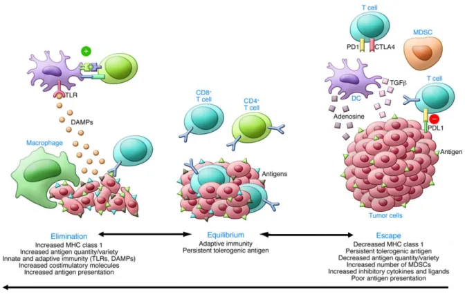
One important concept regarding immune system and tumor development is cancer immunoediting, which intends to describe the host-protecting and tumor-sculpting actions of the immune system that not only prevent disease, by suppressing the formation of nascent tumors, but also shapes tumorigenesis. Cancer immunoediting encompasses three phases: elimination, which relies on immune surveillance, equilibrium, based on immune selection, and escape [60, 61] (Figure 1.4). In the elimination phase, there is involvement of cells from both innate and adaptive immunity. The first cells to arrive to the tumor site are neutrophils, followed by macrophages, which will exert an unspecific phagocytic activity against mutated cells, while releasing several pro-inflammatory cytokines (IL-1β, TNF-α IL-6, IL-8, and IL-12), chemokines, leukotrienes, prostaglandins, and complement proteins. These will regulate the adaptive immunity by recruiting and activating T cells. Macrophages, and particularly DCs, act as antigen presenting cells. At the tumor site, DCs recognize neo- or mutated antigens, become activated, maturate and migrate to peripheral lymph nodes, where they synthesize MHC and express co-stimulatory CD80 and CD86 molecules, in order to effectively present the antigens to naïve T cells. At this time, additional T cell co-stimulatory molecules, CD4 (for CD4+ T cells) or CD8 (for CD8+ T cells), maximize the interaction between the T cell receptor (TCR)-CD3 complex and the MHC-II molecule expressed on the antigen presenting cell surface, strengthening the immunological synapse. Activated T cells may then return to tissues to exert their immune functions. CD4 T cells may differentiate into T helper type I (Th1), producing
11 interferon (IFN)-, TNF-α, IL-2 and activating macrophages, or into T helper type II (Th2) cells, producing IL-4, IL-10 or IL-13 and reducing macrophage pro-inflammatory activity. In the presence of anti-inflammatory mediators, CD4 T cells may differentiate into forkhead box P3 (FOXP3) regulatory T cells (Tregs), creating an immunosuppressive environment. On their turn, CD8 T cells exert cytotoxic activity directly against the tumor cells, inducing perforin/granzyme or Fas/FasL-mediated apoptosis. Finally, the coordinated action between innate and adaptive immune cells will recognize transformed cells ultimately leading to their eradication through the activation of transcription factors and IFN-γ dependent pathways [63].
Figure 1.4. Immunoediting hypothesis encompassing three stages: the elimination, the equilibrium and the escape. The elimination phase usually occurs in the early stages of tumor development and relies on the expression
of neo- or mutated antigens by tumor cells. These will be recognized by dendritic cells and macrophages that will activate the adaptive immune cells, which will ultimately lead to tumor cell killing. If this process is not completely effective, some tumor cells will survive and will be maintained in a dormant state, coexisting with immune cells from both innate and adaptive immune system, in a delicate equilibrium. After a series of adaptations by the tumor cells, namely by decreasing antigen presentation or by releasing immunosuppressive cytokines, they will be able to surpass the immune system, escape their control and eventually proliferate and form malignant disease. Republished with permission of American Society for Clinical Investigation, from Kalbasi A , JCI 2013; permission conveyed through Copyright Clearance Center, Inc [62].
Nevertheless, there is the possibility that some cells might survive and will be kept in a dormant state, in a delicate equilibrium with the immune system. At this stage, as a result of constant selection pressure caused by the presence of immune mediators, or by therapeutic intervention, tumor cells undergo a series of genetic and epigenetic alterations originating tumor variants highly resistant and with the ability to escape the immune attack [64]. Two of the most frequent strategies developed by tumor cells to escape the immune system is the
12
decrease of antigen presentation and the induction of an immunosuppressive environment, namely by expressing proteins such as programmed death-ligand 1 (PD-L1). Finally, in the last phase, these selected cells will be able to proliferate and eventually invade the adjacent tissues and form metastasis [65].
Contrary to what happens with the infiltration of cells involved in chronic inflammation, high number of lymphocytes, namely T cells (CD3+), cytotoxic T cells (CD8+), or memory T cells (CD45RO+), correlate with good prognosis in different tumors namely melanoma, breast and non-small cell lung cancer, among others [66]. Similar results were obtained by Galon and colleagues in CRC. By characterizing the infiltrating immune cells, focusing on markers of inflammation, Th1 adaptive immunity, and immunosuppression, they reported that the type, density and location of immune cells may predict clinical outcome. Moreover, this characterization was more accurate and reliable in predicting patient survival than the classical histophatological methods used, specifically the tumor/node/metastasis (TNM) classification established by the Union for International Cancer Control (UICC) [67]. As a result, an immune-classification named Immunoscore, based on the numeration of two lymphocyte populations (CD3/CD45RO, CD3/CD8 or CD8/CD45RO) quantified within the core of the tumor and on the invasive margin, was created. This was proposed to be introduced in the clinical setting, specifically in CRC, in order to improve prognosis and help to determine therapy response [68-70]. An international consortium is currently ongoing to validate the immunoscore relevance and eventually add this new component to cancer classification, ultimately including other cancer types [71]. Indeed, a meta-analysis by Fridman et al. comprising 124 studies validated the transversal positive association between specific T cells and survival. Conversely, regarding other immune populations, such as B cells, NK cells, myeloid-derived suppressor cells (MDSCs), macrophages and a subset of T-helper populations (Th2, Th17, Treg cells), the prognostic impact varied according to the tumor type [72]. One such example are the FoxP3+ Tregs, which high density was associated with better survival and improved prognosis in CRC [73] whereas, in breast cancer, was correlated with increased relapse and shorter survival [74].
The importance of neoantigen expression and their recognition by the immune system in the prevention of tumor development and progression is well illustrated in the specific case of MSI colorectal tumors. Given the defects in MMR genes, these tumors have an abnormally high mutational load leading to an increased expression of neoantigens. As a result, tumor cells will most likely be detected by the immune system. Interestingly, these tumors also present a strong infiltration of cytotoxic T cells and, when both factors are combined, allow the identification of patients with favorable prognosis [75]. Also in melanoma, high mutational load is associated with improved survival [76]. Besides the number of neoantigens, their quality,
13 meaning their immunogenicity calculated by the probability of TCR recognition, is also extremely important. Recently, Balachandran et al. showed that the combination between highest antigen quality and the most abundant CD8+ T cell infiltrates, stratified patients with the longest survival in pancreatic cancer [77].
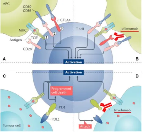
Given the great advances in immuno-oncology, namely the capacity of the immune system to recognize and reject tumors combined with the increased knowledge on the strategies acquired by tumors cells to evade immune destruction, a new era in immunotherapy emerged. Adoptive immune therapy, using either natural host cells with antitumor reactivity, such as tumor-infiltrating lymphocytes (TILs), or genetically engineered ones, specifically chimeric antigen receptors (CAR) T-cells, have shown very positive results in different cancers [78]. As a result, CAR T-cell therapy was recently approved by Food and Drug Administration (FDA) for pediatric and young adult patients with acute lymphoblastic leukemia or advanced lymphoma [79]. On the other hand, the observation that blocking cytotoxic T-lymphocyte protein 4 (CTLA-4), a receptor expressed by T cells involved in their inhibition when bound to CD80 and CD86, enhanced antitumor immunity [80], opened new perspectives in immunotherapy. The introduction of immune-checkpoint inhibitors, targeting not only CTLA-4 but also the PD-L1/Programmed cell death protein 1 (PD1) pathway as a strategy to boost T cell activation, has contributed to revolutionize the field (Figure 1.5). Due to successful results obtained in clinical trials performed with monoclonal antibodies targeting either CTLA-4 or PD-1/PDL-1, these have also been approved by FDA to be used in different cancers [81, 82]. Despite the great enthusiasm in the field, the reality is that the number of patients that respond to such therapies, even in combination with conventional approaches as radio or chemotherapy, is still limited and quite unpredictable. Thus, in this era of personalized medicine, the attention is also directed towards the discovery of new biomarkers that will help to discriminate which patients will truly benefit from such therapeutic regimens [83]. Accordingly, Luksza and colleagues created a fitness model based on immune interactions of neoantigens with T cells, which predicts survival of patients with melanoma and lung cancer using anti-CTLA-4 and anti-PD-1, respectively [84]. In this constantly evolving “omics” period, high-throughput experimental designs and technologies are certainly contributing to increase the knowledge quickly translatable into the clinics. Rivzi et al., by performing whole-genome sequencing of non-cell lung cancers treated with an antibody targeting PD-1, revealed that higher non synonymous mutation burden was associated with improved objective response [85]. In the specific case of melanoma, genomic and transcriptomic analysis of pretreated biopsies enabled the discovery of a transcriptional signature associated with anti-PD-1 resistant tumors. At the same time, patients with increased mutations in the DNA repair gene BRCA2 were the ones with increased response [76]. Furthermore, whole-exome sequencing
14
of melanoma samples, before and after anti-PD-1 treatment, led to the discovery of inactivating mutations of Janus kinase (JAK) 1 and JAK2 associated genes related to acquired resistance to PD-1 blockade [86]. Other strategies currently ongoing involve the study of MHC-associated peptidome aiming at finding predictive neoantigens suitable of being targeted by immunotherapies [87-89]. Finally, genomic, transcriptomic and proteomic analysis of single cells, specifically T cells after cancer treatment, are also expected to contribute to therapy
Figure 1.5. Immune checkpoint blockade in cancer therapy. (A) Normal activation of a T-lymphocyte by an
antigen presenting cell requires binding of the major histocompatibility complex (MHC) presenting an antigen in the APC with T cell receptor, and interaction between CD28 and co-stimulatory molecules CD80 and CD86. CTLA4 in T cells compete with CD28 for CD80/CD86 ligands, preventing the interaction with CD28, and thus inhibiting T cell activation. (B) By neutralizing CTLA4 receptor with a monoclonal antibody, Ipilimumab, it will no longer bind to CD80/CD86, and the latter will be available to interact with CD28 resulting in T cell activation. (C) In order to evade T cell detection, tumor cells start to express the immune checkpoint activator programmed cell death ligand 1 (PD-L1), which will bind to PD1 in T-cell, preventing their activation. (D) Blocking PD1 or PD-L1 using a specific monoclonal antibody, Nivolumab, enables tumor cell detection by T-cells. Adapted by permission from Springer
Nature on behalf of: Byun DJ, Nat Rev Cancer 2017 [90].
decision [91]. Indeed, besides the immunogenicity of these antigens, the immune contexture of each tumor has also proven to be determinant for the success of immunotherapies, particularly of the immune check point inhibitors [92]. In this sense, Chen and Mellman have recently proposed a cancer classification based on the immune phenotype they present: the immune-desert phenotype, the immune-excluded phenotype and the inflamed phenotype. The first two can be considered as non-inflamed tumors, being characterized by the expression of cytokines related with immune suppression or tolerance. What distinguishes both types is that, while the immune-desert has a diminished or absent infiltration of CD8-carrying T cells, the
15 immune-excluded phenotype is abundantly infiltrated by immune cells but these are retained in the stroma, and isolated from the tumor cells by a fibrotic capsule. For these reasons, both tumor types present a very low responsiveness to anti-PD-L1/PD-1 agents. On the other hand, the inflamed type is the one that presents higher infiltration of different immune cells, contains pro-inflammatory cytokines and, despite some exceptions, generally correlates with higher response rates to anti-PD-L1/PD-1 therapy [93]. Moreover, Fridman and colleagues have suggested the integration of immune, vascular and stromal gene-expression signatures of tumors in order to stratify patients for the most appropriate form of immunotherapy [92]. Albeit the fact that immunotherapy is mostly based on T-cells, the reality is that there are other immune populations within the tumor microenvironment, namely macrophages, with crucial roles in the tumorigenic process and hence are also a subject of intensive research [94].
2.5.1 Macrophages 2.5.1.1 Origin
Macrophages are present in virtually all tissues within the body and are key regulators in development, tissue repair, angiogenesis, homeostasis and disease, namely host defense mechanisms, inflammatory diseases and cancer.
Macrophage origin has been a topic that has witnessed great evolution in the last years. In 1970, Furth and colleagues included highly phagocytic cells and their precursors in a system called “mononuclear phagocyte system” (MPS), encompassing promonocytes, monocytes and macrophages. These cells had a common origin, being the promonocytes the most immature cells that differentiated sequentially into monocytes and these into macrophages, and shared specific characteristics, namely morphology and function [95]. The idea that circulating monocytes, originated from hematopoietic progenitors, were continuously replenishing macrophages within tissues lasted for many years and only in the last decade there was a complete shift in this paradigm. Nowadays, it is known that, in fact, tissues have tissue-resident macrophages [96] which, in the majority of cases, are able to self-maintenance independently of circulating monocytes. By using chimeric animals obtained by parabiosis, Ajami and colleagues were able to prove that microglia was maintained in the brain independently of bone-marrow derived progenitors, in both healthy and central nervous system (CNS) degenerative disease. In fact, microgliosis resulted exclusively from the expansion of CNS-resident cells [97]. This was further validated by work from Ginhoux et al who revealed that the adult microglia had a distinct ontogeny than the mononuclear phagocyte system and that these cells derived from primitive yolk sac macrophages [98]. More recently, it was shown that the same was true for other organs, namely lung, splenic red-pulp, peritoneal and bone marrow
16
[99]. Conversely, in the specific case of the intestine, yolk sac-derived macrophages, characterized by being F4/80hiCD11blo, seem to be lost in adulthood and are the circulating monocytes, Ly6Chi, which sustain macrophage populations in this organ, through a mechanism dependent of the CCR2 chemokine receptor. Interestingly, macrophage proliferation is mostly detected in neonatal colon but decreases gradually with age, concomitantly with the arrival of circulating monocytes [100]. Despite the recognized complexity regarding macrophage origin within adult tissues, the most updated model describes three major sources. It all begins early in embryogenesis, being the first macrophages derived from early embryonic progenitors during primitive hematopoiesis in the yolk sac. Once the circulatory system is established, they will colonize different organs in the embryo, namely brain, liver, kidney, spleen, lung, and skin. These yolk sac macrophages will also migrate to the fetal liver where, together with hematopoietic stem cells (HSC), originate myeloid progenitor cells. Once differentiated in fetal liver-derived monocytes, these cells will also populate the previously mentioned organs, contributing to the resident macrophage population, characterized by self-renewal capacity and high proliferative ability when homeostasis is lost. Apart from the CNS, in which microglia is exclusively originated from the primitive yolk sac derived macrophages, in the other tissues the main contributors are the fetal-derived monocytes. Additionally, within the fetal liver, HSC suffer an expansion and colonize the bone marrow. These cells will be the source of circulating monocytes in the blood, which will then be recruited to tissues with high turn-over, such as the intestine, as previously described, and in the case of infection or disease [96, 101-106].
2.5.1.2 Functions and classification
Macrophages were first reported by Élie Metchnifoff and his work won him the Nobel Prize for Physiology or Medicine in 1908. He discovered the process of phagocytosis and named the responsible cells as phagocytes [107]. Only later the term macrophage which, in fact, means big eaters, was adopted. Indeed, macrophages are described as professional phagocytes having a key role in maintaining tissue homeostasis.
Depending on the organ they are in, macrophages present different characteristics, perform specific functions and have precise nomenclatures, e.g. microglia in the brain, Kupffer cell in the liver, osteoclast in the bone, Langerhans cell in the spleen, alveolar macrophage in the lung, etc. [108]. Macrophages are innate immune cells responsible for the clearance of apoptotic or necrotic cells in tissues such as the respiratory system, gastrointestinal tract or central nervous system [108], and for the removal of red blood cells in the liver or spleen [109]. Macrophages are also important cells in bone morphogenesis and in ductal branching during the mammary gland development [110]. These cells benefit from their strategic location in the body, working as sentinels constantly searching for danger signals, particularly pathogens.