FACULDADE DE FARMÁCIA
DEPARTAMENTO DE BIOQUÍMICA
NOVEL ASPECTS OF CARNITINE FUNCTION AND METABOLISM
Sara Liliana Nunes Violante
DOUTORAMENTO EM FARMÁCIA
(Especialidade Bioquímica)
FACULDADE DE FARMÁCIA
DEPARTAMENTO DE BIOQUÍMICA
NOVEL ASPECTS OF CARNITINE FUNCTION AND METABOLISM
Sara Liliana Nunes Violante
Supervisor:
Professora Doutora Maria de Fátima Vieira Ventura
Co-supervisors:
Professor Doutor Ronald J. A. Wanders
Professora Doutora Maria Isabel Ginestal Tavares de Almeida
Doutoramento em Farmácia
Bioquímica
Novos Aspectos da Função e Metabolismo da Carnitina
Dissertação apresentada à Faculdade de Farmácia da Universidade de Lisboa para obtenção do
grau de Doutor em Farmácia (especialidade Bioquímica)
Sara Liliana Nunes Violante
Lisboa
2013
Supervisor:
Professora Doutora Maria de Fátima Vieira Ventura
Co-supervisors:
Professor Doutor Ronald J. A. Wanders
The studies presented in this thesis were performed at the Metabolism and Genetic group, iMed.UL (Research Institute for Medicines and Pharmaceutical Sciences), and at the Department of Biochemistry and Human Biology, Faculdade de Farmácia da Universidade de Lisboa, Portugal, under the supervision of Prof. Maria de Fátima Vieira Ventura and Prof. Maria Isabel Ginestal Tavares de Almeida, and in the Department of Paediatrics and Clinical Chemistry, Academic Medical Center, University of Amsterdam, The Netherlands, under the supervision of Prof. Ronald J. A. Wanders.
This work was finantially supported by Fundação para a Ciência e Tecnologia (FCT), Lisboa, Portugal (SFRH/BD/38074/2007).
De acordo com o disposto no ponto 1 do artigo nº 41 do Regulamento de Estudos Pós-Graduados da Universidade de Lisboa, deliberação nº93/2006, publicada em Diário da Republica – II série nº153 – de 5 julho de 2003, a autora desta dissertação declara que participou na conceção e execução do trabalho experimental, interpretação dos resultados obtidos e redação dos manuscritos.
TABLE OF CONTENTS
Abbreviations ………. ix
Summary ………. xiii
Sumário ………. xv
Chapter 1 – Aims and outline of the thesis ……… 1
Chapter 2 – General introduction ………. 5
Chapter 3 – Carnitine palmitoyltransferase 2: new insights on the substrate specificity and implications for acylcarnitine profiling Biochim Biophys Acta (2010) 1802: 728-732 ……….. 27
Chapter 4 – Substrate specificity of human carnitine acetyltransferase: implications for fatty acid and branched-chain amino acid metabolism Biochim Biophys Acta (2013) in press ………. 35
Chapter 5 – Carnitine palmitoyltransferase 2 and carnitine/acylcarnitine translocase are involved in the mitochondrial synthesis and export of acylcarnitines FASEB J (2013) Jan 15 [Epub ahead of print] ……….. 49
Chapter 6 – Carnitine palmitoyltransferase 1A is not crucial for the translocation of acylcarnitines across the outer mitochondrial membrane Submitted ……… 57
Chapter 7 – Peroxisomes contribute to the acylcarnitine production when the carnitine shuttle is deficient Submitted ……… 69
Chapter 8 – A common X-linked inborn error of carnitine biosynthesis may be a risk factor for non-dysmorphic autism Proc Natl Acad Sci U S A (2012) 109: 7974-7981 ………. 81
Chapter 9 – Concluding remarks and Perspectives ………... 91
List of publications ……….. 97
Curriculum Vitae ………. 101
Abbreviations
3MGH 3-Methylglutaconyl-CoA hydratase 3-OH-C16- 3-Hydroxypalmitoyl-
γ-BB 4-N-Trimethylaminobutyrate; γ-Butyrobetaine γ-BBD γ-Butyrobetaine dioxygenase
ABC ATP-binding cassette ABCD ABC transporter ACOX Acyl-CoA oxidase ACS Acyl-CoA synthetase
ACSL Long-chain acyl-CoA synthetase ADP Adenosine 5’-diphosphate AFLP Acute Fatty Liver of Pregnancy
ALDP Adrenoleukodystrophy protein (or ABCD1) ALDRP ALD-related protein (or ABCD2)
ASD Autism spectrum disorders ASP Acid soluble products ATP Adenosine 5’-triphosphate BCAA Branched-chain amino acid
BCAAO Branched-chain amino acid oxidation BSA Bovine serum albumin
C10:0 Decanoic acid; capric acid C12:0 Dodecanoic acid; lauric acid
C4:1- Trans-2-butenoyl-; trans-2-ene-C4-; crotonyl-
C5:1- Trans-2-methyl-2-butenoyl; trans-2-ene-2-methyl-C4-; tiglyl-
C12:1- Trans-2-dodecenoyl- C16:1- Trans-2-hexadecenoyl-
CACT Carnitine/acylcarnitine translocase
Cn-carnitine Acyl residue with n carbons esterified with carnitine Cn-CoA Acyl residue with n carbons esterified with CoA CoA Coenzyme A
CPT1 Carnitine palmitoyltransferase 1 CPT2 Carnitine palmitoyltransferase 2 CrAT Carnitine acetyltransferase CrOT Carnitine octanoyltransferase CS Citrate synthase
DBP D-bifunctional protein DCA Dicarboxylic acid
x
DHCA Dihydroxycholestanoic acid DMH 2,6-Dimethylheptanoyl- DMN 4,8-Dimethylnonanoyl-
DTNB 5,5’-Dithiobis-(2-nitrobenzoic acid) EDTA Ethylene diamine tetraacetic acid
ESI-MS/MS Electrospray ionization tandem mass spectrometry ETF Electron-transfer flavoprotein
FABPpm Fatty acid binding protein, isoform from plasma membrane FAD Flavin adenine dinucleotide (oxidized)
FADH2 Flavin adenine dinucleotide (reduced)
FAO Fatty acid oxidation FAT/CD36 Fatty acid translocase FATP Fatty acid transport protein FBS Fetal bovine serum
GABA 4-Aminobutyric acid
HELLP Hemolysis, Elevated Liver enzymes, Low Platelets HIBADH 3-Hydroxyisobutyrate dehydrogenase
HIBCH 3-Hydroxyisobutyryl-CoA dehydrogenase HMG 3-Hydroxy-3-methylglutaryl-
HPLC High-performance liquid chromatography HTML 3-Hydroxy-6-N-trimethyllysine
HTMLA HTML aldolase HSA Human serum albumin IBD Isobutyryl-CoA dehydrogenase IMM Inner mitochondrial membrane IMS Intermembrane space
IVD Isovaleryl-CoA dehydrogenase Ki Inhibition constant
Km Michaelis-Menten constant L-AC L-Aminocarnitine
LBP L-Bifunctional protein
LCAD Long-chain acyl-CoA dehydrogenase LCEH Long-chain enoyl-CoA hydratase LCFA Long-chain fatty acid
LCHAD Long-chain 3-hydroxyacyl-CoA dehydrogenase LKAT Long-chain 3-ketoacyl-CoA thiolase
MADD Multiple acyl-CoA dehydrogenation defect MCAD Medium-chain acyl-CoA dehydrogenase
MCFA Medium-chain fatty acid
MCC 3-Methylcronotyl-CoA carboxylase MCT Medium-chain triglycerides
MES 2-[N-morpholino]ethanesulfonic acid mFAO Mitochondrial fatty acid β-oxidation
mFAOD Mitochondrial fatty acid β-oxidation disorders MHBD 2-Methyl-3-hydroxybutyryl-CoA dehydrogenase MKAT Medium-chain 3-ketoacyl-CoA thiolase MOPS 3-[N-morpholino]propanesulfonic acid MS/MS Tandem mass spectrometry
MTP Mitochondrial trifunctional protein
NAD+ Nicotinamide adenine dinucleotide (oxidized) NADH Nicotinamide adenine dinucleotide (reduced) NDA Non-dysmorphic autism
OCTN2 Organic cation/carnitine Na+-dependent transporter OMM Outer mitochondrial membrane
PBS Phosphate buffer saline
PDHc Pyruvate dehydrogenase complex PDK Pyruvate dehydrogenase kinase PEX Peroxisomal biogenesis factor
PMP70 70-kDa Peroxisomal membrane protein (or ABCD3) PMP70R PMP70-related protein
POCA 2-[5-(4-chlorophenyl)pentyl]oxirane-2-carboxylate PPARα Peroxisome proliferator activated receptor alpha PPARδ Peroxisome proliferator activated receptor delta Prist- Pristanoyl-
SBCAD Short branched-chain acyl-CoA dehydrogenase SCAD Short-chain acyl-CoA dehydrogenase
SCHAD Short-chain 3-hydroxyacyl-CoA dehydrogenase SEM Sucrose/EDTA/MOPS
shRNA short hairpin RNA
SKAT Short-chain 3-ketoacyl-CoA thiolase SPC Sterol carrier protein
SPCx SPC-2/3-ketoacyl-CoA thiolase TH Thioesterase
THCA Trihydroxycholestanoic acid TMABA 4-Trimethylaminobutyraldeyde TMABA-DH TMABA dehydrogenase
xii
TML 6-N-Trimethyllysine
TMLD Trimethyllysine dioxygenase TMLHE Trimethyllysine hydroxylase, epsilon
UPLC-MS/MS Ultra-performance liquid chromatography tandem mass spectrometry VDAC Voltage dependent anion channel
VLCAD Very long-chain acyl-CoA dehydrogenase Vmax Maximum velocity
Summary
Normal functioning of fatty acid oxidation and energy metabolism depends on carnitine. Although this compound has been extensively studied over the years, some aspects of carnitine metabolism and function remain unclear. This thesis aimed to: 1) elucidate the reverse action of the carnitine shuttle in the mitochondrial detoxification of intermediates accumulating in mitochondrial fatty acid β-oxidation (mFAO) and branched-chain amino acid (BCAA) oxidation disorders; 2) shed light on how acylcarnitine intermediates cross the outer mitochondrial membrane; 3) clarify the contribution of peroxisomes to the oxidation of medium-chain fatty acids when the carnitine shuttle is impaired, and 4) give a new insight on the importance of carnitine and the carnitine biosynthesis pathway in autism spectrum disorders.
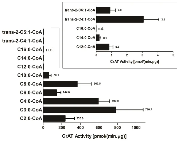
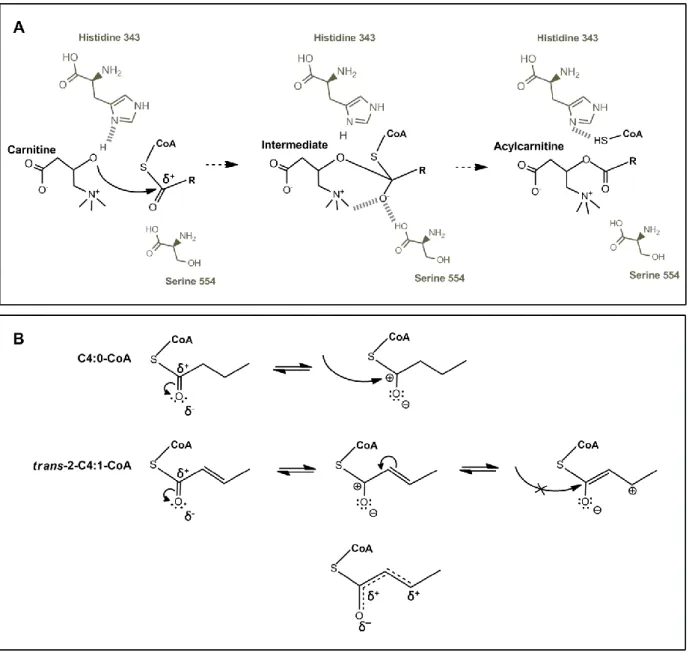
Deficiencies in the mFAO or BCAA oxidation pathways generally lead to the intramitochondrial accumulation of acyl-coenzyme A (CoA) esters. These acyl-CoAs are potentially toxic to the mitochondria and to the cell and are, therefore, readily conjugated with other molecules and converted into compounds that can then undergo mitochondrial and cellular export. The production of acylcarnitines is one of the mechanisms used to detoxify the mitochondria and the cell from the accumulating acyl-CoAs. Using in vitro techniques we have thoroughly studied the substrate specificity of two acyltransferases generally assumed to be responsible for the mitochondrial synthesis of acylcarnitines. We have shown that carnitine palmitoyltransferase 2 (CPT2) and carnitine acetyltransferase are responsible for the production of most of the acylcarnitines commonly found in mFAO disorders or deficiencies affecting the BCAA oxidation. Furthermore, we observed that trans-2-enoyl-CoAs are poor substrates for both acyltransferases and even act as CPT2 inhibitors, possibly by interfering with the catalytic mechanism of the enzyme. This may explain not only the absence of some of the respective trans-2-enoyl-carnitine intermediates from plasma acylcarnitine profiles of patients affected with a mFAO or BCAA oxidation disorder, but also the severity associated with such pathologies, as trans-2-enoyl-CoAs may additionally interfere with the metabolism of other compounds.
It was also important to clarify the mechanism by which acylcarnitnes are exported to the cytosol and out of the cell. Using cell lines with deficiencies in specific proteins associated with the carnitine transport and in the presence and absence of a deficiency in the medium-chain acyl-CoA dehydrogenase enzyme, induced by gene silencing, we have confirmed the in vitro results obtained for recombinant CPT2 and showed that, in intact cells, CPT2 is responsible for the production of medium-chain acylcarnitines. Moreover, we also observed that carnitine/acylcarnitine translocase (CACT) is the protein responsible for the mitochondrial export of medium-chain acylcarnitines (through the inner mitochondrial membrane) and that the cellular export of these intermediates does not seem to rely on the plasma membrane carnitine transporter, OCTN2. In the same context, the mechanism by which acylcarnitines cross the outer mitochondrial
xiv
membrane (OMM) is still far from being elucidated. It has been suggested that carnitine palmitoyltransferase 1 (CPT1) forms oligomeric complexes in the OMM. This would result in the formation of a hexameric structure that could function as a pore, facilitating the transport of acylcarnitines across the OMM. We have demonstrated that CPT1 does not seem to be crucial in the passage of acylcarnitines through the OMM for further oxidation within the mitochondria, or at least this is not the exclusive route for the access of acylcarnitines to the intermembrane space.
It is generally assumed that medium-chain fatty acids (MCFAs) are oxidized in mitochondria independently from carnitine. However, the true contribution of the carnitine shuttle to the oxidation of MCFAs has remained elusive. We show that lauric acid, a MCFA, also depends on the carnitine shuttle to be fully oxidized in the mitochondria. Furthermore, we observed that when the carnitine shuttle is impaired, this MCFA is able to undergo one or two cycles of peroxisomal β-oxidation. This shows that peroxisomes may have a more prominent role than earlier assumed in the oxidation of straight-chain fatty acids which are not typically metabolized in this cellular compartment, potentially acting as a compensatory mechanism when mFAO is compromised.
Finally, we show that carnitine and its biosynthetic pathway may have an important role in neurologic development. In patients with non-dysmorphic autism, we found a common deletion in exon 2 of the
TMLHE gene, encoding the first enzyme of the carnitine biosynthesis pathway, trimethyllysine dioxygenase.
This leads to the absence of protein and accumulation of trimethyllysine in the plasma and urine of patients. We have therefore characterized the first inborn error of carnitine biosynthesis and found that this defect is associated with non-dysmorphic autism. This suggests that TMLHE deficiency may be a risk factor for the development of autism spectrum disorders and that the maintenance of a proper carnitine homeostasis during development may be particularly important in these patients.
Altogether, the results obtained provide new insights on the function and metabolism of carnitine in health and disease. We established the origin and export route of the acylcarnitines that appear in the plasma of patients with mFAO or BCAA oxidation deficiencies and showed that CPT1 does not seem to be crucial for the mitochondrial import of these carnitine derivatives. We have also observed that the carnitine shuttle has a greater contribution to the oxidation of MCFAs than previously expected and that peroxisomes are able to oxidize these fatty acids in cases of mFAO impairment. Moreover, a new inborn error of metabolism is described in the carnitine biosynthesis pathway being its implications in autism spectrum disorders thoroughly discussed.
Keywords: Carnitine shuttle; Mitochondrial fatty acid β-oxidation; Acylcarnitines; Carnitine palmitoyltransferase 1; Carnitine palmitoyltransferase 2; Carnitine/acylcarnitine translocase; Carnitine biosynthesis
Sumário
A carnitina (ou L-carnitina) é um composto crucial no metabolismo energético. No Homem, grande parte da carnitina é proveniente da dieta, nomeadamente através da ingestão de carne e outros produtos de origem animal. No entanto, o organismo tem a capacidade de produzir carnitina endogenamente, um processo do qual indivíduos estritamente vegetarianos dependem para a obtenção de níveis adequados deste composto. A carnitina tem um papel fundamental no transporte de ácidos gordos de cadeia longa do citosol para a mitocôndria, local onde ocorre a β-oxidação.
Apesar da carnitina ser um composto amplamente estudado ao longo dos anos, alguns aspetos relativos ao seu metabolismo e função permanecem pouco claros. Assim, estudos planeados durante esta tese tiveram como objetivos: 1) elucidar a ação do ciclo da carnitina na eliminação de intermediários tóxicos que se acumulam em doenças da β-oxidação mitocondrial dos ácidos gordos e do metabolismo dos aminoácidos de cadeia ramificada, numa função inversa à do seu mecanismo fisiológico; 2) aprofundar o conhecimento acerca do mecanismo de translocação de acilcarnitinas através da membrana externa da mitocôndria; 3) clarificar a contribuição dos peroxissomas para a oxidação de ácidos gordos de cadeia média em casos de deficiência do ciclo da carnitina; 4) proporcionar uma nova perspetiva sobre a importância da carnitina e da sua biossíntese em perturbações do espetro do autismo.
As deficiências na β-oxidação mitocondrial dos ácidos gordos ou no metabolismo dos aminoácidos de cadeia ramificada levam, em geral, à acumulação intramitocondrial de ésteres de acil-coenzima A (acil-CoAs). Estes metabolitos são potencialmente tóxicos e, como tal, são degradados em vias alternativas tais como a ω e ω-1 oxidação, levando à produção de ácidos dicarboxílicos e/ou conjugação com outras moléculas sendo convertidos em acilglicinas ou acilcarnitinas, compostos que podem ser mais facilmente exportados da mitocôndria e da célula. A produção de acilcarnitinas é um dos mecanismos usados para a eliminação mitocondrial e celular dos acil-CoAs que se acumulam devido ao bloqueio enzimático. A especificidade de substrato das duas aciltransferases, tidas geralmente como responsáveis pela síntese mitocondrial de acilcarnitinas, foi extensivamente estudada usando técnicas in vitro. Os resultados obtidos demonstraram que as enzimas carnitina palmitoiltransferase 2 (CPT2) e carnitina acetiltransferase são responsáveis pela produção de grande parte das acilcarnitinas encontradas no plasma de doentes com défices ao nível da β-oxidação mitocondrial dos ácidos gordos e do metabolismo dos aminoácidos de cadeia ramificada. Por outro lado, verificou-se que os intermediários trans-2-enoil-CoAs não são bons substratos para estas aciltransferases, actuando mesmo como inibidores da CPT2. Esta inibição poderá dever-se a uma interferência destes compostos com o mecanismo catalítico da enzima. Estes dados poderão explicar, não só a ausência de trans-2-enoilcarnitinas nos perfis metabólicos em alguns casos de defeitos genéticos ou adquiridos onde se prevê a acumulação dos respetivos ésteres de CoA, mas também a gravidade de
xvi
algumas destas patologias, uma vez que os trans-2-enoil-CoAs poderão ainda interferir com o metabolismo de outros compostos.
De modo a confirmar os estudos in vitro e a clarificar o mecanismo de exportação de acilcarnitinas para o citosol e para o plasma, recorreu-se ao uso de linhas celulares deficientes em proteínas específicas associadas ao transporte de carnitina com e sem silenciamento do gene que codifica para a desidrogenase dos ésteres acil-CoA de cadeia média. Desta forma, foi possível induzir a acumulação intramitocondrial de acil-CoAs, confirmando os resultados obtidos anteriormente para a CPT2. Demonstrou-se que na ausência desta enzima não há produção de acilcarnitinas de cadeia média após a acumulação do respetivo acil-CoA dentro da mitocôndria. Os resultados demonstraram ainda que o transportador carnitina/acilcarnitina translocase (CACT) é responsável pela exportação mitocondrial de acilcarnitinas de cadeia média através da membrana interna da mitocôndria e que a exportação celular destes intermediários não parece depender do transportador de carnitina da membrana plasmática, OCTN2. Do mesmo modo, o mecanismo pelo qual as acilcarnitinas atravessam a membrana externa da mitocôndria permanece por elucidar. Estudos apresentados na literatura sugerem que a enzima carnitina palmitoiltransferase 1 (CPT1) tem a capacidade de se organizar em estruturas oligoméricas que conduzem à formação de complexos hexaméricos na membrana externa da mitocôndria. É ainda sugerido que esta potencial oligomerização da proteína origina uma estrutura semelhante a um poro que poderá facilitar o transporte de acilcarnitinas através da membrana externa da mitocôndria. Os resultados obtidos durante este trabalho revelaram, contudo, que a CPT1 não parece ser crucial na passagem de acilcarnitinas através da membrana externa da mitocôndria para posterior oxidação na matriz mitocondrial ou, pelo menos, esta não parece ser uma via exclusiva para o acesso das acilcarnitinas ao espaço intermembranar.
O papel dos peroxissomas na oxidação de ácidos gordos de cadeia linear média ou longa não é claro. É geralmente aceite que estes substratos são totalmente oxidados na mitocôndria. Em particular, assume-se que os ácidos gordos de cadeia média entram na mitocôndria essencialmente por difusão, ao contrário dos ácidos gordos de cadeia longa que requerem o ciclo da carnitina para aceder à matriz mitocondrial. Os dados obtidos demonstraram que o ácido láurico, normalmente assumido como sendo um ácido gordo de cadeia média, também depende do ciclo da carnitina para ser completamente oxidado na mitocôndria. Adicionalmente foi observado que, em casos de deficiência nas proteínas que compõem o ciclo da carnitina, o ácido láurico é direcionado para os peroxissomas sofrendo pelo menos um ciclo de β-oxidação peroxissomal. Estes resultados sugerem que, ao contrário do que tem sido aceite pela comunidade científica, os peroxissomas têm uma função importante na oxidação de ácidos gordos que não são tipicamente oxidados neste compartimento celular, podendo actuar como um mecanismo compensatório quando a β-oxidação mitocondrial dos ácidos gordos está comprometida.
Por fim, foi demonstrado que a carnitina e a sua biossíntese podem ter um papel relevante em distúrbios neurológicos. Em doentes com autismo não-dismórfico foi encontrada uma deleção no exão 2 do gene
TMLHE, que codifica para a primeira enzima da biossíntese da carnitina, a dioxigenase da trimetil-lisina.
Esta deleção conduz à ausência de proteína, bem como à acumulação de trimetil-lisina e deficiência em γ-butirobetaína no plasma e na urina dos doentes. Deste modo, os resultados obtidos permitiram a caracterização do primeiro erro hereditário da biossíntese da carnitina. Foi ainda observada uma associação desta deficiência genética ao nível do gene TMLHE com o autismo não-dismórfico. Estes dados indicam que a deficiência em TMLHE pode ser um fator de risco para o desenvolvimento de doenças do espetro do autismo e que a manutenção da homeostase da carnitina durante o desenvolvimento pode ser particularmente importante nestes doentes.
De um modo geral, os resultados obtidos durante este trabalho proporcionam uma nova perspetiva sobre a função e o metabolismo da carnitina em situações fisiológicas ou em casos de doença. Foi possível estabelecer a origem das acilcarnitinas presentes no plasma de doentes com alterações genéticas ou adquiridas da β-oxidação mitocondrial dos ácidos gordos ou do metabolismo dos aminoácidos ramificados e demonstrou-se que a CPT1 não parece ser crucial para a importação mitocondrial destes metabolitos. Observou-se ainda que o ciclo da carnitina apresenta um papel mais importante na oxidação dos ácidos gordos de cadeia média do que até agora assumido e que os peroxissomas podem actuar como um mecanismo compensatório nos casos em que a β-oxidação mitocondrial dos ácidos gordos está comprometida, aceitando como substratos ácidos gordos que não podem ser oxidados na mitocôndria. Por fim, a descoberta de um novo erro hereditário da biossíntese da carnitina permite refletir sobre as suas implicações em perturbações do espetro do autismo.
Palavras chave: Ciclo da carnitina; β-oxidação mitocondrial; Acilcarnitinas; Carnitina palmitoiltransferase 1; Carnitina palmitoiltransferase 2; Carnitina/acilcarnitina translocase; Biossíntese da carnitina
Chapter 1
Carnitine is a crucial compound in fatty acid oxidation and energy metabolism. In the oxidation of fatty acids, especially the long-chain species, carnitine and the carnitine shuttle have a pivotal role in the transport of the esterified acyl units into mitochondria in the form of acylcarnitines. Once in the matrix of this organelle, acylcarnitines are reconverted to acyl-coenzyme A esters (acyl-CoAs), the true substrates of β-oxidation. Although formal proof is lacking, some of the reactions of the carnitine shuttle, mediated by carnitine palmitoyltransferase 1 (CPT1), carnitine/acylcarnitine translocase (CACT) and carnitine palmitoyltransferase 2 (CPT2), are generally assumed to be reversible and, simultaneously, responsible for the export of acylcarnitines to the cytosol. These intermediates are further exported across the cell membrane into the plasma compartment. Presently, plasma acylcarnitine profiling is an essential tool for the early diagnosis of inborn errors of metabolism, namely those affecting fatty acid and branched-chain amino acid oxidation. Nevertheless, the origin of these plasma acylcarnitines has never been thoroughly elucidated. Facing this, the main goals of this thesis were to clarify the mechanisms underlying the mitochondrial synthesis of acylcarnitines and their mitochondrial and cellular export routes, as well as their import into the mitochondrial matrix. Moreover, we aimed to clarify the contribution of the carnitine shuttle and peroxisomes to the oxidation of straight medium-chain fatty acids and additionally, give a new insight on the carnitine biosynthesis pathway.
In Chapter 2 a detailed review of the state-of-the-art concerning carnitine metabolism and function is presented. This information is complemented by an overview of the mitochondrial and peroxisomal fatty acid β-oxidation and branched-chain amino acid oxidation pathways, as well as the disorders associated. With this background important questions were raised and motivated our studies: What are the enzymes responsible for the synthesis of acylcarnitines and what is their substrate specificity? Is the plasma acylcarnitine profiling found in patients with a fatty acid β-oxidation or branched-chain amino acid oxidation defect a reflection of the intramitochondrial acyl-CoA accumulation which occurs as a result of the blockage upon these pathways? In Chapters 3 and 4 we aimed to resolve the origin of the acylcarnitines commonly found in these patients. The involvement of CPT2 and carnitine acetyltransferase (CrAT) in this mechanism is generally assumed, but a comprehensive study of the substrate specificity profile of these enzymes has never been performed. With this purpose, detailed in vitro studies were performed on the substrate specificity of these key human acyltransferases using homogenates of Saccharomyces cerevisiae overexpressing human CPT2 and purified recombinant human CrAT obtained from Escherichia coli.
The work reported in Chapter 5 aimed to confirm the in vitro results described in Chapter 3 and to answer the following additional questions: What are the players involved in the mitochondrial and cellular export of acylcarnitines? Is it possible to follow the detoxification path after inducing acyl-CoA accumulation within the mitochondria? To further understand the export route of these acylcarnitines, this chapter describes an
4
intact cell model developed from patient cell lines deficient in specific proteins potentially involved in the mitochondrial synthesis of carnitine derivatives and in their mitochondrial and cellular export. In these cell lines, a knockdown of the medium-chain acyl-CoA dehydrogenase (MCAD) gene was introduced in order to mimic the intramitochondrial accumulation of medium-chain acyl-CoAs, as commonly observed in MCAD deficiency.
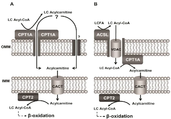
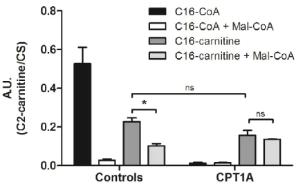
The transport of acylcarnitines into mitochondria across the outer mitochondrial membrane has also remained unclear so far. Carnitine palmitoyltransferase 1 has been proposed to play a role in mediating the transport of long-chain acylcarnitines into the intermembrane space. The work presented in Chapter 6, where a patient cell line not expressing CPT1 was used, aimed to clarify whether this enzyme is indeed the exclusive player in the import of acylcarnitines into the mitochondria, or if these metabolites may gain access to the inner mitochondrial membrane transporter CACT through an alternative route, which facilitates the transport of long-chain acylcarnitine derivatives into the matrix.
Contrary to long-chain fatty acids that rely entirely on the carnitine shuttle for their degradation within the mitochondria, it is generally accepted that the oxidation of medium-chain fatty acids is carnitine independent. Moreover, the role of peroxisomes in the oxidation of medium- and long-chain fatty acids has remained elusive. Using as a model cell lines from patients with deficiencies in the members of the carnitine shuttle or in crucial proteins for peroxisome biogenesis, in Chapter 7 we aimed to understand the contribution of the carnitine shuttle and peroxisomes to the oxidation of medium-chain fatty acids.
In Chapter 8 some light was shed on the importance of the biosynthesis of the carnitine moiety itself. In fact, in situations such as starvation and vegetarian diet, the body totally depends on the adequate function of the proteins involved in the carnitine biosynthetic pathway. Deficiencies in the enzymes contributing to the endogenous formation of carnitine have never been reported and thus, the implications of an impaired carnitine biosynthesis are so far unknown. In this chapter, a novel inborn error of metabolism affecting the biosynthesis of carnitine is described in children with non-dysmorphic autism. Making use of genetic and biochemical studies the work reported in Chapter 8 aimed primarily to find a correlation between this deficiency in the carnitine metabolism and autism spectrum disorders and ultimately to discuss the potential role of carnitine in the mechanisms of neurologic disease.
The final chapter comprises the concluding remarks of the thesis where the results obtained during its development and their impact in carnitine metabolism and function are discussed. Chapter 9 also includes a reflection on the aspects that still demand further attention and some perspectives for future work.
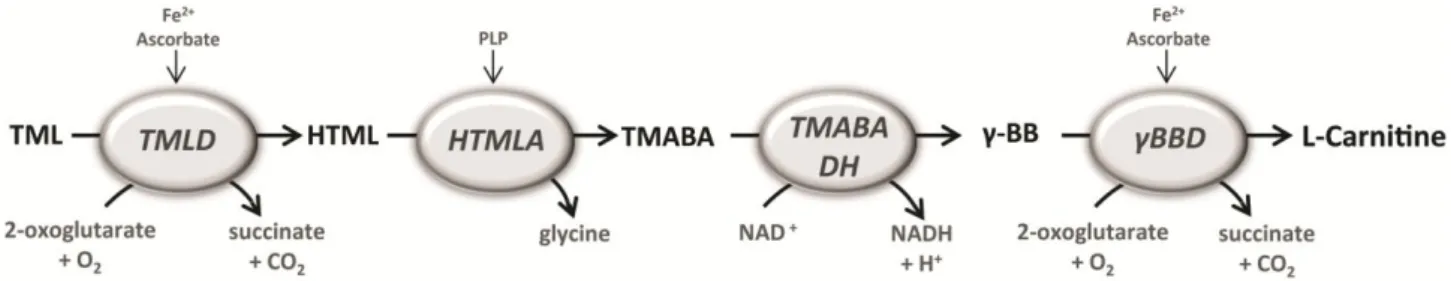
Chapter 2
Figure 1 – Schematic representation of the carnitine biosynthesis pathway. 6-N-trimethyllysine (TML) residues are
hydroxylated by TML-dioxygenase (TMLD) to 3-hydroxy-TML (HTML). HTML is converted to 4-trimethylaminobutyraldeyde (TMABA) by HTML aldolase (HTMLA), which uses pyridoxal 5’-phosphate (PLP) as cofactor. TMABA-dehydrogenase (TMABA-DH) oxidizes TMABA to 4-N-trimethylaminobutyrate (butyrobetaine; γ-BB), which is hydroxylated to carnitine by γ-BB-dioxygenase (γ-BBD).
1. Carnitine function
Carnitine (L-3-hydroxy-4-N,N,N-trimethylamino-butyrate; L-carnitine) is a quaternary ammonium compound crucial in energy metabolism, described as a conditionally essential nutrient [1, 2]. It has a primary role in the transport of activated long-chain fatty acids from the cytosol to the mitochondrial matrix, where β-oxidation takes place [3]. Carnitine is also used in the transfer of peroxisomal β-oxidation intermediates and products to mitochondria to be completely metabolized through β-oxidation and/or the Krebs cycle [4, 5]; in the modulation of the intramitochondrial acyl-CoA/CoA ratio [6, 7]; and as a detoxifying agent facilitating the elimination from the cell of accumulating acyl units in the form of carnitine esters [8]. In humans, about 75% of carnitine is obtained from the diet. L-carnitine, the biologically active stereoisomer, is found essentially in meat and other animal products being very limited in plants [2, 9]. Therefore, in strictly vegetarian individuals, the amount of carnitine derived from the diet is quite low and the body mostly relies on its endogenous synthesis.
1.1 Carnitine biosynthesis
The biosynthesis of L-carnitine occurs in kidney, liver and brain from the amino acids lysine and methionine [9]. The carbon backbone of carnitine is provided by lysine while methionine is the donor of the 4-N-methyl groups [10-12]. The N-methylation of lysine residues on the 6-amino group occurs as a post-translational event in certain proteins [13, 14]. This reaction is catalyzed by specific methyl-transferases that use S-adenosylmethionine as the methyl donor to form 6-N-trimethyllysine (TML)
residues [15]. Protein hydrolysis in lysosomes results in the release of TML, the first metabolite in the biosynthesis of carnitine [16].
After lysosomal degradation, TML residues undergo four enzymatic reactions before the formation of L-carnitine (Fig. 1). The first reaction corresponds to the hydroxylation of TML on the third position by the enzyme trimethyllysine dioxygenase (TMLD), encoded by the TMLHE (trimethyllysine hydroxylase, epsilon) gene, which maps to the long arm of the X chromosome. The resultant 3-hydroxy-6-N-trimethyllysine (HTML) undergoes an aldolytic cleavage to yield 4-trimethylaminobutyraldeyde (TMABA) and glycine in a reaction catalyzed by HTML aldolase (HTMLA). TMABA is oxidized by TMABA dehydrogenase (TMABA-DH) which results in the formation of 4-N-trimethylaminobutyrate (γ-butyrobetaine; γ-BB). Finally, γ-BB is hydroxylated to L-carnitine by the enzyme γ-butyrobetaine dioxygenase (γ-BBD) [16].
1.2 Carnitine homeostasis
In humans, exogenous TML does not function efficiently as a precursor for carnitine biosynthesis. Loading tests have shown that after ingestion of TML, most of the compound (approximately 75%) is excreted unchanged in the urine [9, 17]. Tissues such as the heart and muscle are not able to efficiently absorb TML from the circulation and it is believed that the TML produced intracellularly is converted to BB in the tissue of origin [9]. The γ-BB produced is then secreted into circulation and readily absorbed by the tissues that contain γ-BBD (liver, kidney and brain) [18], where final conversion to carnitine takes place. The carnitine produced is further transported through the blood into the
8
tissues where it is needed for fatty acid oxidation. Its cellular uptake is mediated by the plasmalemmal high affinity organic cation/carnitine transporter (OCTN2) and other low affinity transporters present in the plasma membrane.
Carnitine homeostasis is maintained by a modest rate of endogenous synthesis, absorption from dietary sources, and efficient tubular reabsorption by the kidney. However, urinary carnitine excretion largely depends on the diet and the kidney has been shown to adapt to a higher carnitine intake by reducing the efficiency of carnitine reabsorption [19, 20].
1.3 The cellular uptake of carnitine: the organic cation/carnitine transporter
The plasmalemmal organic cation/carnitine transporter – OCTN2 – is a member of the solute carrier family 22 of sodium-dependent organic cation/carnitine transporters (SLC22A5). The
SLC22A5 gene, which encodes in humans the
OCTN2 transporter, is mapped to chromosome 5 (5q31) [21, 22]. OCTN2 transports carnitine with high affinity in a sodium-dependent manner [22, 23]. This transporter is also responsible for the sodium-independent transport of other organic cations [24, 25]. Human OCTN2 contains 557 amino acids organized in 12 putative transmembrane domains with the N- and C-termini on the cytoplasmic side [21]. In humans, OCTN2 is expressed in several tissues including placenta, skeletal muscle, heart and kidney. In kidney, it is localized on the apical membrane of renal tubular epithelial cells, which demonstrates the importance of OCTN2 for reabsorption of carnitine after glomerular filtration [26]. OCTN2 has a marked homology to the low affinity sodium-independent carnitine transporter OCTN1, as well as with OCTN3 which is only known from rat and mouse [22, 27]. The OCTN2 transporter presents a higher affinity for carnitine and short-chain acylcarnitines as substrates but it is also efficient in facilitating the cellular uptake of betaine [28, 29]. The nuclear receptor peroxisome proliferator activated receptor alpha (PPARα) regulates the expression of several genes involved in metabolism and has been described to up-regulate as well the usually low OCTN2 expression in mouse liver, leading to an
increase in the hepatic transport of carnitine and of its precursor γ-BB [30, 31].
1.4 The mitochondrial uptake of carnitine: the carnitine/acylcarnitine transporter
The carnitine/acylcarnitine translocase (CACT) is an inner mitochondrial membrane (IMM) transporter, mediating the electroneutral exchange between acylcarnitine esters and free carnitine [32-34], as well as the unidirectional transport of free carnitine into the mitochondrial matrix [35]. This transporter, with 301 amino acid residues, is a member of the solute carrier family 25 (SLC25A20) and the human gene, cloned and sequenced by Huizing et al [36] is located on chromosome 3 (3p21.31) [37]. Expression of CACT in mouse liver is up-regulated by PPARα and PPARδ [38]. Similarly to other mitochondrial carriers, CACT displays three repeated homologous regions, being its monomer likely organized into six transmembrane alpha-helixes linked asymmetrically by five hydrophilic loops with the N-terminus, the C-terminus and two small loops facing the intermembrane space and three extensive loops exposed to the matrix side [39-41]. Functional properties of CACT have been investigated in isolated rat liver mitochondria after purification from living tissues [34, 42] or as a recombinant protein reconstituted in liposomes [43-45]. Both rat and human transporters accept acylcarnitines from 2 to 18 carbon atoms with higher affinity for the long-chain species [42-45]. Differences in the Km values on the cytosolic and on the matrix side imply that the preferential reaction is the transport of acylcarnitines from the intermembrane space into the matrix [46, 47]. Despite the information gathered towards the rat CACT, knowledge on the structure and the kinetic properties of the human homologue is still limited [44, 45].
1.5 The carnitine acyltransferases
Carnitine acyltransferases are a group of enzymes that catalyze the transfer of acyl groups between coenzyme A (CoA) and carnitine, being crucial in the metabolism of fatty acids. The carnitine acyltransferases’ family includes four members: carnitine acetyltransferase, carnitine octanoyltrans-ferase, carnitine palmitoyltransferase 1 and carnitine palmitoyltransferase 2.
1.5.1 The carnitine acetyltransferase
Carnitine acetyltransferase (CrAT) is a soluble enzyme present in peroxisomes, mitochondria and in the endoplasmic reticulum [7, 48, 49]. It is active with acyl groups with a chain-length of 2 to 10 carbon atoms [50-53]. The mitochondrial and peroxisomal enzymes contain about 600 amino acids and differ only in the initial mitochondrial targeting signal [54]. CrAT is widely distributed in different tissues [55, 56] and the corresponding gene (CRAT) is localized to the chromosome 9 (9q34.1) [57]. In mitochondria, CrAT has an important role in the maintenance of the acyl-CoA/CoA pool [7, 8] and in the regulation of the mitochondrial energetic status [56]. The production of acetylcarnitine by CrAT has been associated with positive effects in several models of neuronal dysfunction [58-62], namely through the redirection of excess carbon equivalents to tissues with less ability to use long-chain fatty acids (such as the brain) [56]. Furthermore, it has been recently described that CrAT has an impact in whole body glucose homeostasis due to its role in allowing tissue efflux of acetylcarnitine derived from the pyruvate dehydrogenase complex (PDHc) in a mouse muscle-specific CrAT knockout model [55]. The structural studies performed with the human CrAT and the resolution of the 3D structure of this enzyme were major advances in the understanding of the catalytic mechanism of acyltransferases as a whole [54, 63].
1.5.2 The carnitine octanoyltransferase
Carnitine octanoyltransferase (CrOT) is found specifically in the peroxisome [64, 65] and this 612 amino acid protein is encoded by the gene CROT, which has been localized to the chromosome 7 (7q21.1) [66]. It has preference for substrates with medium-chain length (between 6 and 10 carbon atoms) but it also handles long-chain acyl-CoAs, although to a lesser extent [51]. Very long-chain, branched-chain and long-chain dicarboxylic fatty acids undergo β-oxidation in the peroxisomes. The medium-chain acyl-CoAs that result from this oxidation are converted to carnitine esters by CrOT and exported to the cytosol. These intermediates are further imported into the mitochondria for complete oxidation [7]. CrOT has 36% amino acid sequence identity with CrAT, and like CrAT its
crystal structure has been determined [67]. Significant differences are observed in the acyl-CoA binding site of these two enzymes, which in CrOT is larger and thus more suited for medium-chain acyl groups [67]. CrOT has been implicated in the regulation of ketogenesis due to its role in the transfer of shortened fatty acids from peroxisomes to mitochondria [68]. Furthermore, it has been recently suggested that CrOT may regulate very long-chain fatty acid metabolism through alteration of the amount of medium-chain fatty acids produced in the peroxisome [69].
1.5.3 The carnitine palmitoyltransferase 1 Integrated in the outer mitochondrial membrane (OMM), carnitine palmitoyltransferase 1 (CPT1) catalyzes the transesterification reaction between long-chain acyl-CoA and acylcarnitine esters. Three isoforms are known to date: CPT1A (773 amino acids) expressed ubiquitously with high levels in liver and kidney, CPT1B (772 amino acids) mainly present in cardiac and skeletal muscle, and CPT1C (798 amino acids) expressed in brain [7, 70]. These isoforms are encoded by different genes localized on the chromosome 11 (11q13), 22 (22qter) and 19 (19q13), respectively for CPT1A, CPT1B and CPT1C [70, 71]. The reaction catalyzed by CPT1 is considered the rate limiting step of mitochondrial fatty acid β-oxidation and its activity is strongly regulated by malonyl-CoA [72], the first intermediate of the fatty acid biosynthesis. CPT1 has two transmembrane domains connected by a loop protruding to the intermembrane space (IMS) and both N- and C-termini appear to be projected to the cytosol [73], although this remains a matter of discussion [74]. Interactions between the N- and the C-terminus help to stabilize the catalytic site [74, 75] and seem to modulate malonyl-CoA sensitivity, even though the mechanism is still poorly understood [76-78].
It has been suggested that the transmembrane domain 2 of CPT1A is responsible for the oligomerization of the enzyme, which would result in the formation of hexameric complexes potentially arranged as a pore within the OMM [79, 80]. Moreover, CPT1A has recently been implicated in the formation of hetero-oligomeric complexes in the OMM with long-chain acyl-CoA synthetase (ACSL) and the voltage dependent anion channel
10
(VDAC) [74]. However, the functional relevance of such complexes remains unknown.
1.5.4 The carnitine palmitoyltransferase 2 Carnitine palmitoyltransferase 2 (CPT2) contains 658 amino acid residues and is associated with the inner face of the IMM having only one isoform, ubiquitously expressed [7]. The gene encoding this homotetrameric protein locates in humans on chromosome 1 (1p32) [81]. This enzyme is malonyl-CoA insensitive and its main function is to catalyze
the conversion of long-chain acylcarnitines into the respective acyl-CoA esters in the presence of CoA, providing the substrates for β-oxidation. Studies on the crystal structure of rat CPT2 have shown an insert that serves as a membrane anchor for the enzyme [82]. It is also suggested that this insert could physically interact with CACT, allowing the direct channeling of acylcarnitine substrates to the active site of CPT2 [82-84]. Studies on human CPT2 show higher activity towards medium and long-chain substrates, its specificity ranging from C8 to C20 carbon chain [85]. Furthermore, CPT2 has been
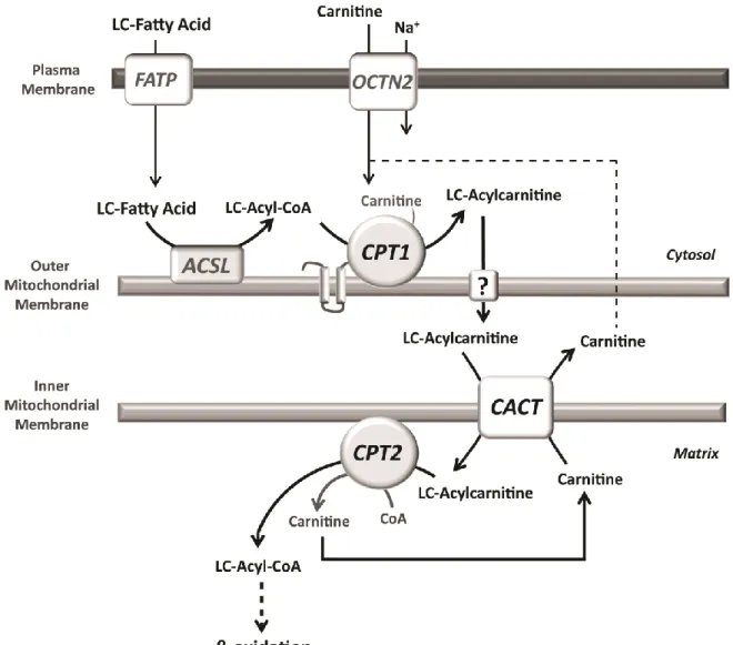
Figure 2 - Schematic representation of the carnitine shuttle. Long-chain fatty acids (LCFA) enter the cell via various
plasmalemmal transporters namely the fatty acid transport protein (FATP), being activated to coenzyme A (CoA) esters in the cytosol by the long-chain acyl-CoA synthetase (ACSL). Carnitine palmitoyltransferase 1 (CPT1) converts the long-chain (LC) acyl-CoAs into the respective acylcarnitines, which are transported into the mitochondrial matrix by the carnitine/acylcarnitine translocase (CACT) in exchange with free carnitine. In the matrix, carnitine palmitoyltransferase 2 (CPT2) reconverts the LC-acylcarnitines into acyl-CoA esters, the true substrates for β-oxidation.
described to reverse its physiological mechanism in
vitro and accept acyl-CoA esters as substrates
converting them into the corresponding acylcarnitines [85, 86]. Pharmacological up-regulation of CPT2 might be achieved through the action of PPAR agonists such as bezafibrates [87]. These compounds seem to enhance CPT2 gene expression and CPT2 residual activity, restoring long-chain fatty acid oxidation capacities in mild cases of CPT2 deficiency [88].
1.6 The carnitine shuttle and regulation of carnitine transfer
In order to be metabolized in the mitochondria, long-chain fatty acids must undergo activation by long-chain acyl-CoA synthetases prior to its transport into the mitochondrial matrix [89-91]. Transport of activated long-chain fatty acids (long-chain acyl-CoAs) into the mitochondrial matrix is carried out by the carnitine shuttle (Fig. 2). This system requires the presence of L-carnitine and is composed of the acyltransferases CPT1 and CPT2 as well as the mitochondrial carrier CACT. Long-chain acyl-CoAs are firstly converted into the corresponding carnitine esters by CPT1. The resulting acylcarnitines are then transported across the IMM by CACT, in exchange with free carnitine. The final step of this cycle, catalyzed by CPT2, reconverts the acylcarnitines into the respective acyl-CoA esters that can then undergo β-oxidation. Carnitine returns to the cytosol as free carnitine for another cycle or it can be exported out of the cell in the form of acylcarnitines [3], potentially through the reversible activity of CPT2 and CACT.
2. Mitochondrial fatty acid β-oxidation
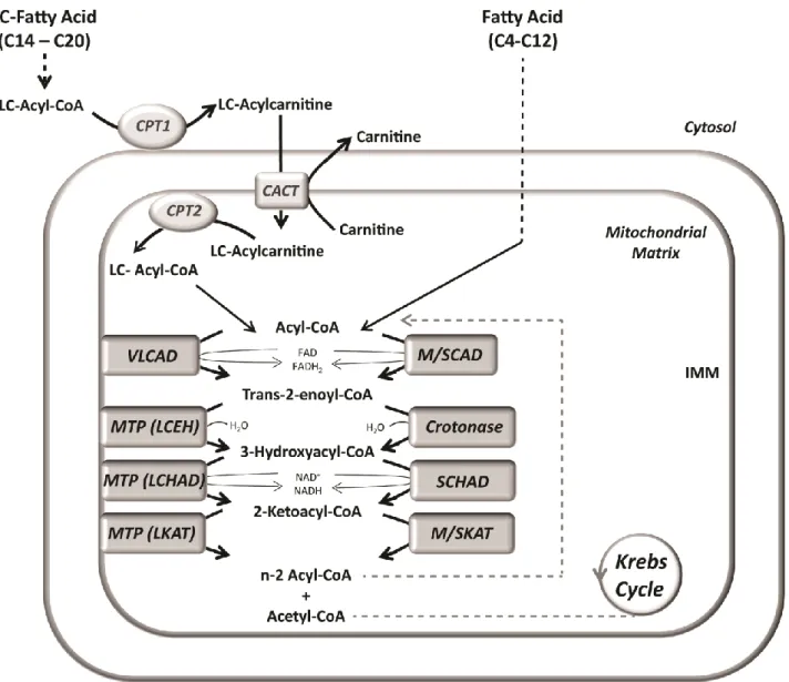
Once in the mitochondria, acyl-CoA esters enter the β-oxidation pathway (mFAO), a four-step mechanism where the β-carbon of the acyl unit is oxidized, being the acyl-CoA shortened in two-carbon atoms which are released as acetyl-CoA. Each of these steps is catalyzed by chain-length specific enzymes [3] (Fig. 3). In the first step the acyl-CoA is dehydrogenated, yielding a trans-2-enoyl-CoA intermediate. Depending on the length of the carbon chain, this reaction is catalyzed by specific acyl-CoA dehydrogenases: short- (SCAD for C4-C6 CoAs), medium- (MCAD for C4-C12 acyl-CoAs), long- (LCAD for C8-C20 acyl-CoAs) or very
long-chain acyl-CoA dehydrogenase (VLCAD for C12-C24 acyl-CoAs). However, the exact role of LCAD in mitochondrial β-oxidation is still quite unclear. It has been shown that this enzyme is not only capable of handling straight-chain fatty acids, but also branched-chain substrates such as 2,6-dimethylheptanoyl-CoA [92] and several un-saturated fatty acids [93, 94]. All acyl-CoA dehydrogenases are localized in the mitochondrial matrix, with exception of VLCAD which is loosely bound to the inner mitochondrial membrane. VLCAD is also distinguishable from the other enzymes in that it is a dimer, whereas the other acyl-CoA dehydrogenases are homotetramers [95]. Each subunit contains one molecule of non-covalently bound flavin adenine dinucleotide (FAD) which accepts the electrons involved in the dehydrogenation reaction and is further reoxidized by the electron-transfer flavoprotein (ETF), via which these electrons are transferred to the respiratory chain [96, 97]. The second step of the β-oxidation pathway is the hydratation of the trans-2 double bond of the enoyl-CoA intermediate, leading to the formation of a 3-hydroxyacyl-CoA. This step is catalyzed by two different enoyl-CoA hydratases: short-chain enoyl-CoA hydratase (crotonase) and long-chain enoyl-CoA hydratase (LCEH). Crotonase is active with fatty acyl units of chain-length up to 10 carbons and LCEH handles the long-chain enoyl-CoA intermediates [98]. LCEH is part of the mitochondrial trifunctional protein (MTP), a heterooctamer associated with the IMM also carrying long-chain hydroxyacyl-CoA and 3-ketoacyl-CoA thiolase activities [98, 99]. A second dehydrogenation step is catalyzed by two different NAD+-dependent enzymes with 3-hydroxyacyl-CoA dehydrogenase activity (specific for short- and long-chain substrates) yielding 3-ketoacyl-CoA intermediates and NADH, the latter being further reoxidised by the complex I of the respiratory chain [100]. Short-chain 3-hydroxyacyl-CoA dehydro-genase (SCHAD) has broad substrate specificity (C4 to C16) but shows optimal activity with C6 substrates. The other enzyme, long-chain 3-hydroyacyl-CoA dehydrogenase (LCHAD), also part of the MTP complex, has higher activity towards long-chain intermediates [101]. In the final step of the β-oxidation cycle the 3-ketoacyl-CoA undergoes a thiolytic cleavage catalyzed by short- (SKAT),
12
Figure 3 - Schematic representation of the mitochondrial fatty acid β-oxidation. The first step of the β-oxidation
pathway is catalyzed by specific acyl-CoA dehydrogenases, depending on the chain-length of the substrate: very long- chain acyl-CoA dehydrogenase (VLCAD) for long-chain (LC) substrates and medium- or short-chain acyl-CoA dehydrogenase (M/SCAD), for medium- and short-chain acyl-CoA esters. The next reaction is catalyzed by long-chain enoyl-CoA hydratase (LCEH) or short-chain enoyl-CoA hydratase (crotonase) for long- and short-chain substrates, respectively. LCEH is part of the mitochondrial trifunctional protein (MTP), a heterooctamer associated with the inner mitochondrial membrane (IMM also including long-chain 3-hydroxyacyl-CoA dehydrogenase (LCHAD) and long-chain 3-ketoacyl-CoA thiolase (LKAT) activities. The third reaction is catalyzed by LCHAD or short-chain 3-hydroxyacyl-CoA dehydrogenase (SCHAD). Finally, an acyl-CoA with the chain shortened by 2 carbon atoms and acetyl-CoA are produced by either long-, medium- or, short-chain 3-ketoacyl-CoA thiolase (LKAT, MKAT and SKAT, respectively) and the new acyl-CoA can enter another cycle of β-oxidation. FAD, flavine adenine dinucleotide (oxidized); FADH2, flavine
adenine dinucleotide (reduced); NAD+, nicotinamide adenine dinucleotide (oxidized); NADH, nicotinamide adenine dinucleotide (reduced).
medium- (MKAT) or long-chain 3-ketoacyl-CoA thiolase (LKAT, which is also part of MTP), forming an acyl-CoA, with the chain shortened by two carbon atoms, and acetyl-CoA. The resultant acyl-CoA will undergo a new cycle of β-oxidation and the acetyl-CoA can either enter the Krebs cycle or be used to produce ketone bodies in the liver or kidney [100].
2.1 Mitochondrial fatty acid β-oxidation disorders Mitochondrial fatty acid β-oxidation disorders (mFAOD) are a clinically and biochemically heterogeneous group of rare inherited metabolic diseases, with an incidence of approximately 1:10,000 to 1:50,000 births [102, 103]. Defects in one or more enzymes of the β-oxidation pathway frequently lead to severe deficiencies. The first
inborn error of long-chain fatty acid oxidation reported was CPT2 deficiency and only about ten years later, in 1982, MCAD deficiency was identified [104]. Since then, inherited defects of several enzymes directly involved in this process have been identified in humans. Affected patients usually present hypoketotic hypoglycemia with hepatic, cardiac and/or muscular symptoms. In specific cases, such as MTP and CPT2 deficiencies, patients might also develop peripheral neuropathy. Biochemical manifestations of these disorders comprise deficient production of acetyl-CoA and ketone bodies, and consequently an energy deficit, and accumulation of free fatty acids, acyl-CoA and acylcarnitine intermediates upstream of the enzymatic block [3], which contribute to the pathogenesis of the various mitochondrial fatty acid β-oxidation disorders.
2.1.1 Defects of fatty acid transport
These disorders include primary systemic carnitine deficiency, CPT1, CPT2 and CACT deficiencies. Primary systemic carnitine deficiency: This pathology has been associated with a defect at the level of the plasma membrane carnitine transporter OCTN2 [105]. Symptoms usually present in infancy and include muscle weakness and cardiomyopathy. This deficiency is characterized by low total free carnitine in plasma, typically less than 5 µmol/L (normal values range from 25 to 50 µmol/L) [106]. The frequency reported is approximately 1:50,000, with about 1% of the population being a carrier for this disease [106, 107]. Treatment with oral carnitine (starting at 50 mg/kg/day) reverses the symptoms and prevents acute crises and cardiomyopathy [106, 108].
CPT1 deficiency: Deficiency at the level of CPT1 generally presents between birth and 18 months of age with altered mental status and low levels of long-chain acylcarnitines. Biochemical findings include non-ketotic hypoglycemia, mild hyperammonemia, elevated liver function tests and elevated free fatty acids, among others. Furthermore, in contrast with other mFAOD, CPT1 deficiency is consistently associated with elevated plasma carnitine levels [109]. This is a rare disorder with only about 30 cases known to date [110, 111], which is consistent with the crucial role of the CPT1
enzyme in fatty acid metabolism. Despite the small number of cases reported, several mutations have been identified in CPT1 deficient patients (for review see [112]), some of which show a fair correlation between the severity of the enzymatic impairment caused by the mutation and the clinical presentation [113]. However, only the deficiency of the liver type (CPT1A) has been reported in humans, likely because the importance of CPT1 in heart and brain might turn deficiencies in the CPT1B and CPT1C isoforms incompatible with life. Treatment includes the maintenance of high glucose levels in plasma through glucose infusions during the neonatal period and in acute metabolic crisis, as well as a medium-chain triglycerides (MCT) enriched diet [109, 112].
CPT2 deficiency: This is one of the most common inherited disorders of fatty acid metabolism [114, 115]. Three clinical phenotypes can be distinguished including neonatal, infantile and adult presentations. The adult form, firstly identified in 1973 [116], is the most common and usually presents in young adults with recurrent myoglobinuria. The infantile form generally presents in early childhood with fasting-induced hypoketotic hypoglycemia, liver failure, cardiomyopathy, and peripheral neuropathy. CPT2 deficiency is treatable if diagnosed early, but otherwise it is potentially fatal. The perinatal form is the least common clinical presentation of CPT2 deficiency and is usually fatal [115, 117]. Elevated long-chain acylcarnitine species and low carnitine levels are observed. Current treatment involves dietary fat restriction and increased intake of carbohydrates [118]. Pharmacological treatment with bezafibrates in patients with the muscular form of CPT2 deficiency has proved beneficial with significant improvements in exercice tolerance [88]. CACT deficiency: Deficiency of the mitochondrial carnitine transporter CACT is a severe and very rare disorder characterized by the accumulation of long-chain acylcarnitine esters (identical to CPT2 deficiency) and severe secondary carnitine deficiency. These acylcarnitine intermediates are the most relevant findings in plasma and bile of suspected patients. Severe cardiomyopathy, muscle weakness and hypoketotic hypoglycemia are the most common symptoms [119]. When diagnosis is
14
performed at an early stage the chances of a gradual improvement are higher. Nevertheless, these patients are extremely vulnerable and the long-term prognosis is not always encouraging. Avoidance of fasting, maintenance of a high carbohydrate intake during illness and ammonia detoxification help in the management of acute episodes [41, 119].
2.1.2 Defects of the β-oxidation enzymes
Short-chain acyl-CoA dehydrogenase deficiency (SCAD): The first case of SCAD deficiency was reported in 1984 [120] and since then an increasing number of cases have been described. Even though, the clinical phenotype remains uncertain to this day. Neonatal features may include feeding difficulties, hypotonia, lethargy, hypoglycemia, and brain malformations [121]. Biochemical findings include elevated levels of butyrylcarnitine and urinary excretion of ethylmalonic acid. The clinical heterogeneity of this deficiency makes its characterization still rather unclear and difficult. Potential treatment includes riboflavin, the precursor of the SCAD co-factor (FAD) [122]. In addition, it is generally believed that patients should avoid fasting although there are currently no studies to support this assumption.
Medium-chain acyl-CoA dehydrogenase deficiency (MCAD): MCAD deficiency is the most common defect of the β-oxidation pathway [123]. Non-ketotic hypoglycemia, abnormal urinary and plasma metabolites, C6-C10 dicarboxylic acids and medium-chain acylcarnitines, are biochemical findings usually triggered by prolonged fasting, intercurrent infection and intense physical exercise. Nevertheless, many patients may remain asymptomatic through life being only diagnosed subsequently to family studies. With treatment consisting of fasting avoidance, dietary restriction of fat, and L-carnitine therapy, morbidity and mortality can be prevented [123].
Very long-chain acyl-CoA dehydrogenase deficiency (VLCAD): The first cases reported with VLCAD deficiency were in fact described as LCAD deficient patients because the VLCAD enzyme had not yet been discovered [124]. Only later VLCAD deficiency was correctly recognized and
characterized [125, 126], being the most common mitochondrial long-chain fatty acid oxidation defect with 1:50,000 to 1:100,000 individuals affected [127]. This pathology has a severe phenotype characterized by early onset, high mortality rate and cardiomyopathy in most of the patients [128]. A milder phenotype is also described showing a delayed disease onset, lower mortality rate, hypoketotic hypoglycemia and absence of cardiomyopathy [129]. Avoidance of fasting, reduction of long-chain fat intake and supplementation with MCT generally avoids metabolic crisis and long-term recovery can be successful.
Short-chain 3-hydroxyacyl-CoA dehydrogenase deficiency (SCHAD): SCHAD catalyses the penultimate step in β-oxidation of short-chain fatty acids. SCHAD deficiency is a rare disorder with less than 20 cases described and is characterized by an urinary organic acid profile with elevated medium-chain dicarboxylic and 3-hydroxydicarboxylic metabolites and lowered enzyme activity [130, 131]. Patients present hyperinsulinemia in association with hypoglycemia [132-136] and the mechanism of hyperinsulinemia has been defined as a regulatory effect of the SCHAD on glutamate dehydrogenase, independent from the enzyme activity [137]. Treatment includes frequent feeding and therapy with diazoxide, which targets the ATP-sensitive potassium channel in the pancreatic β-cell [138, 139].
Isolated long-chain 3-hydroxyacyl-CoA dehydrogenase (LCHAD) and mitochondrial trifunctional protein (MTP) deficiencies: LCHAD is part of the MTP protein complex associated with the inner face of the IMM. Besides LCHAD, MTP also carries LCEH and LKAT activities. Thus, MTP deficiency can be classified into two categories: isolated LCHAD deficiency and complete MTP deficiency with the compromise of all the three enzyme activities [140]. Most commonly, patients have isolated LCHAD deficiency [141]. The distinction between LCHAD and MTP deficiencies is not clear, and also the acylcarnitine profiles are identical [140]. Symptoms associated with deficiency in MTP/LCHAD include hypoglycemia, cardiomyopathy, acute liver dysfunction and sudden death [142, 143]. In addition, progressive
peripheral neuropathy and pigmentary retinopathy are also observed, which among β-oxidation pathologies seem to be unique features of MTP and LCHAD deficiencies [143, 144]. Moreover, in addition to the ophthalmologic manifestations mentioned above, serious pregnancy complications such as HELLP (Hemolysis, Elevated Liver enzymes, Low Platelets) syndrome and AFLP (Acute Fatty Liver of Pregnancy) have been associated with carriers of LCHAD deficiency [145, 146]. Treatment of these deficiencies, like for the other long-chain fatty acid oxidation disorders, generally consists of avoidance of fasting with frequent meals and MCT supplementation to prevent activation of long-chain fatty acid oxidation [147, 148]. Carnitine supplementation is also often recommended but its benefit/risk ratio is often debated as it might lead to the accumulation of likely deleterious long-chain acylcarnitines [143]. Although treatment may improve long-term prognosis in many cases, it is not sufficient to prevent the progression of the disease. 2.2 Pathogenesis of mitochondrial long-chain
fatty acid β-oxidation
Deficiencies affecting enzymes specific for long-chain fatty acyl substrates generally lead to more severe phenotypes than the ones with specificity for short- and medium-chain substrates. Pathologies concerning long-chain fatty acid oxidation are complex and clinically heterogeneous. Additionally to the symptoms usually observed in mFAOD, defects affecting long-chain mFAO tend to be more severe and present unusual features. Such complications may be associated with accumulation of toxic intermediates from anomalous β-oxidation of long-chain fatty acids [143]. As described above, deficiencies in the mFAO pathway result in the intramitochondrial accumulation of acyl-CoA metabolites. Acyl-CoA esters and particularly long-chain fatty acyl-CoAs and their β-oxidation intermediates have been shown to be potent inhibitors of several enzymes and transport systems namely ATP synthase, ATP/ADP carrier and the dicarboxylate carrier [149-152]. An inhibitory effect upon these proteins will contribute to further compromise energy metabolism including the normal function of the respiratory chain, responsible for the cellular reoxidation of the reducing equivalents FADH2 and NADH formed
during mFAO. Furthermore, as a consequence of the accumulation of acyl-CoA intermediates, CoA depletion is also observed. Altogether, it is expected to occur an elevation of the acyl-CoA/CoA, ADP/ATP and NADH/NAD+ ratios, essential factors in the regulation of certain enzymes, particularly the pyruvate dehydrogenase complex (PDHc). When elevated, these ratios will activate the pyruvate dehydrogenase kinase (PDK) leading to an inactivation of the PDHc by phosphorylation and consequent elevation of pyruvate and lactate [153, 154]. In fact, elevated lactate is one of the frequent findings in long-chain mFAOD such as MTP/LCHAD and VLCAD deficiencies (both in vivo and in vitro) [143, 147], which may contribute to the severity of these disorders. Nevertheless, high lactate levels are accompanied by elevated lactate/pyruvate (L/P) ratio in MTP/LCHAD deficiency but not in VLCAD deficiency where L/P ratios are within the normal range [155]. This fact may explain the apparent specificity of the signs and symptoms of MTP/LCHAD deficient patients, mentioned above when compared with other similarly severe long-chain mFAODs. Furthermore, it may also suggest a differential effect of the intermediates that specifically accumulate in MTP/LCHAD deficiencies (trans-2-enoyl-CoA, 3-OH-acyl-CoA and respective acylcarnitines) upon the enzymes and transport systems involved in energy metabolism, namely pyruvate transport and/or PDHc. Long-chain acylcarnitines have been also recognized as potentially deleterious for the cells and ischemic damage, arrhythmias, and impaired postischemic cardiac function have been associated with high tissue levels of these intermediates [156, 157]. However, it has been also proposed that these ischemic changes result from the inhibition of long-chain fatty acid β-oxidation and reduced energy production, and not from the accumulation of tissue acylcarnitines [158]. Therefore, as mentioned above, the benefit of carnitine supplementation in patients with defects of long-chain mFAO is still controversial, as it may induce the production of potentially toxic acylcarnitine species. In fact, in vivo studies with the VLCAD knockout mouse model show increased acylcarnitine levels induced by carnitine supplementation, as well as in vitro cytotoxic effects of palmitoylcarnitine in hepatic cells [159]. Although the toxicity of the