MESTRADO EM ONCOLOGIA
ESPECIALIZAÇÃO EM ONCOLOGIA LABORATORIAL
Cell-free DNA methylation as a
biomarker for early detection of the
three major cancers in males
Vera Salvado Constâncio
M
2019Vera
Sa
lva
do
Co
ns
tâ
nci
o
C
ell-fre
e D
N
A
m
et
hy
lat
io
n a
s a
b
io
m
ar
ke
r fo
r e
ar
ly d
et
ec
tio
n o
f t
he
th
re
e m
ajo
r c
an
ce
rs
in
m
ale
s
M
.IC
B
C
ell-fre
e D
N
A
m
et
hy
lat
io
n a
s a
b
io
m
ar
ke
r fo
r e
ar
ly d
et
ec
tio
n o
f t
he
th
re
e m
ajo
r c
an
ce
rs
in
m
ale
s
Vera
Sa
lva
do
Co
ns
tâ
nci
o
IN STI TU TO D E C IÊ N C IA S B IO MÉ DI CA S A BE L S A LA Z A RVera Inês Salvado Constâncio
Cell-free DNA methylation as a biomarker for early detection of
the three major cancers in males
Dissertação de Candidatura ao grau de Mestre em Oncologia – Especialização em Oncologia Laboratorial submetida ao Instituto de Ciências Biomédicas
de Abel Salazar da Universidade do Porto
Orientadora: Professora Doutora Carmen de Lurdes Fonseca Jerónimo Professora Associada Convidada com Agregação Departamento de Patologia e Imunologia Molecular
Instituto de Ciências Biomédicas Abel Salazar - Universidade do Porto
Investigadora Auxiliar e Coordenadora do Grupo de Epigenética e Biologia do Cancro Centro de Investigação
Instituto Português de Oncologia do Porto Francisco Gentil, E.P.E
Coorientador: Professor Doutor Rui Manuel Ferreira Henrique Professor Catedrático Convidado Departamento de Patologia e Imunologia Molecular
Instituto de Ciências Biomédicas Abel Salazar - Universidade do Porto
Assistente Graduado de Anatomia Patológica Investigador Sénior do Grupo de Epigenética e Biologia do Cancro
Centro de Investigação
“We can only see a short distance ahead, but we can see plenty there that needs to be done.”
This study was funded by a grant of the Research Centre of
Portuguese Oncology Institute of Porto (CI-IPOP-74-2016)
AGRADECIMENTOS
Quando, há dois anos, cheguei ao Grupo de Epigenética e Biologia do Cancro (GEBC) do IPO-Porto para realizar a minha dissertação de mestrado era impossível imaginar o quão gratificante esta etapa da minha vida iria ser. Foram dois anos de um enorme crescimento pessoal e científico, que só foi possível com a contribuição de várias pessoas que sempre me acompanharam, e às quais não podia deixar de agradecer.
Em primeiro lugar, tenho de agradecer à minha orientadora, Professora Doutora Carmen Jerónimo, por me ter aceite no GEBC e pelo voto de confiança e incentivo para realizar este projeto, assim como todos os outros desafios que me têm sido propostos. Obrigada, sobretudo, por ter sempre a porta aberta quando mais é preciso! E ao meu coorientador, Professor Doutor Rui Henrique, por todas as sugestões e conhecimento científico partilhado em cada lab meeting.
Gostaria também de agradecer ao Professor Doutor Manuel Teixeira, na qualidade de Diretor do Centro de Investigação do IPO-Porto, por me ter permitido realizar este projeto no Centro de Investigação, assim como, ao Engenheiro Luís Antunes do Serviço de Epidemiologia pela disponibilidade, em todos os momentos, para o esclarecimento de dúvidas relativamente à análise estatística realizada nesta dissertação.
Mais ainda, um enorme obrigada a todos os médicos, enfermeiros e auxiliares das Clínicas do Pulmão, Urologia e Digestivos, Central de Colheitas e Imuno-hemoterapia pela disponibilidade e colaboração na colheita de amostras. Assim, como a todos os pacientes e dadores de sangue pela enorme generosidade em participar neste projeto. Muitíssimo obrigada, sem vocês esta dissertação não seria possível!
À Sandra, uma página de agradecimentos seria muito pouco para expressar toda a ajuda imprescindível ao longo deste trabalho! Agradeço-te por tudo o que ensinaste, pela tua disponibilidade e, sobretudo, pela tua amizade. A nossa falta de destreza torna qualquer dia passado na sala de extrações muito mais épico! Que as nossas barrigas ainda nos deixem comer muitas tapas juntas!
À Daniela, por nunca recusar um pedido de ajuda por mais “tolo” que seja, e à Lameirinhas pela ajuda indispensável na formatação desta dissertação. A elas, à Sandra e aos meus colegas e companheiros de horas de almoço, Catarina Teixeira, Carina, Cláudia, Gonçalo, Mariana, Rita, e Zé por todas as partilhas e por contribuírem com tantos risos e bons momentos de descontração, tão importantes para a manutenção da nossa sanidade mental! São a prova de que um bom ambiente de trabalho torna tudo mais fácil. À Cláudia “Belga” e à Irís, a nossa espanhola, por me relembrarem o quão boas, importantes e divertidas são as partilhas culturais.
Aos restantes membros do GEBC: Catarina Macedo, Filipa, Helena, João, Nair, Sara, Sofia, Vânia e Vera, obrigada pela disponibilidade em todos os momentos em que uma pequena (grande) ajuda era essencial.
À Mafalda, a minha conterrânea lisboeta, e ao Augusto, o nosso aluno "Erasmus” da Madeira, por terem sido uns amigos incríveis durante esta jornada e por estarem sempre disponíveis para os nossos jantares pouco saudáveis do ponto de vista alimentar, mas tão necessários psicologicamente. Este mestrado foi muito melhor por vos ter conhecido!
À minha família, e em especial, aos meus pais, que são, indiscutivelmente, os melhores do Mundo, por me apoiarem em todos os desafios a que me tenho proposto, mesmo que impliquem ficar fisicamente longe deles. Obrigada por todo o vosso amor, carinho, e sobretudo por me deixarem seguir os meus sonhos...sempre, sabendo que tenho sempre um porto de abrigo para onde voltar! E à Fox, pela receção maravilhosa sempre que volto a casa, que me faz esquecer o cansaço das horas de viagem que faço até lá!
Obrigada por me fazerem tão feliz!
E por fim, ao Pedro, pela paciência infinita para me aturar. Obrigada por acreditares em mim mesmo quando eu própria começo a duvidar! Obrigada por me tirares “a tampa da panela de pressão” quando mais é preciso! Obrigada por me ensinares que a vida não pode ser só trabalho. Obrigada por tudo e muito mais!
RESUMO
Introdução: Os cancros do pulmão (CaPl), próstata (CaP) e coloretal (CaCr) são os três
mais incidentes no sexo masculino a nível mundial. Apesar dos avanços científicos, a procura de novos métodos ideais para rastreio de cancro para a população em geral continua a ser uma necessidade atual. A metilação aberrante de promotores de genes é um evento precoce no desenvolvimento do cancro, sendo uma alteração específica da tumorigénese passível de ser identificada em fluídos corporais, constituindo, uma potencial ferramenta minimamente invasiva para a deteção precoce de cancro. Assim, o principal objetivo deste trabalho consistiu em desenvolver um teste minimamente invasivo baseado na metilação de DNA circulante extraído de biópsias líquidas para deteção simultânea de CaPl, CaP e CaCr em homens.
Métodos: O DNA circulante foi extraído de amostras de plasmas provenientes de doentes
com CaPl, CaP, CaCr, e de dadores assintomáticos. De seguida, o referido DNA foi sujeito a modificação bissulfito e amplificação. Os níveis de metilação dos genes APCme,
FOXA1me, GSTP1me, HOXD3me, RAR2me, RASSF1Ame, SEPT9me e SOX17me foram
determinados por PCR quantitativo específico de metilação em multiplex.
Resultados: Os genes SEPT9me e SOX17me foram os únicos biomarcadores partilhados
pelos três tipos de cancro, embora tenham detetado CRC com baixa sensibilidade. O painel “PanCancer” (FOXA1me, RAR2me e RASSF1Ame) detetou CaPl e CaCr com 64%
sensibilidade e 70% especificidade, sendo complementado com o painel “CancerType” (GSTP1me e SOX17me) que distinguiu estes dois cancros com alta especificidade (superior
a 90%), embora com sensibilidade limitada. Um painel constituído pelo HOXD3me e
RASSF1Ame discriminou cancro do pulmão de pequenas células de cancro do pulmão de
não pequenas células com 75% sensibilidade e 88% especificidade. Além disso, o painel
APCme e RASSF1Ame demonstrou ser um preditor independente de sobrevivência
específica de doença em doentes com CaPl.
Conclusões: Um teste baseado em metilação de DNA em biópsias líquidas pode permitir
o rastreio de CaPl e CaP de forma minimamente invasiva, tendo o potencial de aumentar a adesão dos doentes a programas de rastreio e reduzir os custos dos sistemas de saúde. Além disso, pode igualmente permitir a sub-tipagem e avaliação prognóstica em CaPl.
ABSTRACT
Background: Lung (LC), prostate (PCa) and colorectal (CRC) cancers are the most
incident in males worldwide. Despite advances, optimal population-based cancer screening methods remains an unmet need. Due to its early onset, cancer specificity and accessibility in body fluids, aberrant DNA promoter methylation might be a valuable minimally invasive tool for early cancer detection. Herein, we aimed to develop a minimally invasive methylation-based test for simultaneous early detection of LC, PCa and CRC in males, using liquid biopsies.
Methods: Circulating cell-free DNA was extracted from 102 LC, 121 PCa and 100 CRC
patients and 136 asymptomatic donors’ plasma samples. Sodium-bisulfite modification and whole-genome amplification was performed. Promoter methylation levels of APCme,
FOXA1me, GSTP1me, HOXD3me, RAR2me, RASSF1Ame, SEPT9me and SOX17me were
assessed by multiplex quantitative methylation-specific PCR.
Results: SEPT9me and SOX17me were the only biomarkers shared by all three cancer types,
although they detected CRC with limited sensitivity. A “PanCancer” panel (FOXA1me,
RAR2me and RASSF1Ame) detected LC and PCa with 64% sensitivity and 70% specificity,
complemented with “CancerType” panel (GSTP1me and SOX17me) which discriminated
between LC and PCa with high specificity (over 90%), but with modest sensitivity. Moreover, a HOXD3me and RASSF1Ame panel discriminated small cell lung carcinoma from non-small
cell lung carcinoma with 75% sensitivity and 88% specificity. An APCme and RASSF1Ame
panel independently predicted disease-specific survival in LC patients.
Conclusions: We concluded that DNA methylation-based test in liquid biopsies might
enable minimally invasive screening of LC and PCa, improving patient compliance and reducing healthcare costs. Moreover, it might assist in LC subtyping and prognostication.
TABLE OF CONTENTS
I. INTRODUCTION ... 1
1. Cancer Biomarkers ... 3
2. Liquid Biopsies ... 3
2.1. Circulating Cell-free DNA ... 4
3. Epigenetics ... 5
3.1. DNA Methylation ... 6
4. Cell-Free DNA Methylation Based Biomarkers ... 7
4.1. Lung Cancer ... 8
4.1.1. Screening and Diagnosis ... 9
4.1.2. Prognosis, Prediction and Monitorization ... 13
4.2. Prostate Cancer ... 14
4.2.1. Screening and Diagnosis ... 14
4.2.2. Prognosis, Prediction and Monitorization ... 17
4.3. Colorectal Cancer ... 18
4.3.1. Screening and Diagnosis ... 19
4.3.2. Prognosis, Prediction and Monitorization ... 25
5. Cell-free DNA Methylation as a Candidate “PanCancer” Screening Biomarker ... 27
6. Selected Genes Under Study ... 29
II. AIM ... 31
III. MATERIAL AND METHODS ... 35
1. Clinical Samples ... 37
1.1. Patients and Samples Collection ... 37
2. Cell-Free DNA Extraction ... 37
3. Sodium-Bisulfite Modification ... 39
4. Whole-Genome Amplification ... 39
5. Nucleic Acid Quantification ... 40
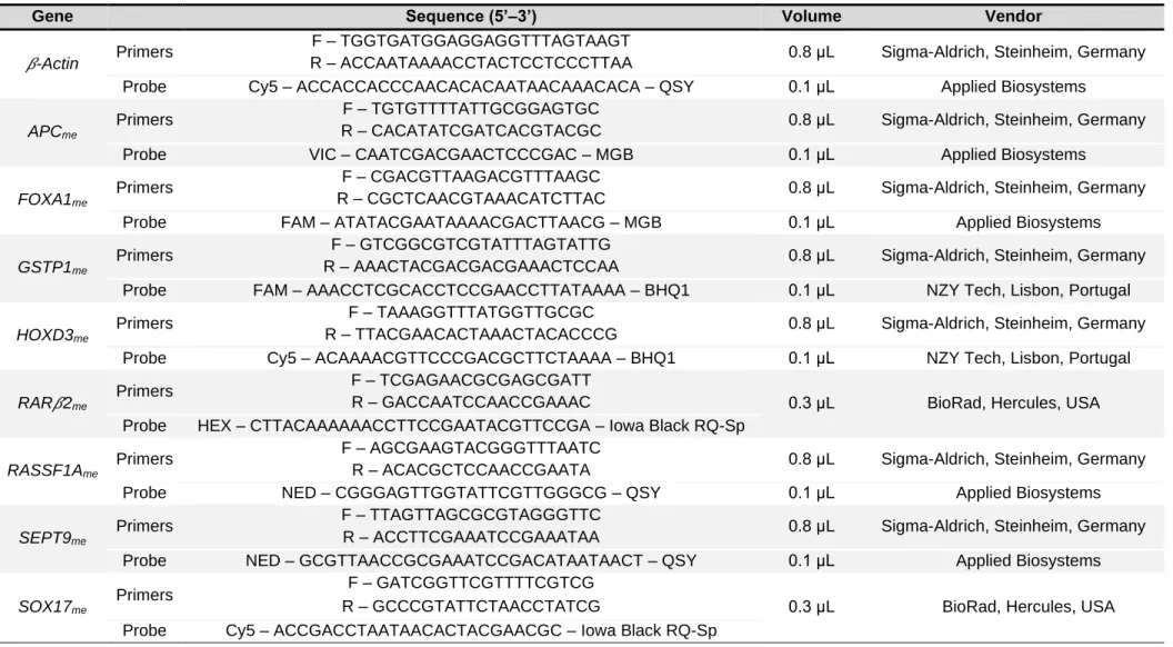
6. Multiplex Quantitative Methylation Specific PCR ... 40
7. Statistical Analysis ... 43
IV. RESULTS ... 45
1. Clinical and Pathological Data ... 47
2. Gene Promoter Methylation Levels in ccfDNA ... 49
2.2. Association Between Promoters’ Methylation Levels and Clinicopathological
Features ... 54
2.3. Prognostic Biomarker Performance of ccfDNA ... 58
V. DISCUSSION ... 63
VI. CONCLUSION & FUTURE PERSPECTIVES... 69
VII. REFERENCES ... 73
VIII. APPENDIX ... I
APPENDIX I ... III
Methylated genes’ nomenclature ... III
APPENDIX II ... VI
TNM Staging ... VI
APPENDIX III ...XIII
Distribution of Promoters’ Methylation Levels ...XIII
APPENDIX IV ... XV
Association between Promoters’ Methylation Levels and Clinicopathological Features in ccfDNA ... XV
APPENDIX V ... XIX
Association between Clinicopathological Features and Promoters’ Methylation Levels and Disease-Specific Survival in ccfDNA ... XIX
FIGURES INDEX
Figure 1. Blood-based liquid biopsy. Circulating tumour cells (CTC), circulating cell-free
DNA (ccfDNA) [including circulating tumour DNA (ctDNA)], circulating cell-free RNA (ccfRNA) and exosomes are released from tumour cells to the bloodstream. Hence, blood can be collected and analysed in the context of a liquid biopsy. Constâncio, V. unpublished. ... 4
Figure 2. Major studied epigenetic mechanisms involved in gene expression regulation.
DNA methylation consists in the addition of a methyl group to a cytosine present in a cytosine-phosphate-guanine (CpG). Histone post-translational modifications refer to the addition of biochemical modifications on histone tails, such as methylation, acetylation, phosphorylation, ubiquitylation and SUMOylation, that regulate gene expression. Histone variants differ a few amino acids from canonical histones and regulate chromatin remodelling and histone post-translational modifications. Chromatin remodelling complexes regulate the nucleosome structure by removing, relocate and shifting histones. Constâncio, V. unpublished. ... 5
Figure 3. DNA methylation within a gene promoter region. Unmethylated CpG island enable
gene transcription. When CpG island is methylated, gene transcription is repressed. Constâncio, V. unpublished. ... 6
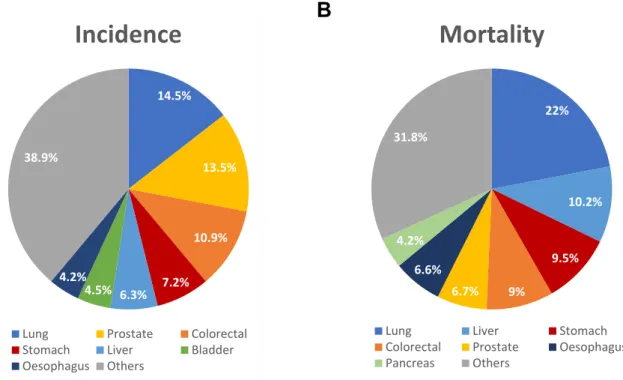
Figure 4. Estimated percentage of cancer-related incidence (A) and mortality (B) in males,
worldwide, in 2018. Adapted from [26]. ... 8
Figure 5. Circulating cell-free DNA methylation-based biomarkers described in literature for
cancer detection common to at least two cancer types [Lung Cancer (blue box), Prostate Cancer (yellow box), Colorectal Cancer (orange box)]. Constâncio, V. unpublished. ... 28
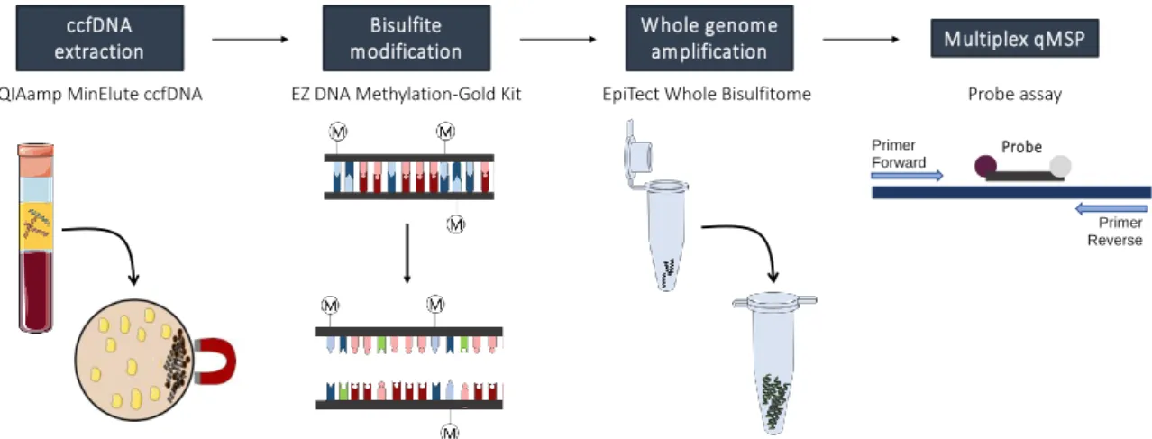
Figure 6. Overview of the techniques performed. Circulating cell-free DNA (ccfDNA) was
extracted from plasma samples using a kit based on the concentration of ccfDNA onto magnetic beads. Then, sodium-bisulfite modification was performed to convert unmethylated cytosines into uracil, while maintaining methylated cytosines unchanged, and whole genome amplification (WGA) was performed to increase modified-DNA quantity. Lastly, promoters’ methylation levels were assessed by multiplex quantitative methylation specific PCR (qMSP) with TaqMan probes. (Kindly provided by S.P. Nunes, unpublished) ... 38
Figure 7. Distribution of (A) APC, (B) FOXA1, (C) GSTP1, (D) HOXD3, (E) RAR2, (F) RASSF1A, (G) SEPT9 and (H) SOX17 relative methylation levels of asymptomatic controls (AC) (n=136), lung cancer (LC) (n=102), prostate cancer (PCa) (n=121) and colorectal cancer (CRC) (n=100) samples. Mann-Whitney U Test between AC and each cancer type,
n.s. p>0.05, *p<0.05, **p<0.01, ***p<0.001, ****p<0.0001. Red horizontal lines represent median methylation levels. (Raw data available in Appendix III: Supplementary Table 9) ... ... 50
Figure 8. Percentage of cases identified by “PanCancer” panel in cancer samples (64%
Positive, 36% Negative) and in asymptomatic controls (AC) (30% Positive, 70% Negative). ... 53
Figure 9. (A) FOXA1 and (B) RAR2 promoter’s methylation levels according with T stage [T1 (n=12) and T2-4 (n=82)] in lung cancer patients. Mann-Whitney U Test, *p<0.05. Red horizontal lines represent median methylation levels. (Raw data available in Appendix III: Supplementary Table 9). ... 54
Figure 10. (A) APC, (B) FOXA1, (C) HOXD3 and (D) RAR2 promoter’s methylation levels according with node status, node-negative (N0) (n=25) and node-positive (N+) (n=72) in lung cancer patients. Mann-Whitney U Test, *p<0.05. Red horizontal lines represent median methylation levels. (Raw data available in Appendix III: Supplementary Table 9) ... 55
Figure 11. Distribution of methylation levels in lung (LC) (A) and in colorectal (CRC) (B)
cancer patients according with metastatic dissemination. (A)-(1) APC, (2) FOXA1, (3)
HOXD3 and (4) RASSF1A promoter’s methylation levels in non-metastatic (M0) (n=47) and
metastatic (M+) (n=55) LC patients. (B)-(1) RAR2, (2) SEPT9, (3) SOX17 promoter’s methylation levels in non-metastatic (M0) (n=82) and metastatic (M+) (n=18) CRC patients. Mann-Whitney U Test, *p<0.05, **p<0.01, ****p<0.0001. Red horizontal lines represent median methylation levels. (Raw data available in Appendix III: Supplementary Table 9). ... 56
Figure 12. Scatter plot of (A) RASSF1A promoter methylation levels between Clinical Stage
I & II (n=17) and III & IV (n=85) lung cancer patients. (B) GSTP1 promoter methylation levels between Clinical stage I (n=31), II (n=55) and III & IV (n=35) prostate cancer patients. (C) (1) RAR2 and (2) SEPT9 promoters’ methylation levels between Clinical Stage I & II (n=39) and III & IV (n=61) CRC patients. Mann-Whitney U Test for A and C and Mann-Whitney U test with Bonferroni’s correction for B, n.s. p<0.05, *p<0.05. Red horizontal lines represent median methylation. (Raw data available in Appendix III: Supplementary Table 9) ... 57
Figure 13. Scatter plot of (A) HOXD3, (B) RASSF1A, and (C) SOX17 promoter’s
methylation levels according with Histological Subtype [Non-Small Cell Lung Carcinoma (NSCLC) (n=86) and Small Cell Lung Carcinoma (SCLC) (n=16)]. Mann-Whitney U Test, *p<0.05, **p<0.01, ****p<0.0001. Red horizontal lines represent median methylation levels. (Raw data available in Appendix III: Supplementary Table 9). ... 58
Figure 15. Disease-Specific Survival curves according to panel (APCme and RASSF1Ame)
TABLES INDEX
Table 1. Circulating cell-free DNA methylation-based biomarkers for lung cancer detection.
... 11
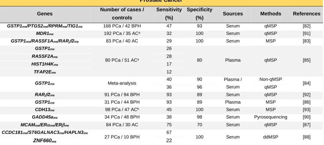
Table 2. Circulating cell-free DNA methylation-based biomarkers for prostate cancer
detection. ... 16
Table 3. Circulating cell-free DNA methylation-based biomarkers for colorectal cancer
detection. ... 21
Table 4. Component mix (QIAmp MinElute ccfDNA Mini Kit). ... 38 Table 5. Primers and probes sequences with respective fluorochrome and quencher. ... 41 Table 6. Gene combinations for multiplex qMSP. ... 42 Table 7. Formulas for biomarker performance calculations. ... 43 Table 8. Clinical and pathological features of lung, prostate and colorectal cancer patients
and asymptomatic controls included in this study. ... 47
Table 9. Biomarker performance of each gene promoter methylation for lung (LC), prostate
(PCa) and colorectal (CRC) cancer detection in circulating cell-free DNA. ... 52
Table 10. Biomarker performance of “PanCancer” panel (FOXA1me, RAR2me and
RASSF1Ame) for simultaneous lung and prostate cancer detection in circulating cell-free
DNA. ... 53
Table 11. Biomarker performance of “CancerType” panel (GSTP1me and SOX17me) for
discrimination among lung and prostate cancer in circulating cell-free DNA. ... 54
Table 12. Biomarker performance of HOXD3, RASSF1A and SOX17 promoters’
methylation levels for discrimination among Small Cell Lung Carcinoma (SCLC) and Non-Small cell Lung Carcinoma (NSCLC) in circulating cell-free DNA... 58
Table 13. Cox-regression models assessing the potential of clinical variables and detection
of circulating methylation levels of APC, FOXA1, GSTP1, HOXD3, RAR2, RASSF1A, SEPT9 and SOX17 and prognostic panel (APC, RASSF1A) in the prediction of disease-specific survival in lung cancer patients. ... 61
LIST OF ABBREVIATIONS
5mC 5-methylcytosine
AC Asymptomatic control
ADT Androgen deprivation therapy
AJCC American Joint Committee on Cancer ALK ALK receptor tyrosine kinase
APC APC regulator of WNT signalling pathway
APCme Methylated APC regulator of WNT signalling pathway
BPH Benign prostatic hyperplasia
BRAF B-Raf proto-oncogene, serine/threonine kinase
BrC Breast Cancer
ccfDNA Circulating cell-free DNA
ccfRNA Circulating cell-free RNA
CE Conformité Européenne
CEA Carcinoembryonic antigen
CI Confidence interval
CIMP CpG island methylator phenotype
CIN Chromosomal instability
CpG Cytosine-phosphate-guanine
CRC Colorectal Cancer
CRPC Castration-resistant prostate cancer
CTCs Circulating tumour cells
ctDNA Circulating tumour DNA
ddMSP Droplet digital methylation-specific PCR
DFS Disease-free survival
DNMTs DNA methyltransferases
DRE Digital rectal examination
DSS Disease-specific survival
EAU European Association of Urology
EGFR Epidermal growth factor receptor
FAP Familial adenomatous polyposis
FDA United States Food and Drug Administration
FIT Faecal immunochemical test
FOBT Faecal-occult blood test
FOXA1me Methylated forkhead box A1
GSTP1 Glutathione-S transferase pi 1
GSTP1me Methylated glutathione-S transferase pi 1
HDAC Histone deacetylases
HGNC HUGO Gene Nomenclature Comittee
HNPCC Hereditary non-polyposis colon cancer
HOXD3 Homeobox D3
HOXD3me Methylated homeobox D3
HR Hazard ratio
ISUP International Society of Urological Pathology
IVD In Vitro Diagnostic
LC Lung Cancer
LCLC Large-cell lung carcinoma
LD-CT Low-dose computed tomography
LUAD Lung adenocarcinoma
LUSC Lung squamous cell carcinoma
MBP Methyl-CpG-binding proteins
MDA Multiple displacement amplification
MMR Mismatch repair
MSI Microsatellite instability
NPV Negative predictive value
NSCLC Non-small cell lung carcinoma
OS Overall survival
PCa Prostate Cancer
PD-L1 Programmed death ligand 1
PD1 Programmed cell death protein 1
PFS Progression-free survival
PPV Positive predictive value
PSA Prostate-specific antigen
qMSP Quantitative methylation-specific PCR RAR2 Retinoic acid receptor beta 2
RAR2me Methylated retinoic acid receptor beta 2
RASSF1A Ras association domain family 1 isoform A
RASSF1Ame Methylated Ras association domain family 1 isoform A
SCLC Small cell lung carcinoma
SEPT9 Septin 9
SEPT9me Methylated septin 9
SOX17 SRY-box transcription factor 17
SOX17me Methylated SRY-box transcription factor 17
TKI Tyrosine kinase inhibitor
TRUS Transrectal ultrasound
TSG Tumour suppressor gene
VEGF Vascular endothelial growth factor
1. CANCER BIOMARKERS
During the last decades efforts have scaled up worldwide to develop more effective biomarkers as an approach to reduce cancer mortality [1]. A cancer biomarker can be defined as an objectively measurable biomolecule, such as a protein, metabolite, RNA, DNA or an epigenetic alteration, found in body fluids or tissues, that indicates the presence of cancer or provides information on cancer’s expected future behaviour [2, 3]. Depending on their purpose in the clinical setting, they can be classified as: “Susceptibility/Risk Biomarkers” designed to identify individuals predisposed to develop cancer; “Screening Biomarkers” used to depict early-stage cancer in asymptomatic individuals; “Diagnostic Biomarkers”, used to identify and categorize a patient’s cancer; “Prognostic Biomarkers” if the aim is to inform the likely patient clinical outcome, including recurrence or disease progression; “Predictive Biomarkers” if predict the likely response to specific therapeutic interventions; or “Disease monitoring Biomarkers” if indicate the development of disease relapse during follow-up [1, 3].
2. LIQUID BIOPSIES
Thus far, tissue biopsy sampling has been the gold-standard approach for patients‘ diagnosis and prognostication, however several drawbacks have been pointed out over the years to this approach [4, 5]. Firstly, and mainly, tissue samples might not fully represent the tumour heterogeneity, constituting a limitation for accurate outcome prediction and treatment efficacy [5, 6]. Moreover, early-stage tumour, residual disease and early recurrence detection might be difficult, since tissue biopsy sampling requires a highly invasive intervention with possible risk of complications, and, depending on the tumour anatomical location, can be extremely difficult to obtain [4, 7].
Recently, liquid biopsies obtained from easily assessable body fluids, including blood, urine or sputum, have raised up as a viable alternative to overcome these challenges. Liquid biopsies, mainly based on circulating cell-free DNA (ccfDNA), circulating tumour cells (CTCs), circulating cell-free RNA (ccfRNA) and exosomes [8] are a fast, reliable, cost-effective and minimally invasive approach [9] (Figure 1). Hence, owing to these features they may allow for a real time monitorization of the cancer evolution, while better representing the heterogenous genetic profile of all tumour sub clones [8, 10].
Figure 1. Blood-based liquid biopsy. Circulating tumour cells (CTC), circulating cell-free DNA
(ccfDNA) [including circulating tumour DNA (ctDNA)], circulating cell-free RNA (ccfRNA) and exosomes are released from tumour cells to the bloodstream. Hence, blood can be collected and analysed in the context of a liquid biopsy. Constâncio, V. unpublished.
2.1. Circulating Cell-free DNA
CcfDNA was firstly described in 1948 when extracellular nucleic acids were found in human blood from healthy individuals by Mandel and Métais [11]. Later on, it was found that, in cancer patients, circulating tumour DNA (ctDNA) fragments between 150 and 1000 base pairs could also be detected due to their release into the bloodstream, either by cell death (apoptosis or necrosis) or active secretion by the release of extracellular vesicles, such as exosomes [10, 12]. Depending on several factors, including, tumour burden, metastatic sites or cellular turnover, ctDNA might account for 0.01 to 90% of the total ccfDNA in the blood of cancer patients [12]. Owing to the fact that ctDNA might represent tumour-specific genetic and epigenetic alterations of all tumour’s sub clones present, ccfDNA is an ideal candidate for blood-based liquid biopsies by offering the possibility to test for the presence of cancer and the discrimination of lethally aggressive cancer [13]. Moreover, it gathers also the advantage of representing tumour real-time dynamic changes due to its short half-life (from 15 minutes to few hours), since it is rapidly cleared by the liver and kidney [13].
3. EPIGENETICS
Although the study of tumour mutations were the focus of biomarker research for a long time, their wide diversity has been a challenge for the development of effective diagnostic biomarkers since very large proportions of the genome would need to be examined in order to provide adequate sensitivity [14]. Contrarily, epigenetic alterations seem to be more stable and homogenous in cancer, representing a good alternative for biomarker development [15]. The definition of epigenetics, firstly coined in 1941 by Conrad Waddington, evolved over time and, currently, it is described as the study of gene expression modifications, carried during cell division, that do not change the primary DNA sequence [16, 17]. These alterations are, indeed, an alternative to genetic changes, by mimicking their effect. The main studied epigenetic mechanisms are DNA methylation, histone post-translational modifications, histone variants and chromatin remodelling complexes (Figure 2) [16]. Although these mechanisms are crucial for normal cell development and regulation of specific gene expression patterns, epigenetic processes dysregulation often lead to inappropriate activation or inhibition of several signalling pathways, which can trigger the development of several pathologies, including cancer [18].
Figure 2. Major studied epigenetic mechanisms involved in gene expression regulation. DNA
methylation consists in the addition of a methyl group to a cytosine present in a cytosine-phosphate-guanine (CpG). Histone post-translational modifications refer to the addition of biochemical modifications on histone tails, such as methylation, acetylation, phosphorylation, ubiquitylation and SUMOylation, that regulate gene expression. Histone variants differ a few amino acids from canonical histones and regulate chromatin remodelling and histone post-translational modifications. Chromatin remodelling complexes regulate the nucleosome structure by removing, relocate and shifting histones. Constâncio, V. unpublished.
3.1. DNA Methylation
DNA methylation, the most widely studied epigenetic modification in humans, was also the first to be identified in cancer [18, 19]. This epigenetic mechanism consists in a covalent addition of a methyl group, donated by S-adenosylmethionine (SAM), to the 5-position carbon of a cytosine ring to form 5-methylcytosine (5mC) [20, 21]. This modification is catalysed by DNA methyltransferase enzymes (DNMTs), namely, DNMT3a and DNMT3b that catalyse de novo DNA methylation during embryonic development, stablishing tissue-specific DNA methylation, and DNMT1 that is often associated with maintenance of methylation patterns during replication [21]. Typically, this process occurs on cytosine residues present at CpG dinucleotides (cytosine followed by a guanine) commonly found in large clusters named CpG islands, which are predominantly located at the 5’ end of genes, occupying approximately 60% of human gene promoter regions [18, 21, 22]. Although gene promoter’s hypermethylation is associated with transcription repression of the nearby gene (Figure 3), depending on DNA methylation genomic location, it can display different functions [20, 23]. For instance, gene body methylation is correlated with its transcriptional activation [23]. Epigenetic gene silencing by DNA promoter methylation can happen either directly, by blocking transcription factors binding to target sites in or near the promoter, or indirectly, by the binding of methyl-CpG-binding proteins (MBP), which can recruit other enzymes like DNMTs and histone deacetylases (HDAC), leading to chromatin conformation changes that further repress gene transcription [18, 20].
Figure 3. DNA methylation within a gene promoter region. Unmethylated CpG island enable gene
transcription. When CpG island is methylated, gene transcription is repressed. Constâncio, V.
unpublished.
methylated CpG unmethylated CpG
Unmethylated CpG island Active gene transcription
Methylated CpG island Inactive gene transcription
Although, in normal cells, CpG island promoters are usually found unmethylated, physiological hypermethylation associated with promoters is observed on the silenced copy of the X chromosome in females, on imprinted genes, and in a tissue-specific manner [20]. Moreover, CpG dinucleotides within repetitive genomic sequences throughout the genome, retrotransposons and parasite sequences are also highly methylated to prevent their transcription, and to maintain genomic stability [16].
As aforementioned, DNA methylation is crucial for multiple cellular processes, thus it is understandable that its deregulation has been linked to cancer. Indeed, normal and cancer cells display different methylomes. Usually, a global hypomethylation pattern, which contributes to genomic instability and activation of silenced oncogenes, is observed in cancer [21, 24]. Alongside, tumour suppressor genes (TGS) frequently undergo inactivation due to focal promoter’s hypermethylation [21, 24]. Currently, the latter process is considered a major contributor of neoplastic transformation [25].
Interestingly, aberrant DNA methylation is thought to occur at very early stages of cancer development and specific genes seem to be methylated at different tumour stages [21]. Moreover, since these alterations can be assessed in several body fluid samples [21], it is widely accepted that DNA methylation-based liquid biopsies are a promising approach, not only for premalignant/early cancer detection but also for prognostic assessment. Furthermore, since some genes seem to acquire tissue-specific DNA methylation, it may be possible to discriminate between different cancer types in the context of metastatic tumours [21] or in liquid biopsies.
4. CELL-FREE DNA METHYLATION BASED BIOMARKERS
According to GLOBOCAN 2018, Lung Cancer (LC), Prostate Cancer (PCa), and Colorectal Cancer (CRC) were estimated to account for 39% of all cancers diagnosed in males worldwide (Figure 4A), or 46% if only European countries were considered [26], representing major public health issues. Therefore, for the purposes of this work, a detailed literature review was conducted to assess the current state of art of the ccfDNA methylation blood-based biomarkers for cancer screening, diagnosis, prognostication, prediction and monitorization of the three most diagnosed cancers in males worldwide. On a PubMed database search, the keywords “Lung Cancer / Prostate Cancer / Colorectal Cancer”, “DNA methylation” and “Serum / Plasma” were used.
Figure 4. Estimated percentage of cancer-related incidence (A) and mortality (B) in males,
worldwide, in 2018. Adapted from [26].
4.1. Lung Cancer
LC is the most incident and lethal cancer worldwide (Figure 4) [26], having a 5-year survival rate of about 15% [27]. The most well-established risk factor for this cancer is tobacco, being estimated that roughly 85% of all LC cases are attributed to cigarette smoking history [28].
LC is a heterogeneous disease, being classified into two major histological subtypes based on the prognostic and therapeutic implications: the small cell lung carcinoma (SCLC) and the non-small cell lung carcinoma (NSCLC) [29, 30].
NSCLC accounts for 85-90% of all cases, and is further classified into adenocarcinoma (LUAD), squamous cell carcinoma (LUSC) and large-cell lung carcinoma (LCLC) [30]. LUAD, the predominant LC subtype, accounts for nearly 40% of all LC, and is an epithelial tumour with glandular differentiation, being mostly located at the peripheral areas of the lung [30]. Although all LC subtypes are strongly associated with tobacco, LUAD is the most common subtype in never-smokers [30]. LUSC accounts for 20% of all LC cases and is usually present in a central location [30]. Accounting for only 3% of all diagnosed LC, LCLC is defined as an undifferentiated NSCLC [30].
SCLC revels neuroendocrine differentiation and arises in a central location [31]. This 14.5% 13.5% 10.9% 7.2% 6.3% 4.5% 4.2% 38.9%
Incidence
Lung Prostate Colorectal Stomach Liver Bladder Oesophagus Others 22% 10.2% 9.5% 9% 6.7% 6.6% 4.2% 31.8%
Mortality
Lung Liver Stomach
Colorectal Prostate Oesophagus Pancreas Others
95% of all patients having a heavy tobacco exposure history) and the most aggressive LC subtype [32], being characterized by rapid doubling time, early development of widespread metastases and worse survivals [33]. In fact, 60-65% of SCLC patients display metastatic disease at diagnosis, whereas the remaining often present locally advanced disease, which is also not amenable to surgical removal [31, 33].
4.1.1. Screening and Diagnosis
Despite advancements in new treatment options over the years, the high mortality rate observed in LC patients is mainly related with the fact that more than 75% of LC patients are diagnosed with advanced stage disease [34]. Hence, effective screening options aiming to shift LC diagnosis from advanced to curative early stages are crucial to change the fate of this disease [35]. Currently, low-dose computed tomography (LD-CT) is considered the best LC screening method available [36]. Nonetheless, despite The National Lung Screening Trial showed a 20% decrease in LC-related mortality rate among high-risk smokers with LD-CT screening comparing to chest X-ray, which was corroborated by the largest European trial (Dutch-Belgian Lung Cancer Screening Trial), from the 24% positive test results in this trial, 96.4% were classified as false-positive results [37, 38]. Hence, due to the risks related with LD-CT screening, namely, overdiagnosis, radiation exposure, and false positive results leading to unnecessary anxiety and costs [35], the development of specific and accurate screening tools is urgently needed to improve LC survival.
LC diagnosis procedures combine imaging exams and evaluation of histological or cytological specimens, collected either by bronchoscopy, sputum cytology or fine-needle aspiration [29, 30].
In 2002, Usadel et al. and Bearzatto et al. described for the first time, APCme
(Appendix I: Supplementary Table 1) and p16INK4ame, respectively, in ccfDNA as putative
minimally-invasive biomarkers for LC detection [39] [40]. Thenceforth, numerous other methylated gene promoters detected in ccfDNA have been purposed for LC detection either individually or in panel (Table 1). RASSF1Ame and p16INK4ame represent the two most
reported genes in blood-based liquid biopsies, displaying, between 22-66% sensitivity and 57-100% specificity for LC detection, individually [40-42].
After the commercialization in Europe of a test based on SHOX2me assessment in
bronchial aspirates, the methylation of this gene was also evaluated as a LC detection biomarker in plasma. Indeed, plasma SHOX2me distinguished LC from control samples with
60% sensitivity and 90% specificity, even though higher sensitivity was found in stages II (72%), III (55%) and IV (83%) compared with stage I patients (27%) [43]. In line with these
results, another study reported that SHOX2me discriminated LC in subjects undergoing
bronchoscopy with 81% sensitivity and 79% specificity [44]. Later on, Weiss et al. reported that SHOX2me and PTGER4me panel distinguished LC patients with 67% sensitivity for a
fixed specificity of 90%, and with 73% specificity for a fixed sensitivity of 90% [45]. Remarkably, in the end of 2017, the “Epi proLung®” assay, developed by Epigenomics AG, based on these two genes received the Conformité Européenne (CE) mark for In Vitro Diagnostic (IVD) test. According to their validation study comprising 360 patients from the US and Europe, of which 152 were diagnosed with LC, depending on the Epi proLung test score threshold chosen, 85% sensitivity was achieved for 50% specificity, whereas sensitivity decreased to 59% if 95% specificity was considered [46, 47].
Although the majority of these studies have been focused on detection of NSCLC (the most diagnosed LC subtype), different detection frequencies of genes’ methylation have been reported among the different subtypes. Indeed, SHOX2me detected with higher
sensitivity SCLC (80%) and LUSC (63%) than LUAD (39%) [43]. Similarly, DCLK1me was
more frequent in SCLC than NSCLC [48], and our research team recently reported that higher APCme and RAR𝛽2me levels were observed in SCLC compared to LUAD in females
[42]. Conversely, SEPT9me was more frequent in NSCLC (53%) than in SCLC (26%) [49].
A serum-based gene panel (MARCH11me, HOXA9me, CDO1me, UNCXme, PTGDRme and
AJAP1me) detected stage I LUAD and LUSC with 71% specificity and 72% and 60%
Table 1. Circulating cell-free DNA methylation-based biomarkers for lung cancer detection.
Lung Cancer
Genes Number of cases /
controls
Sensitivity (%)
Specificity
(%) Sources Methods References
APCme 89 LC / 50 AC 47 100 Serum/Plasma qMSP [39] p16INK4a me 35 NSCLC / 15 AC 34 100 Plasma F-MSP [40] MGMTme/p16INK4ame/RASSF1Ame/ DAPKme/RAR𝛽me 91 LC / 109 BPD 50 85 Serum MSP [51] p16INK4a me/CDH13me 61 NSCLC / 15 BPD 39 100 Serum MSP [52] RASSF1Ame 80 LC / 50 ACa 34 100 Serum MSP [53] CDH13me/p16INK4ame/FHITme/
RAR𝛽me/RASSF1Ame/ZMYND10me
63 NSCLC / 36 BPD 73 83 Plasma Two-step MSP [54]
KLK10me 78 NSCLC / 50 ACa 38 96 Plasma MSP [55]
SFRP1me 78 NSCLC / 50 ACa 28 96 Plasma MSP [56]
DLEC1me 78 NSCLC / 50 ACa 36 96 Plasma MSP [57]
Kif1ame/DCCme/RAR𝛽2me/NISCHme 70 LC / 80 BPD 73 71 Plasma qMSP [58]
APCme/RASSF1Ame/CDH13me/ KLK10me/DLEC1me 110 NSCLCb / 50 ACa 84 74 Plasma MSP [59] APCme/CDH1me/MGMTme/DCCme RASSF1Ame/AIM1me 76 LC / 30 AC 84 57 Serum qMSP [60]
SHOX2me 188 LC / 155 ACa,c 60 90 Plasma qMSP [43]
TMEFF2me 316 NSCLC / 50 AC 9 100 Serum Two-step MSP [61]
RAR𝛽2me
60 NSCLC / 32 AC 72 62 Plasma qMSP [41]
RASSF1Ame 66 57
p14ARFme 107 NSCLC / 20 BPD 25 95 Plasma Two-step MSP [62] DCLK1me 65 LC / 95 AC 49 92 Plasma qMSP [48] SOX17me 48 Operable NSCLC / 49 AC 56 98 Plasma qMSP [63] 74 Advanced NSCLC / 49 AC 36 SHOX2me 38 LC / 31 BPD 81 79 Plasma qMSP [44] SHOX2me/PTGER4me 50 LC / 122 ACa 67 90 90 73 Plasma Multiplex qMSP [45]
CDO1me/TAC1me/SOX17me 150 NSCLCb / 60 AC 93 62 Plasma qMSP [64]
MARCH11me/HOXA9me/CDO1me/
UNCXme/PTGDRme/AJAP1me 43 LUADd / 42 AC 72 71 Plasma qMSP [50] 40 LUSCd / 42 AC 60 NID2me 46 NSCLC / 30 BPD 46 80 Plasma qMSP [65] APCme 73 LCe / 103 ACe 36 94 Plasma Multiplex qMSP [42] FOXA1me 72 74 RAR𝛽2me 25 95 RASSF1Ame 22 98 SOX17me 38 95 CDKN2Ame/DLEC1me/CDH1me/ DAPKme/RUNX3me 42 NSCLC / 10 AC 95 100 Plasma Two-step MSP [66]
RASSF1Ame/CDKN2Ame/DLEC1me/
CALCAme/CDH13me/PITX2me/
HOXA1me/WT1me
39 NSCLCd / 11 BPD 72 91 Plasma qMSP [66]
aIncluded Benign pulmonary Diseases; bOnly stage I/II; cIncluded other cancer types; dOnly included stage I; eOnly included females; Abbreviations: AC – Asymptomatic Controls;
BPD – Benign Pulmonary Diseases; F-MSP – Fluorescent methylation-specific PCR; LC – Lung Cancer; LUAD – Lung Adenocarcinoma; LUSC – Lung Squamous Cell Carcinoma; MSP – Methylation-specific PCR; NSCLC – Non-Small Cell Lung Cancer; qMSP – Quantitative methylation-specific PCR
4.1.2. Prognosis, Prediction and Monitorization
Besides LC histological subtype, TNM prognostic stage groups of the American Joint Committee on Cancer (AJCC), based on tumour size and local invasion (T), and presence of nodal (N) and distant (M) metastasis (Appendix II: Supplementary Tables 2-3), remains the most important prognostic feature to predict recurrence and survival, followed by tumour histological grade, gender and age [33, 67]. Recently, molecular subtypes have also emerged in order to provide more personalized genetic information that can improve prognostic estimates and prediction to treatment response [33]. LC treatment options include surgery for early diagnosed cancers, chemotherapy and/or radiotherapy, specific tyrosine kinase inhibitors (TKI) in molecularly-defined NSCLC presence of epidermal growth factor receptor (EGFR) mutation, and ALK receptor tyrosine kinase (ALK) or ROS proto-oncogene 1, receptor tyrosine kinase (ROS1) rearrangements, as well as, immunotherapy with programmed cell death protein (PD1) and programmed death-ligand 1 (PD-L1) antibodies [29, 68].
Thus far, only few small-scaled studies reported the prognostic, predictive and follow-up potential of ccfDNA methylation for LC, being most of them performed in patients with advanced disease.
DCLK1me and SOX17me levels were associated with reduced overall survival (OS) in
advanced LC and NSCLC, respectively [48, 63]. Similarly, higher SHP1P2me levels
observed in advanced NSCLC associated with reduced progression-free survival (PFS) and OS [69], whereas BRMS1me with both reduced disease-free survival (DFS) and OS in
operable NSCLC, and reduced PFS and OS in advanced NSCLC [70]. Moreover, RASSF1Ame associated with presence of regional node metastasis [42] and advanced
clinical stage [53], whereas SOX17me associated with distant metastasis [42].
Interestingly, after neoadjuvant chemotherapy and surgery with intraoperative radiation therapy, NSCLC patients showed decreased RASSF1Ame and RAR𝛽2me levels,
similar to levels in healthy subjects. Moreover, methylation levels’ increase of at least one of these genes, up to the levels detected before treatment, was observed in all the 5 patients that showed evidence of disease progression [41]. Increased APCme and/or RASSF1Ame
levels within 24h after cisplatin-based chemotherapy also associated with increased OS [71].
Remarkably, advanced LC patients who clinically responded to chemo/radiotherapy demonstrated a decrease in plasmatic SHOX2me levels, observable at 7-10 days after
therapy beginning were indicative of shorter OS [72]. Contrarily, 14-3-3σme levelsin stage
IV NSCLC patients before treatment with cisplatin-gemcitabine associated with longer survival [73]. Stage IV NSCLC patients with unmethylated CHFR showed longer OS when treated with EGFR-TKI compared to those treated with chemotherapy as second-line therapy [74].
4.2. Prostate Cancer
PCa was estimated to be the second most incident cancer in men in 2018, worldwide (Figure 4A), and the first in Europe [26]. Approximately 95% of all PCa are adenocarcinomas mainly found in the peripheral zone of the prostate [75]. Currently, only nonmodifiable risk factors are well-established for PCa development, namely, advanced age, black race and positive family history [76]. Nonetheless, other factors such as diet, obesity, physical inactivity, smoking and chronic inflammation have also been associated, although with inconsistent results [76].
4.2.1. Screening and Diagnosis
PCa is mostly asymptomatic, therefore, patients might be diagnosed with advanced disease, resulting in worse patient outcomes and limited treatment options [75]. Digital rectal examination (DRE) in combination with serum prostate-specific antigen (PSA) remain the gold standard PCa screening tools [77], however both methods present drawbacks. A DRE positive result is dependent on clinicians’ expertise and the majority of cancers detected by this method are at an advanced stage [78], besides compliance is rather low. The widespread adoption of PSA screening since late 1980s has facilitated the shift to detection of PCa at early stages [78]. Nonetheless, despite being highly sensitive, since benign prostatic hyperplasia (BPH) and other benign conditions can also cause PSA elevation, the lack of cancer-specificity of this approach entails a high false-positive rate and overdiagnosis of non-life threatening PCa [77, 79]. Indeed, only less than one third of the patients undergoing transrectal ultrasound-guided (TRUS) biopsy (standard diagnostic approach) due to elevated PSA levels or abnormal DRE are diagnosed with cancer [80]. In parallel, a negative result does not completely rule out the existence of cancer, leading to a large number of unnecessary invasive tissue biopsies that might be repeated due to the uncertainty of diagnosis if elevated PSA levels persist [80]. Thus, the introduction of more specific alternatives is urgently sought.
exfoliated cells and ccfDNA from diverse sites of the urinary system [81]. Hence, several studies have been addressed using this source [81]. Nonetheless, in PCa, higher sensitivities are achieved after manipulation of the prostate, either by massage and DRE, increasing the invasiveness of this procedure [81]. Therefore, blood-based liquid biopsies might represent the most minimally invasive procedure for PCa detection. A summary of the currently reported blood-based DNA methylation PCa detection biomarkers is depicted on Table 2.
GSTP1me is the most described epigenetic alteration in ccfDNA of PCa patients due
to its remarkably high specificity for PCa [82-86]. Indeed, a meta-analysis of 10 studies published until 2010, evaluating GSTP1me PCa detection performance in plasma/serum, reported a pooled specificity of 90% non-quantitative methylation specific PCR qMSP) and 96% (qMSP-based detection), although with a modest sensitivity of 40% (non-qMSP) and 36% (qMSP-based detection) [84].
Since epigenetic alterations are usually multiple and not necessarily overlapped, multigene panels are pivotal to increase the modest sensitivities of individual genes. Indeed, Ellinger et al. reported that using a gene panel comprising GSTP1me, PTGS2me, RPRMme
and TIG1me, PCa diagnostic coverage increased from 42% (GSPT1me alone) to 47%
(panel), maintaining 93% specificity [82]. Furthermore, Sunami et al. reported that GSTP1, RASSF1A and RAR𝛽2 were hypermethylated in 13%, 24% and 12% of serum samples from PCa patients, respectively, whereas the three gene panel increased the detection rate to 29%, with 100% specificity [83]. More recently, other panels without comprising GSTP1me
have also been tested. Indeed, MCAMme, ERme and ERme panel demonstrated 75%
sensitivity and 70% specificity for early PCa detection [87]. Likewise, ZNF660me,
CCDC181me, ST6GALNAC3me and HAPLN3me in serum displayed 22%, 26%, 31% and 44%
sensitivity, respectively, and 100% specificity for PCa. Remarkably, the best gene panel (ST6GALNAC3me, CCDC181me and HAPLN3me) increased sensitivity to 67%, while keeping
100% specificity [88].
Interestingly, given the modest sensitivities obtained with ccfDNA methylation even in panels, studies have also been addressed to understand if ccfDNA might have a better performance by complementing it with serum PSA levels. Indeed, in a Mexican cohort with biopsy-confirmed PCa, a panel comprising GSTP1me and RASSF1Ame allowed cancer
detection with 73% positive predictive value (PPV) and 59.6% negative predictive value (NPV), increasing to 81% and 66%, respectively, when serum PSA was also considered [89]. Similarly, serum GADD45ame increased its sensitivity from 38% to 94% when PSA and
free circulating DNA levels were also considered, even though the 98% specificity decreased to 88% [90].
Table 2. Circulating cell-free DNA methylation-based biomarkers for prostate cancer detection.
Prostate Cancer
Genes Number of cases /
controls
Sensitivity (%)
Specificity
(%) Sources Methods References
GSTP1me/PTGS2me/RPRMme/TIG1me 168 PCa / 42 BPH 47 93 Serum qMSP [82]
MDR1me 192 PCa / 35 ACa 32 100 Serum qMSP [91]
GSTP1me/RASSF1Ame/RAR𝛽2me 83 PCa / 40 AC 29 100 Serum MSP [83]
GSTP1me 80 PCa / 51 ACa 26 80 Plasma qMSP [85] RASSF2Ame 28 HIST1H4Kme 17 TFAP2Eme 12 GSTP1me Meta-analysis 40 90 Plasma / Serum Non-qMSP [84] 36 96 qMSP
RAR𝛽2me 91 PCa / 94 BPH 93 89 Serum qMSP [92]
GSTP1me 31 PCa / 44 BPH 93 89 Plasma MSP [86]
CDH13me 98 PCa / 47 ACb 45 100 Serum MSP [93]
GADD45ame 34 PCa / 48 BPH 38 98 Serum Pyrosequencing [90]
MCAMme/ERme/ERme 84 PCa / 30 AC 75 70 Serum qMSP [87]
CCDC181me/ST6GALNAC3me/HAPLN3me
27 PCa / 10 BPH
67
100 Serum ddMSP [88]
ZNF660me 22
aBiopsy negative; bIncluded BPH; Abbreviations: AC – Asymptomatic Controls; BPH – Benign Prostatic Hyperplasia; ddMSP – Digital droplet methylation-specific PCR; MSP –
4.2.2. Prognosis, Prediction and Monitorization
PCa is a very heterogenous disease, ranging from small, low grade clinical indolent to large, lethally aggressive tumours [94]. Thus, the main goal after establishing the presence of the disease is to evaluate its extension and aggressiveness through staging, in order to assess prognosis and plan the treatment course. Currently, PCa prognostic stage groups are based on nomograms combining TNM classification, preoperative serum PSA levels, and histological International Society of Urological Pathology (ISUP) Grade Group (Appendix II: Supplementary Tables 4-6) [95, 96], which is based on the evaluation of the two most common differentiation patterns in a tumour [97]. PCa is mostly asymptomatic and organ-confined, amenable to curative intent treatment options, namely, radical prostatectomy and radiotherapy (external beam radiotherapy or brachytherapy) or conservative approaches (monitoring PCa progression while not undergoing definitive therapy) by means of active surveillance and watchful waiting [98]. Moreover, androgen-deprivation therapy (ADT) and chemotherapy are available for locally advanced or metastatic disease [98]. Nonetheless, the median duration of response to ADT is 18-24 months, after which most patients develop a more aggressive form of disease named castration-resistant prostate cancer (CRPC) [99]. Furthermore, it is estimated that 30-50% patients curatively treated may show a rising of serum PSA levels (biochemical recurrence) within 10 years after treatment, with disease progression in up to 40% of these [99, 100]. Thus, considering these uncertainties, there is an urgent need for development and implementation of more trustworthy PCa biomarkers to assist clinicians and patients in decision-making.
Besides the study of its detection value, the potential of methylation-based biomarkers in ccfDNA to predict disease progression and therapy response have also been tackled. Namely, GSTP1me [91, 101], MDR1me, EDNRBme and RAR𝛽2me [91] were reported
to be more frequent in CRPC patients than in early-stage PCa patients. GSTP1me levels
also associated with Gleason score and presence of metastasis [101] and with reduced disease-specific survival (DSS) along with APCme [102] in CRPC patients. Furthermore,
GSTP1me and RASSF2Ame were more frequently detected in men with non-organ confined
compared to organ-confined disease and both associated with increased Gleason score [85]. Interestingly, preoperative serum GSTP1me was also reported as an independent
predictor of biochemical recurrence following radical prostatectomy [103]. Sunami et al. reported that GSTP1me, RASSF1Ame and RAR𝛽2me associated with Gleason score and
serum PSA levels, whereas GSTP1me and RAR𝛽2me also associated with advanced stages
also associated with advanced clinical stage, higher preoperative serum PSA, as well as lymph node metastasis and shorter biochemical recurrence free survival [104-106]. Besides being also associated with Gleason score, advanced tumour stage and high PSA, CDH13me
was further associated with shorter OS [93].
Remarkably, a recent phase III multicentre trial comprising 600 CRPC patients showed that detectable serum GSTP1me levels prior and after two cycles of chemotherapy
were both independently associated with decreased OS [107]. In the same study, undetectable serum GSTP1me after two cycles of docetaxel further associated with longer
time to PSA progression [107].
4.3. Colorectal Cancer
CRC is the third most incident cancer worldwide (Figure 4), being estimate to account for over 1 million new cases detected and almost half a million deaths in males in 2018 [26]. Several risk-factors, such as family history, inflammatory bowel diseases, and lifestyle (western-type diet, sedentarism, smoking and excessive alcohol consumption) are associated with the development of this disease [108, 109].
Thirty five percent of all CRC cases might be attributable to hereditary syndromes including, the autosomal dominant disorders: familial adenomatous polyposis (FAP), characterized by APC regulator of WNT signalling pathway (APC) mutation, and Lynch syndrome, the most common hereditary non-polyposis colon cancer (HNPCC) characterized by germline mutations at the mismatch repair (MMR) genes, resulting in microsatellite instability (MSI) [109, 110]. Nevertheless, most CRC cases are sporadic and develop for over 10 years through the accumulation of multiple genetic and epigenetic alterations that lead to loss of genomic stability [109, 111]. Usually, small (<1 cm) polypoid alterations (adenomas) with low-grade dysplasia emerge from normal cells that transform into hyperproliferative cells of the colon epithelium, and develop into high-grade dysplasia and potentially in in situ cancers, that can eventually metastasize [111].
The most frequent pathway resulting in genomic instability is chromosomal instability (CIN) which is present in about 85% of sporadic CRC and arises through activating mutations of oncogenes and inactivating mutations in TSG [109, 111]. MSI pathway due to deficiency of DNA MMR genes is characterized by the accumulation of mutations across the whole genome, and occur in 15-20% of sporadic CRC [109, 111]. Alternatively, the CpG island methylator phenotype (CIMP) is characterized by a global hypermethylation of tumour suppressor genes and can be found in 15% of all CRC cases [111].
4.3.1. Screening and Diagnosis
Due to its quite slow progression time and the opportunity to easily remove precancerous and early stage cancerous lesions, if caught in time, CRC entail minimal risk to the patient with about 90% long-term survival [112, 113]. Therefore, CRC screening programs are of great interest. Currently, analysis of trace blood in stool by faecal-occult blood test (FOBT) / faecal immunochemical test (FIT), and internal imaging of the colon by colonoscopy are the principal screening options available for CRC early detection [112, 114]. Additionally, biopsy samples during colonoscopy are mandatory to histological diagnosis [109]. Nevertheless, faecal screening tests have limited sensitivity to detect precancerous lesions, whereas although colonoscopy is very precise and can be used to remove the lesion during the examination, it is a costly and highly invasive procedure with low patients’ compliance [115]. Hence, despite being recognized that screening reduces CRC incidence and mortality, the availability and compliance to the current screening tests remain suboptimal [111].
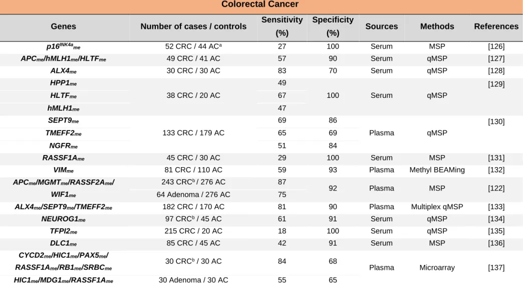
The increasing knowledge of the influence of epigenetic alterations in malignant transformation in the gut, gave rise to an opportunity for development of sensitive and specific minimally invasive epigenetic-based biomarkers for CRC. Hence, plentiful studies have investigated the detection value of these biomarkers (Table 3). SEPT9me is the main
described methylated gene in blood from CRC patients. Remarkably, this marker was the first blood-based IVD assay for detection of occult cancer based on an epigenetic alteration approved by the US Food and Drug Administration (FDA), in 2016, under the name of “Epi ProColon® 2.0” (Epigenomics AG) [46]. Additionally, this CE-IVD marked test is also commercially available in Europe and China [116]. A meta-analysis published in 2017 reported that SEPT9me sensitivity for CRC detection varies between 73-78% depending on
the algorithm used to consider a positive result, while specificity varies between 84-96% [117]. Nevertheless, when the biomarker performance of this gene’s methylation was assessed in a multicentre screening setting (PRESEPT clinical trial) with asymptomatic individuals older than 50 years old, the results from 53 CRC cases and 1457 subjects without CRC yielded 48% sensitivity and 92% specificity [118].
Given the importance to detect pre-malignant conditions, several studies have not only studied the CRC detection performance, but also the capability to detect adenomas. Disappointingly, SEPT9me performance to detect advanced adenomas ranged between
8-31% [119-121], being reported to be 11% in the previously mentioned screening setting study [118]. Thus, the usefulness of this gene for population-based screening is questionable.
As expected, gene panels have improved the performance to detect both adenomas and CRC. Remarkably, APCme, MGMTme, RASSF2Ame and WIF1me panel discriminated
adenomas and early stage CRC with 75% and 87% sensitivity, respectively, and 92% specificity [122], whereas SFRP1me, SFRP2me, SDC2me and PRIMA1me panel detected
adenomas with 89% sensitivity and 87% specificity, and CRC with 92% sensitivity and 97% specificity [123]. Nevertheless, the performance of these panels in a large screening setting remains to be elucidated. Interestingly, BCAT1me and IKZF1me panel performance has been
evaluated in large multicentre studies, displaying 62-66% sensitivity and 92-95% specificity for CRC detection, although with a limited 6-9% sensitivity for adenomas detection [124, 125].
Table 3. Circulating cell-free DNA methylation-based biomarkers for colorectal cancer detection.
Colorectal Cancer
Genes Number of cases / controls Sensitivity (%)
Specificity
(%) Sources Methods References
p16INK4a me 52 CRC / 44 ACa 27 100 Serum MSP [126] APCme/hMLH1me/HLTFme 49 CRC / 41 AC 57 90 Serum qMSP [127] ALX4me 30 CRC / 30 AC 83 70 Serum qMSP [128] HPP1me 38 CRC / 20 AC 49 100 Serum qMSP [129] HLTFme 67 hMLH1me 47 SEPT9me 133 CRC / 179 AC 69 86 Plasma qMSP [130] TMEFF2me 65 69 NGFRme 51 84 RASSF1Ame 45 CRC / 30 AC 29 100 Serum MSP [131]
VIMme 81 CRC / 110 AC 59 93 Plasma Methyl BEAMing [132]
APCme/MGMTme/RASSF2Ame/
WIF1me
243 CRCb / 276 AC 87
92 Plasma MSP [122]
64 Adenoma / 276 AC 75
ALX4me/SEPT9me/TMEFF2me 182 CRC / 170 AC 81 90 Plasma Multiplex qMSP [133]
NEUROG1me 97 CRCb / 45 AC 61 91 Serum qMSP [134]
TFPI2me 215 CRC / 20 AC 18 100 Serum qMSP [135]
DLC1me 85 CRC / 45 AC 42 91 Serum MSP [136]
CYCD2me/HIC1me/PAX5me/
RASSF1Ame/RB1me/SRBCme
30 CRCb / 30 AC 84 68
Plasma Microarray [137]
![Figure 1. Blood-based liquid biopsy. Circulating tumour cells (CTC), circulating cell-free DNA (ccfDNA) [including circulating tumour DNA (ctDNA)], circulating cell-free RNA (ccfRNA) and exosomes are released from tumour cells to the](https://thumb-eu.123doks.com/thumbv2/123dok_br/15132161.1010910/29.892.103.748.113.458/figure-circulating-circulating-including-circulating-circulating-exosomes-released.webp)


![Figure 5. Circulating cell-free DNA methylation-based biomarkers described in literature for cancer detection common to at least two cancer types [Lung Cancer (blue box), Prostate Cancer (yellow box), Colorectal Cancer (orange box)]](https://thumb-eu.123doks.com/thumbv2/123dok_br/15132161.1010910/53.892.102.766.104.517/circulating-methylation-biomarkers-described-literature-detection-prostate-colorectal.webp)