1044
|
wileyonlinelibrary.com/journal/rda © 2019 Blackwell Verlag GmbH Reprod Dom Anim. 2019;54:1044–1049.1 | INTRODUCTION
Primary ovarian neoplasms account for 6.25% of the neoplasms in intact bitches, but only 0.45%–1.2% of all canine neoplasms (Pires, Seixas, Palmeira, & Payan‐Carreira, 2010; Saba & Lawrence, 2012). Teratomas are the rarest ovarian tumours (2–10% of all ovarian tu‐ mours) in dogs and affect mainly adult and geriatric patients (Rota, Tursi, Zabarino, & Appino, 2013). Arising from totipotent germ cells, they are characteristically composed of tissue derived from any of the three different embryonic germ layers (Nagashima et al., 2000). When only one germ layer component is present, they are classified as monophasic (Rota et al., 2013).
In general, well‐differentiated teratomas display a benign biological behaviour, remaining unnoticed through large peri‐ ods of time. Conversely, malignant variants (teratocarcinomas) present proliferation of poorly differentiated cells from multi‐ ple origins, with high metastatic potential (rates of 30%–50%). Metastases have been reported in the peritoneum, kidney, ab‐ dominal lymph nodes, lungs, adrenal glands and bones (Saba & Lawrence, 2012). Initially, teratomas evolve silently, but show clinical signs after a given size or in malignant conditions, such as a palpable abdominal mass, abdominal discomfort, fever, ap‐ athy, lameness, vomiting, weight loss and anorexia (Greenlee & Patnaik, 1985).
Received: 15 November 2018
|
Accepted: 7 March 2019 DOI: 10.1111/rda.13430S H O R T C O M M U N I C A T I O N
Co‐existing monophasic teratoma and uterine adenocarcinoma
in a female dog
Maria dos Anjos Pires
1| José Carlos Catarino
2| Hugo Vilhena
1,3,4| Susana Faim
5|
Tiago Neves
5| Andreia Freire
5| Fernanda Seixas
1| Leonor Orge
1,6|
Rita Payan‐Carreira
1,71Animal and Veterinary Research Centre (CECAV), University of Trás‐os‐Montes and Alto Douro (UTAD), Vila Real, Portugal 2Universidade Lusofona de Humanidades e Tecnologias, Medicina Veterinária, Lisboa, Portugal
3Baixo Vouga Veterinary Hospital, Águeda, Portugal
4Department of Veterinary Medicine, Centre for Investigation Vasco da Gama (CIVG), Vasco da Gama University School, Coimbra, Portugal
5University Veterinary Hospital of Coimbra, Coimbra, Portugal
6Pathology Laboratory, UEISPSA, Instituto Nacional de Investigação Agrária e Veterinária (INIAV), I.P., Oeiras, Portugal 7Department of Veterinary Medicine, School of Sciences and Technology, University of Évora, Évora, Portugal
Correspondence
Maria dos Anjos Pires, Animal and Veterinary Research Centre (CECAV), University of Trás‐os‐Montes and Alto Douro (UTAD), Vila Real, Portugal. Email: apires@utad.pt
Contents
Ovarian teratomas are occasionally reported in dogs; the rarest type is the monopha‐ sic teratoma, composed of tissues originating from only one germ layer. Canine endo‐ metrial adenocarcinomas are also rare in dogs and mainly affect geriatric females. This report describes the case of co‐existing ovarian teratoma and uterine adenocar‐ cinoma in a 10‐year‐old nulliparous female Boxer presented with lethargy, anorexia and purulent vaginal discharge. Abdominal ultrasonography evidenced pyometra and a mass in the left ovary. This was composed of a uniform whitish tissue with multiple cystic structures. The histology revealed an atrophy of the ovarian parenchyma, com‐ pressed by a proliferation of well‐differentiated nervous tissue staining positively to vimentin, S100 and neuronal specific enolase (NSE), and negatively to keratin and inhibin. The left uterine horn, whose diameter was markedly increased, showed foci of endometrial cellular atypia, evident nucleoli and mitoses, at light microscopy. To our best knowledge, this is the first report of a co‐existing ovarian monophasic tera‐ toma and endometrial adenocarcinoma, two rare reproductive neoplasia in dogs.
K E Y W O R D S
Uterine tumours are also uncommon in dogs (prevalence of 0.3%–0.4%), affecting mainly middle‐aged to geriatric females. They can be classified as epithelial or mesenchymal in origin. Epithelial tu‐ mours may be separated in adenoma and adenocarcinoma, which are the rarest type (Pires et al., 2010). Leiomyoma is the most common uterine tumour, comprising 85%–90% of the cases described (Saba & Lawrence, 2012). The clinical signs of uterine adenocarcinoma resem‐ ble those of other uterine diseases, namely purulent or haemorrhagic vaginal discharge, dysuria/hematuria, anorexia, abdominal pain, ab‐ dominal distension and anorexia (Pires et al., 2010).
Albeit ovarian and uterine masses may be identified using imag‐ ing diagnostic, only histopathology provides a conclusive diagnosis (Coggeshall, Franks, Wilson, & Wiley, 2012). Ovariohysterectomy is recommended as treatment in most cases and is curative if no meta‐ static disease is present (Saba & Lawrence, 2012).
This report describes two rare reproductive neoplasms co‐ex‐ isting in a Boxer female dog. To our best knowledge, this is the first report of concomitant ovarian monophasic teratoma and uterine ad‐ enocarcinoma described in the dog.
2 | CASE REPORT
A 10‐year‐old nulliparous female Boxer was admitted to the University Veterinary Hospital of Coimbra due to lethargy, anorexia and puru‐ lent vaginal discharge. Anamnesis highlighted existing regular oes‐ trous cycles at 6‐month intervals and inexistence of contraceptive treatments. Last oestrus was recorded 5 months ago. The clinical
and gynaecologic examinations were completed with haematology (showed mild neutrophilia 13.66 × 10³/µl; Supporting Information Table S1), serum biochemistry (glucose, serum total protein, BUN and creatinine within normal values; Supporting Information Table S2), urinalysis (decreased urine density [1.020]) and abdominal ultra‐ sonography (showing uterine distension by fluid and a mass in the left ovary). Ovariohysterectomy was recommended for suspected pyometra. Recovery from the surgery was uneventful, and the dog was discharged 48 hr after surgery with large spectrum anti‐ biotherapy (amoxicillin and clavulanic acid, 20 mg/kg, q12h, 10 days) and non‐steroidal anti‐inflammatory (meloxicam, 0.1 mg/kg, q24h, 5 days) medication. The post‐surgical follow‐up appointments were uneventful. Clinical controls, including abdominal ultrasonography, were performed at 1, 3, 6, 9 and 12 months after surgery. The animal was disease‐free at the 12‐month post‐surgery control.
The resected genital tract was fixed in 10% buffered forma‐ lin and submitted to the histopathological evaluation. At inspection, an enlarged and irregularly shaped left ovary (Figure 1) was found, with 8 × 5 × 4 cm (length × width × thickness), but the right ovary (2.5 × 1.5 × 1.5 cm) was morphologically normal. On cross section, the left ovary presented a uniform, compact mass tissue affecting the ma‐ jority of the parenchyma, which was reduced to a margin with 0.3 mm, and multiple cystic structures, the biggest with 3 cm in diameter. The uterus presented an irregular morphology and a haemorrhagic content (right horn: 22 × 2 cm; left horn: 30 × 3.5 cm in length and diameter, re‐ spectively). Besides the marked dilation, the left uterine horn showed multiple adhesions between its cranial end and the ovarian bursa, dis‐ placing the ovary to the mesometrial side of the uterine horn.
F I G U R E 1 (a–c) Macroscopic aspect
of the left ovary. The characteristic canine ovarian morphology disappeared, presenting an irregular and vascularized surface. (c) In the cross section, the gonad showed a uniform, solid and dense mass of firm whitish tissue with cystic structures, which occupied almost the entire structure, reducing the ovarian parenchyma to a thin margin. (d) Right ovary with normal structure and corpus luteum. Barr = 1 cm
(a) (b)
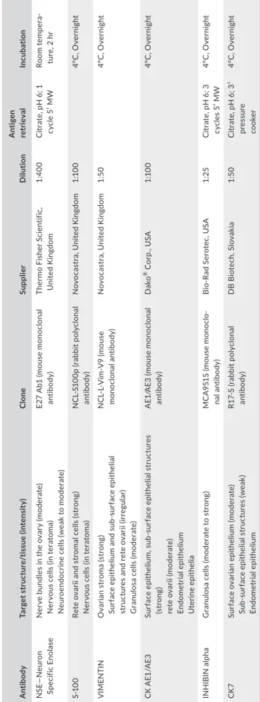
T A B LE 1 A nt ib od ie s u se d t o c ha ra ct er iz e t he ov ar ia n a nd e nd om et ria l l es io ns A nt ib ody Ta rg et s tr uc tu re /t is sue (i nt en si ty ) Cl one Su pp lier D ilut io n A nt ig en re tr ie val Inc ub at io n N SE — N eu ro n Sp ecif ic E no las e N er ve b un dl es i n t he o va ry ( m od er at e) N er vo us c el ls (i n t er at om a) N eu ro en do cr in e c el ls (w ea k t o m oder at e) E2 7 A b1 ( m ou se m on oc lo na l an tib od y) Th er m o F is her S ci en tif ic , U nit ed K in gd om 1: 40 0 C itr at e, p H 6 ; 1 cy cl e 5 ′ M W Ro om te mp er a‐ tu re , 2 h r S‐ 10 0 Re te o va rii a nd s tr om al c el ls ( st ro ng ) N er vo us c el ls (i n t er at om a) N C L‐ S1 00 p ( ra bb it p ol yc lo na l an tib od y) N ov oc as tr a, U ni te d K in gd om 1:1 00 4° C , O ve rn ig ht V IME N TIN O va ria n s tr om a ( st ro ng ) Su rf ac e ep ith el iu m a nd s ub ‐s ur fac e ep ith el ia l st ru ct ur es a nd re te o va rii (ir reg ula r) G ra nu lo sa c el ls (m oder at e) N C L‐ L‐ V im ‐V 9 ( m ou se m on oc lo na l a nt ib od y) N ov oc as tr a, U ni te d K in gd om 1: 50 4° C , O ve rn ig ht C K AE 1/ AE 3 Su rf ac e ep ith el iu m , s ub ‐s ur fac e ep ith el ia l s tr uc tu re s (s tr on g) re te o va rii (m od er ate ) En do me tr ia l ep ith el iu m U ter in e ep ith el ia A E1 ⁄A E3 ( m ou se m on oc lo na l an tib od y) D ako ® C or p. , U SA 1:1 00 4° C , O ve rn ig ht IN H IB IN al ph a G ra nu lo sa c el ls ( m od er at e t o s tr on g) M C A 95 1S (m ou se m on oc lo ‐ na l a nt ib od y) B io ‐R ad S er ot ec , U SA 1: 25 C itr at e, p H 6 ; 3 cy cl es 5 ′ M W 4° C , O ve rn ig ht CK 7 Su rf ac e o va ria n ep ith el iu m (m oder at e) Su b‐ su rf ac e e pi th el ial s tr uc tu re s ( w ea k) En do me tr ia l ep ith el iu m R1 7‐ S ( ra bb it p ol yc lo na l an tib od y) DB Bi ot ec h, S lo va ki a 1: 50 C itr at e, p H 6 ; 3 ′ pre ss ure co oke r 4° C , O ve rn ig ht
Ovarian and uterine sections were routinely processed, par‐ affin‐embedded and stained with haematoxylin–eosin (H‐E). Immunohistochemistry was performed with specific antibodies against neuronal specific enolase (NSE), S‐100, vimentin, broad‐ spectrum cytokeratin (CK) AE1‐AE3, CK7 and inhibin (Table 1), with 3,3‐Diaminobenzidine (DAB) as chromogen and Gill’s haematoxylin for counterstaining.
Histologically, the parenchyma of the right ovary presented multiple small surface cysts and Call‐Exner‐like bodies in the me‐ dulla. Atrophy of the left ovary parenchyma was confirmed, and the compression was exerted by a mass composed of a prolif‐ eration of well‐differentiated nervous tissue (Figure 2a–d), sur‐ rounded by a dense connective tissue. Neoplastic cells showed
strong cytoplasmic immunoreactivity against both S‐100 and NSE, occasional positivity to vimentin, but were negative to tested CKs and inhibin (Figure2e–h). The surface epithelium showed strong immunoreactivity to CK AE1‐AE3 and CK7, while the stroma was positive to vimentin and interstitial glands posi‐ tive to inhibin.
The uterus displayed cystic glandular hyperplasia, with a muco‐ purulent content in the glands and lumen, contaminated with cocci bacteria. In the cranial segment of the left horn, it was observed pro‐ liferation of the endometrium showing cellular atypia, aberrant cells with evident nucleoli and mitosis in a papillary disposition, invading the muscular layer. The neoplastic cells were positive to large spec‐ trum keratin and CK7 (Figure 3).
F I G U R E 2 Left ovary. At microscopy,
the parenchyma was reduced to a thin margin (a and b) with interstitial glands, small cysts of sub‐surface epithelial structures and dense capsule of connective tissue (arrow). The proliferation of well‐differentiated nervous tissue is notorious in the images (a, c and d. Haematoxylin and eosin stain. e–f) Immunohistochemical positive labelling of teratoma nervous tissue with S100 (e), NSE (f) and vimentin (g) and negative to labelling to inhibin (h). Counterstain with Gill’s haematoxylin
(a) (b)
(c) (d)
(g) (h)
Supported by the histopathological findings, the diagnosis was mature ovarian monophasic teratoma co‐existing with an endome‐ trial adenocarcinoma and septic pyometra.
Ethical approval was not deemed necessary for this study; the informed consent of the owners was granted to publicly describe the current case.
3 | DISCUSSION
Ovarian and endometrial tumours are considered rare in dogs, al‐ beit some bias may result from the customary practice of spaying in early ages (Saba & Lawrence, 2012). Ovarian teratomas are the less common type of ovarian neoplasms in female dogs. They are charac‐ terized by proliferation of any tissue of embryonic origin (e.g., skin, sebaceous glands, hair follicles, adipocytes or nervous tissue, among others). In the case described herein, a proliferation of only one type of germinative cell (nervous tissue) was observed, and consequently, a diagnosis of monophasic mature teratoma was established. To the authors’ knowledge, only one other case of was previously reported in dogs, in an ovarian remnant of a 5‐years‐old mixed bred bitch (Rota et al., 2013).
The morphological appearance of the neoplastic cells in the H‐E staining clearly evidenced the neural origin of tumour cells, as further confirmed by the positive immunoreactivity against NSE, S‐100 and vimentin. On the atrophic ovarian parenchyma, positiv‐ ity to vimentin, CK A1/A3, CK7 and inhibin confirmed the presence of typical ovarian components. The negative labelling for keratin and inhibin in the neoplastic cells excluded the presence of tissues from other origins in the teratoma and other ovarian tumours.
Adenocarcinomas are considered rare neoplasm in the canine uterus (Maya‐Pulgarin, Gonzalez‐Dominguez, Aranzazu‐Taborda,
Mendoza, & Maldonado‐Estrada, 2017), contrasting to other do‐ mestic species (Kennedy et al., 1998), including the queen (Saraiva et al., 2015). Dogs tend to develop degenerative and inflammatory conditions of the endometrium (e.g., cystic hyperplasia and pyome‐ tra) with age and in response to irritative stimuli (Payan‐Carreira & Pires, 2016).
Clinical signs associated with endometrial adenocarcinomas de‐ pend upon the tumour size and the presence of metastatic disease or other comorbidities. They are often clinically masked by signs com‐ mon to most genital tract diseases (Payan‐Carreira & Pires, 2016), such as vaginal discharge. Other clinical signs include anorexia and abdominal discomfort or enlargement. Adenocarcinomas might be underdiagnosed in dogs due to an inadequate post‐surgical or post‐ mortem evaluation of the genital tract. As the clinical signs are often misinterpreted as pyometra or abortion, the histopathological evalu‐ ation of genital apparatus is seldom required.
In the present case, the same patient presented two rare tu‐ mours: a monophasic teratoma and an endometrial adenocarcinoma. This diagnosis was only possible after a careful and detailed histo‐ pathological evaluation, emphasizing the importance of this ex‐ amination. Immunohistochemistry confirmed the presence of only neuronal tissue on the teratoma and determined the endometrial origin of the uterine lesion, proving the usefulness to differentiate and describe different tumour cell populations.
ACKNOWLEDGEMENTS
The authors acknowledge the technical support of Ligia Lourenço, Gustavo Paixão for language editing and to CECAV for the use of the digital image acquisition software Nikon NIS‐Elements D (found by PEst‐OE/AGR/UI0772/2014) and also the owners of the female dog for granting permission to publish this case.
F I G U R E 3 Cranial segment
of the uterine horn showing the endometrium with cellular atypia, aberrant cells, with evident nucleoli, arranged in a papillary disposition (a–d). Immunohistochemical positive labelling to broad‐spectrum keratin (c) and keratin 7 (d). Haematoxylin and eosin (a and b). Immunohistochemistry counterstain with Gill’s haematoxylin (c and d)
(a) (b)
CONFLIC T OF INTERESTS
The authors have no conflict of interests to declare.
AUTHOR CONTRIBUTIONS
MAP and FS performed the histopathological diagnose; JC and MAP performed the immunohistochemistry; RP‐C, MAP and FS evaluated the immunohistochemistry; LO was the consultant in neuropathol‐ ogy; MAP, RP‐C and JC contributed to the manuscript writing and the reviewing of the literature. All the authors read and approved the content of the MS.
ORCID
Maria dos Anjos Pires https://orcid.org/0000‐0001‐9473‐5283
Hugo Vilhena https://orcid.org/0000‐0003‐3976‐1619
Fernanda Seixas https://orcid.org/0000‐0002‐1912‐1457
Leonor Orge https://orcid.org/0000‐0002‐2673‐2758
Rita Payan‐Carreira https://orcid.org/0000‐0001‐5225‐4510
REFERENCES
Coggeshall, J., Franks, J., Wilson, D., & Wiley, J. (2012). Primary ovarian teratoma and GCT with intra‐abdominal metastasis in a dog. Journal of the American Animal Hospital Association, 48(6), 424–428. https:// doi.org/10.5326/JAAHA‐MS‐5809
Greenlee, P., & Patnaik, A. (1985). Canine ovarian tumors of germ cell origin. Veterinary Pathology, 22, 117–122. https://doi. org/10.1177/030098588502200204
Kennedy, P., Cullen, J., Edwards, J., Goldschmidt, M., Larsen, S., Munson, L., & Nielsen, S. (1998). Histological classification of tumors of genital system of domestic animals, 2nd ed. Vol. IV. (p. 32). Washington, DC: World Health Organization and Armed Forces Institute of Pathology. Maya‐Pulgarin, D., Gonzalez‐Dominguez, M., Aranzazu‐Taborda, D.,
Mendoza, N., & Maldonado‐Estrada, J. (2017). Histopathologic findings in uteri and ovaries collected from clinically healthy dogs
at elective ovariohysterectomy: A cross‐sectional study. Journal of Veterinary Science, 18(3), 407–414. https://doi.org/10.4142/ jvs.2017.18.3.407
Nagashima, Y., Hoshi, K., Tanaka, R., Shibazaki, A., Fujiwara, K., Konno, K., … Yamane, Y. (2000). Ovarian and retroperitoneal teratomas in a dog. Journal of Veterinary Medicine Science, 62(7), 793–795. https:// doi.org/10.1292/jvms.62.793
Payan‐Carreira, R., & Pires, M. A. (2016). The value of a routine histo‐ pathological examination of uterine specimens in dogs and cats. In Histopathology (pp 1–14). Dover, DE: SM Group Open Access eBooks. Pires, M. A., Seixas, F., Palmeira, C., & Payan‐Carreira, R. (2010).
Histopathologic and Immunohistochemical exam in one case of ca‐ nine endometrial adenocarcinoma. Reproduction in Domestic Animals, 45, 545–549. https://doi.org/10.1111/j.1439‐0531.2008.01243.x Rota, A., Tursi, M., Zabarino, S., & Appino, S. (2013). Monophasic ter‐
atoma of the ovarian remnant in a bitch. Reproduction in Domestic Animals, 48(2), e26–e28. https://doi.org/10.1111/rda.12090 Saba, C., & Lawrence, J. (2012). Tumors of the female reproductive sys‐
tem. In S. J. Withrow, & D. M. Vail (Eds.), Withrow and MacEwen’s small animal clinical oncology, 5th ed. (pp. 610–618). St. Louis, MO: Saunders Elsevier.
Saraiva, L., Payan‐Carreira, R., Gartner, F., Santana, I., Rêma, A., Lourenço, L., & Pires, M. A. (2015). Immunohistochemical expression of cyclooxygenase‐2 (COX‐2) in feline endometrial adenocarcinoma and in normal and hyperplastic endometria. Reproduction in Domestic Animals., 50(2), 333–340. https://doi.org/10.1111/rda.12497
SUPPORTING INFORMATION
Additional supporting information may be found online in the Supporting Information section at the end of the article.
How to cite this article: Pires MDA, Catarino JC, Vilhena H, et
al. Co‐existing monophasic teratoma and uterine adenocarcinoma in a female dog. Reprod Dom Anim. 2019;54:1044–1049.