DIVERSITY, DISTRIBUTION AND
PHYLOGENETIC RELATIONSHIPS
OF ROCK SPONGES (PORIFERA,
'LITHISTID' DEMOSPONGIAE) OF
THE MACARONESIAN ISLANDS
AND NORTHEAST ATLANTIC
SEAMOUNTS
Francisca Correia de Carvalho
Dissertação de Mestrado apresentada à
Faculdade de Ciências da Universidade do Porto em
Biodiversidade, Genética e Evolução
DIVERSITY, DISTRIBUTION AND
PHYLOGENETIC RELATIONSHIPS OF
ROCK SPONGES (PORIFERA,
'LITHISTID' DEMOSPONGIAE) OF THE
MACARONESIAN ISLANDS AND
NORTHEAST ATLANTIC
SEAMOUNTS
Francisca Correia de Carvalho
Biodiversidade, Genética e Evolução
Departamento de BiologiaOrientadora
Doutora Joana R. Xavier, Post-Doc., CIBIO-Açores
Coorientador
Todas as correções determinadas pelo júri, e só essas, foram efetuadas. O Presidente do Júri,
ACKNOWLEDGEMENTS
My work was only possible with the help of many people that during this period of time supported me in many ways.
I am deeply thankful to Joana Xavier, my supervisor. For making me believe. For all the hours spent sharing your knowledge, strength, support and friendship. For all the enthusiastic hours spent on the microscope. For all the forgiveness when I was late. For supporting me since the first day and make my confidence come back even when it seemed to be lost. It was an amazing experience to work and learn with you. Thank you so much!!
I also wish to thank James Harris for accepting to co-supervise me and dedicate so much of his time and patience to a very important part of this work. It wouldn´t be possible without your support. Thank you for all the kindness and availability when I needed the most. To the Spongelab team, a very special thanks. It was really an enriching experience to work with all of you. To Francisco Pires for being a friend, and a teacher at the lab. And for converting a simple boring lab room into a nice and cool place to work. To Raquel Pereira for the endless patience helping me doing the maps. And for the help whenever I needed. I am going to miss all these times.
I am also very thankful to Raquel Xavier for receiving me so well and helping me in the most difficult hours during the molecular work. Thank you for teaching me and for always clarifying my doubts. All the people of CIBIO´s lab for being there, always available when I needed.
This work also wouldn´t be possible without the help of Jorge Medeiros with the SEM microscopy. Thank you for all the hours spent in search of microscleres and for your great work. To Isadora Moniz for helping me to finish the huge amount of preparations that seemed to be endless!
To Joana Xavier once again, for providing me the chance to meet exceptional researchers and to participate in the "International Workshop on Taxonomy of Atlanto-Mediterranean Deep-Sea Sponge Fauna". It was an incredible and fascinating experience where I could learn more and more about sponge taxonomy. To Andrzej Pisera for sharing his expert knowledge with me. To Shirley Pomponi for providing so many interesting samples to complete my work and for all her kindness. It was very nice to meet and get to
work with you. To Paco Cárdenas for his help every time I needed during my thesis and to work with me.
To Dr. Filipe Porteiro for making the Azorean samples available for this study. To all the people, technicians and researchers who have done an incredible work on collecting samples and take a good care of them in the collections.
To Joana Vilela, Íris Sampaio and Susana Freitas. A huge thank you. For being more than just colleagues and partners. For the friendship, all the good conversations in beer's company. For being so kind, helpful and for preventing my "panic attacks" to be more serious. Thank you!
To my Mom and Dad. Always present in the most difficult moments. Always supporting - and sponsoring - my decisions and making my act of dreaming possible. For helping me whenever I needed and giving me wings since ever.
To my brothers and sisters for being themselves. Simply for being themselves. My life wouldn't be the same and I wouldn't be the same without you. My dreams have grown with you. They wouldn't be possible without your love, support and care. And the courage you all give me. Obrigada Susana, Bábá, Zé e Gui. :)
To my three grandmothers. Vó Zecas, Vó Glória e Zairinha. I can't tell how much your love fulfills me and makes me a better person each day. Your embrace and your smile every time I see you warms and deeply encourages me.
To my uncle Maria Rui, for passing me the enthusiasm for Biology and for always supporting my decisions.
To you, for making part of my life.
To all my friends and family. For making me so happy. It feels so good to have people like you on my side.
This thesis is dedicated to all of you. Obrigada!!!
ABSTRACT
'Lithistid' Demospongiae sensu Pisera & Lévi, 2002 is a much understudied polyphyletic group of sponges usually found at bathyal or bathyal-like environments, such as caves, in tropical and warm temperate regions.
In this study we investigated the diversity, distribution and phylogenetic relationships of the lithistid sponge fauna of the northeast Atlantic with a focus on the Macaronesian Islands (Azores, Madeira, Selvagens and Canaries) and neighbouring seamounts (of the Great Meteor group). Specimens were collected in the course of several research campaigns conducted by the Azores University in the Azores EEZ, by the MNHN of Paris in the seamounts southern of the Azores (the Seamount-2 campaign) and by the Harbor Branch Oceanographic Institute (HBOI) in Madeira, Selvagens and Canary islands.
A total of 162 specimens were analyzed and assigned to 11 taxa representing five families and nine genera. Eight species were recorded for the first time on the seamounts southern of the Azores, of which seven are shared with this archipelago. Three species constitute new records for Madeira Island (Neophrissospongia nolitangere, Discodermia verrucosa and Racodiscula clava), two for Selvagens (N. nolitangere and Macandrewia azorica), and two for the Canaries, of which one is a new species for science of the genus Isabella. Of the 14 species found to occur in the Macaronesian islands, only three viz. N. nolitangere, Leiodermatium pfeifferae and R. clava are shared among all archipelagos, the remainder being restricted to one or two island groups. Similarly, only four species seem to be shared between the Mediterranean Sea and these Atlantic archipelagos: N. nolitangere (with the Azores, Madeira, Selvagens and the Canaries) Neoschrammeniella bowerbankii (with Madeira); Siphonidium ramosum (with the Azores) and L. lynceus (with the Azores and the Canaries). Furthermore, several changes of taxonomic significance were proposed based on the new findings such as re-definition of the genera Neophrissopongia and Isabella, and the species N. bowerbankii. A discussion concerning the validity of Topsent’s Racodiscula clava is also presented.
Phylogenetic reconstructions, by means of Bayesian analyses, of the mtDNA COI and the rDNA 28S genes confirmed the polyphyly of this group. The astrophorid affinity of the families Corallistidae, Macandrewiidae and Theonellidae was shown. Species and genera were always retrieved as well-supported monophyletic clades. The COI analysis has revealed for the first time the monophyly of the family Corallistidae and a sister-taxa
relationship was found between the genera Neoschrammeniela/Neophrissospongia and Herengeria/Isabella within this family. The family Theonellidae (Discodermia, Theonella and Racodiscula) was revealed non-monophyletic in both analysis. Lastly the family Azoricidae, here represented by Leiodermatium, exhibits a basal position in relation to the astrophorid/lithistid clade, but its phylogenetic affinity remains unclear.
This study greatly expands the knowledge on the diversity, distribution and phylogenetic relationships of the lithistid sponges associated with the bathyal environment of the Macaronesian Islands and the northeast Atlantic seamounts.
Keywords: Lithistids, Macaronesian Islands, seamounts, diversity, distribution,
RESUMO
'Lithistid' Demospongiae sensu Pisera & Lévi, 2002 é um grupo polifilético de esponjas muito pouco estudado que normalmente se encontra na zona batial ou em ambientes semelhantes, como grutas, em regiões tropicais e temperadas quentes.
Neste estudo investigou-se a diversidade, distribuição e relações filogenéticas da fauna de esponjas lithistidas do nordeste Atlântico com foco nas ilhas da Macaronésia (Açores, Madeira, Selvagens e Canárias) e nas montanhas submarinas adjacentes (do grupo do Great Meteor). Os exemplares foram recolhidos no decurso de várias expedições científicas realizadas pela Universidade dos Açores na ZEE dos Açores, pelo MNHN de Paris nas montanhas submarinas localizadas a sul dos Açores (a campanha Seamount-2) e pelo Harbor Branch Oceanographic Institute (HBOI) nas ilhas da Madeira, Selvagens e Canárias.
Um total de 162 espécimes foram analisados, tendo sido identificados 11 taxa representantes de cinco famílias e nove géneros. Pela primeira vez, foram registadas oito espécies de lithistidas nas montanhas submarinas a sul dos Açores, das quais sete são partilhadas com este arquipélago. Na Madeira, foram encontrados três novos registos (Neophrissospongia nolitangere, Discodermia verrucosa e Racodiscula clava), dois nas Selvagens (N. nolitangere and Macandrewia azorica) e dois nas Canárias, dos quais um representa uma espécie nova para a ciência do género Isabella. Das 14 espécies registadas para as ilhas da Macaronésia, apenas três são partilhadas entre todos os arquipélagos, N. nolitangere, Leiodermatium pfeifferae e R. clava, sendo as restantes espécies restritas a um ou dois grupos de ilhas. Similarmente, apenas quatro espécies parecem ser partilhadas entre o mar Mediterrâneo e estes arquipélagos do Atlântico: N. nolitangere (com os Açores, Madeira, Selvagens e Canárias), Neoschrammeniella bowerbankii (com a Madeira); Siphonidium ramosum (com os Açores) e L. lynceus (com os Açores e as Canárias). Para além disso, várias alterações de significância taxonómica foram aqui propostas, com base nos resultados obtidos, tais como a re-definição dos géneros Neophrissospongia e Isabella, e da espécie Neoschrammeniella bowerbankii. A validade da espécie Racodiscula clava descrita por Topsent, também é aqui discutida.
Reconstruções filogenéticas, por métodos de inferência Bayesiana, dos genes mtDNA COI e do rDNA 28S confirmam a polifilia deste grupo. A afinidade das famílias Corallistidae, Macandrewiidae e Theonellidae com as astrophoridas foi verificada. Espécies
e géneros foram sempre obtidos em clados monofiléticos bem suportados. Na análise do COI verificou-se, pela primeira vez, a monofilia da família Corallistidae e foi encontrada uma relação de grupo-irmão entre os géneros Neoschrammeniela/Neophrissospongia e Herengeria/Isabella. A família Theonellidae (Discodermia, Theonella e Racodiscula) revelou-se não monofilética em ambas as análises. Por último, a família Azorecidae, representada nas análises pelo género Leiodermatium, demonstra uma posição basal em relação ao clade astrophoridas/lithistidas, mas as suas afinidades filogenéticas continuam por esclarecer.
Este estudo expande significativamente o nosso conhecimento da diversidade, distribuição e relações filogenéticas das esponjas lithistidas associadas ao ambiente batial das ilhas da Macaronésia e das montanhas submarinas do nordeste Atlântico.
Palavras chave: Lithistidas, Ilhas da Macaronésia, montanhas submarinas, diversidade,
TABLE OF CONTENTS
ACKNOWLEDGEMENTS VII ABSTRACT IX Keywords X RESUMO XI Palavras-chave XII INDEX OF FIGURES XVINDEX OF TABLES XXI
LIST OF ACRONYMS XXIII
CHAPTER 1. INTRODUCTION 25
1.1. Phylum Porifera 27
1.2. Lithistid sponges 29
1.3. Goals 32
CHAPTER 2. MATERIAL AND METHODS 33
2.1. Samples and study area 35
2.2. Taxonomy 36
2.3. Phylogenetic relationships 37
2.3.1. DNA extraction, amplification and sequencing 37
2.3.2. Data analysis 38
CHAPTER 3. RESULTS 41
3.1. Diversity and distribution of lithistids on the northeast Atlantic Ocean 43
3.2. Systematics 46
'Lithistid' Demospongiae sensu Pisera & Lévi, 2002 46
Family Corallistidae Sollas, 1888 46
Family Siphonidiidae Lendenfeld, 1903 71
Family Azoricidae Sollas, 1888 74
Family Macandrewiidae Schrammen, 1924 78
3.3. Phylogenetic relationships 81
CHAPTER 4. DISCUSSION 89
4.1. Lithistid sponges of the North Atlantic Ocean and the Mediterranean Sea 91
4.2. Phylogenetic relationships of lithistids 95
CHAPTER 5. REFERENCES 99
GLOSSARY 113
APPENDIX I 119
APPENDIX II 127
INDEX OF FIGURES
Fig. 1 - Study area: the Macaronesian archipelagos of the Azores, Madeira, Selvagens
and Canaries, and the northeast Atlantic seamounts located southern of the Azores.
36
Fig. 2 - Distribution of lithistids in Macaronesian islands and neighboring seamounts:
white-filled circles represent records from the literature and red-filled circles represent the specimens analyzed in this study.
43
Fig. 3 - Corallistes masoni (Bowerbank, 1869). A, specimen from Madeira in situ; B,
specimen collected in Madeira, photo ex situ (scale 1 cm); C, ectosomal skeleton showing a layer of dichotriaenes (scale 200 µm); D, choanosomal and ectosomal skeleton (scale 200 µm); E, dicranoclone desmas (scale 200 µm); F, dichotriaenes (scale 200µm); G, oxeas (scale 200 µm); H, two types of spirasters: type I with thin, short and numerous rays, and type II with thin, long and few rays (scale 50 µm).
48
Fig. 4 - Neophrissospongia nolitangere (Schmidt, 1870). A, adult individual from the
Canaries, in situ; B, young specimen collected in the Azores, ex situ (scale 1 cm); C, ectosomal skeleton with a compact layer of dichotriaenes and microscleres (scale 200 µm); D, choanosomal skeleton (scale 200 µm).
51
Fig. 5 - Neophrissospongia nolitangere (Schmidt, 1870). A, dicranoclone desmas
(scale 100 µm); B, detail of dicranoclones where one can observe the circular-shape elevated tubercles (scale 100 µm); C, ectosomal dichotriaene (scale 50 µm); D, cladome of dichotriaene of an adult individual, view from below (scale 50 µm); E, smooth cladome of dichotriaene of a young individual, view from below (scale 50 µm);
F, microstyles (scale 10 µm); G, spirasters/streptasters with thick and spiny arms
(scale 5 µm); H, spirasters/streptasters with long and smooth arms (scale 5 µm).
52
Fig. 6 - Neoschrammeniella bowerbankii (Johnson, 1863). A, specimen collected in
Madeira Island, in situ; B, specimen collected in Hyeres seamount, ex situ (scale 1 cm); C, top view of ectosomal skeleton where one can observe a layer of dichotriaenes and abundant microscleres (scale 200 µm); D, transversal view of choanosomal skeleton (scale 200 µm).
Fig. 7 - Neoschrammeniella bowerbankii (Johnson, 1863). A, overview of
dicranoclone desmas (scale 100 µm); B, detail of dicranoclone desmas, note the tubercles divided into small tubercles and the oxea with blunt tip (scale 10 µm); C, dichotriaene (scale 100 µm); D, smooth cladome of a dichotriaene (scale 100 µm); E, spirasters of type I, with short, thick and blunt arms (scale 5 µm); F, spirasters of type II, with long, thin and pointed arms (scale 10 µm); G, spirasters of type III found in this present work, with short, thick and pointed arms (scale 5 µm).
56
Fig. 8 - Isabella harborbranchi Carvalho, Pomponi & Xavier (in prep.). A, specimens
collected in the Canaries, ex situ (scale 1 cm); B, top view of the ectosome where one can observe the huge quantity of microscleres and some dichotriaenes and triaenes (scale 200 µm); C, choanosomal skeleton and dicranoclone desmas (scale 200 µm);
D, overview of dicranoclone desmas (scale 100 µm); E, detail of dicranoclone desmas,
note the abundant microacanthoxeas and part of an oxea (in the lower-left corner) (scale 100 µm).
59
Fig. 9 - Isabella harborbranchi Carvalho, Pomponi & Xavier (in prep.). A, dichotriaene
(scale 100 µm); B, short-shafted triaene (scale 100 µm); C, long-shafted triaene with a small arm in the rhabdome (scale 100 µm); D, microacanthoxeas (scale 10 µm); E, centrotylotes (scale 10 µm); F, detail of the extremity of a microacanthoxea (scale 10 µm); G, detail of the extremity of a centrotylote (scale 10 µm); H-J, spirasters with long, thin and thorny arms (scale 5 µm); K-N, streptasters with short, thick and thorny arms (scale 5 µm).
60
Fig. 10 - Discodermia ramifera Topsent, 1892. A, specimen collected in the Azores,
ex situ (scale 1 cm); B, ectosome, where one can observe the compact layer of discotriaenes (scale 100 µm); C, choanosomal skeleton (scale 100 µm).
63
Fig. 11 - Discodermia ramifera Topsent, 1892. A, overview of tetraclone desmas
(scale 500 µm); B, detail of tetraclones; C, top view of discotriaenes (scales 50 and 100 µm, respectively); D, bottom view of discotriaenes, note the variability of shapes (scales 100 µm); E, long oxeas (scales 50 and 100 µm, respectively); F, microxeas
(scales 10 µm); G, detail of the extremity of microxeas (scale 1 µm); H, acanthorhabds (scales 5 µm).
Fig. 12 - Discodermia verrucosa Topsent, 1928. A, specimens collected in Madeira
Island, ex situ (scale 1 cm); B, ectosome with a compact layer of discotriaenes (scale 200 µm); C, transversal image of choanosomal skeleton (scale 200 µm); D, tetraclone desmas (scale 200 µm); E, discotriaene (scale 200 µm); F, long oxeas (scale 200 µm);
E, microxeas (bigger) and acanthorhabds (smaller)(scale 100 µm).
66
Fig. 13 - Racodiscula clava sensu Topsent, 1892. A, specimens collected in the
Hyeres seamount, ex situ (scale, 1cm); B, ectosome, where one observes a layer of phyllotriaenes and abundant microscleres (scale 200 µm); C, choanosomal skeleton composed of tetraclones (scale 200 µm).
68
Fig. 14 - Racodiscula clava sensu Topsent, 1892. A, general view of tetraclone
desmas (scale 100 µm); B, detail of the tetraclones’ shapes, note the smooth and large tubercles (scale 50 µm); C, bottom view of phyllotriaenes, where is clear the variability of shapes (scale 100 µm); D, oxea (scale 100 µm); E, acanthoxeas (scale 5 µm); F, acanthorhabds (scale 5 µm); G, spirasters (scale 5 µm).
70
Fig. 15 - Siphonidium ramosum (Schmidt, 1870). A, specimens collected in the
Canaries, ex situ (scale 1 cm); B, image of the ectosomal (lower part) and choanosomal skeleton (scale 200 µm); C, choanosomal skeleton formed by extremely compacted rhizoclones (scale 200 µm); D, rhizoclones desmas (scale 200 µm); E, exotylostyle (scale 200 µm).
73
Fig. 16 - Leiodermatium sp.. A, specimen collected in Madeira, in situ; B, specimen
collected in the Azores, ex situ (scale 1 cm); C, ectosome, composed by desmas, bundles of oxeas and some raphides (scale 200 µm); D, choanosome, showing rhizoclone desmas surrounding a pore (scale 200 µm).
76
Fig. 17 - Leiodermatium sp.. A, overview of rhizoclone desmas (scale 100 µm); B,
detail of the sculpture and terminations of rhizoclones, note the multifurcating spines
of the terminations (scale 10 µm); C, oxeas (scale 100 µm); D, detail of an oxea's extremity (scale 10 µm); E, raphide (scale 100 µm).
Fig.18 - Macandrewia azorica Gray, 1859. A, specimens collected on the Tyro
seamount, ex situ, note the diversity of shapes in different stages (no scale); B, specimen collected in Azores, ex situ (scale 1 cm); C, ectosome showing a layer of abundant phyllotriaenes and microscleres around the pores (scale 200 µm); D, choanosomal skeleton formed by rhizoclones desmas (scale 200 µm).
80
Fig. 19 - Macandrewia azorica Gray, 1859. A, general view of rhizoclone desmas
(scale 100 µm); B, detail of articulation and shape of the rhizoclones (scale 50 µm);
C, phyllotriaene (scale 50 µm); D, bottom view of phyllotriaene showing the extremely
indented cladome (scale 100 µm); E, small phyllotriaene in development (scale 50 µm); F, oxea (scale 50 µm); G, fusiform microxeas (scales 10 µm).
81
Fig. 20 - Macandrewia robusta Topsent, 1904. A, Specimens collected on the Hyères
seamount, ex situ (scale 1 cm); B, ectosome composed of abundant phyllotriaenes and microscleres (scale 200 µm); C, transversal view of choanosomal skeleton showing rhizoclones and part of the ectosome (200 µm).
83
Fig. 21 - Macandrewia robusta Topsent, 1904. A, overview of rhizoclone desmas
(scale 100 µm); B, detail of rhizoclones (scale 100 µm); C, phyllotriaenes (scales 50 and 100 µm, from left to right); D, oxea (scale 100 µm); E, fusiform microxeas (scale 10 µm).
84
Fig. 22 - Bayesian phylogeny of lithistid demospongiae based on mtDNA COI gene
sequences. Lithistids are represented by: Discodermia verrucosa (D_v); Discodermia ramifera (D_r); Discodermia polydiscus (D_p); Racodiscula clava (R_c); Macandrewia azorica (M_a); Corallistes masoni (C_m); Neoschrammeniella bowerbankii (N_b); Neophrissospongia nolitangere (N_n); Herengeria vasiformis (H_v); Isabella mirabilis (I_m); and Isabella harborbranchi (I_h). For further information on sequences codes and accession nos. see Table I in Appendix II.
Fig. 23 - Bayesian phylogeny of lithistid Demospongiae based on rDNA 28S (D3-D5)
gene fragment. Lithistids are represented by: Discodermia ramifera (Dr); Discodermia dissolute (Ds); Racodiscula clava (Rc); Tsp as Theonella sp. Cm as Corallistes masoni; Nb as Neoschrammeniella bowerbankii; Nn as Neophrissospongia nolitangere; Lsp as Leiodermatium sp.; Di as Desmanthus incrustans.
INDEX OF TABLES
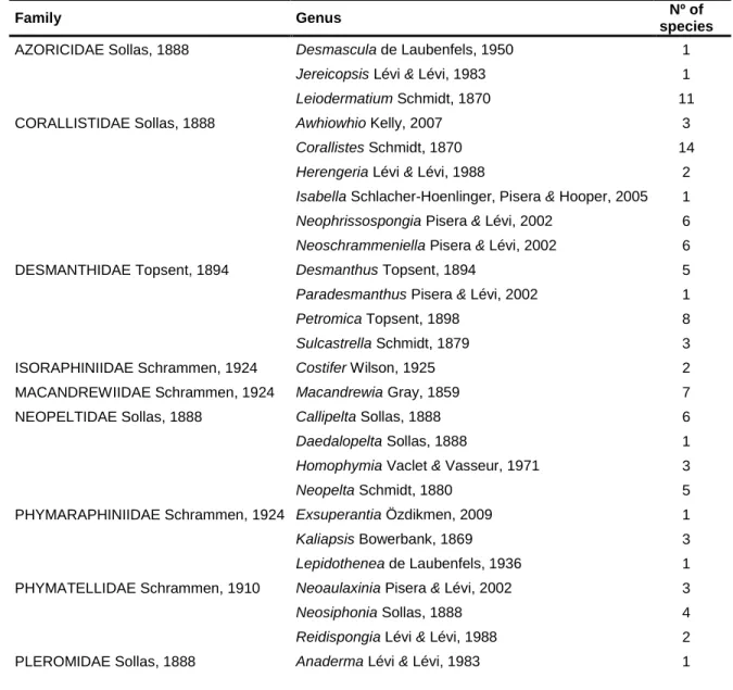
Tab. I - Current classification and diversity account of 'Lithistid' Demospongiae sensu Pisera & Lévi, 2002.
30
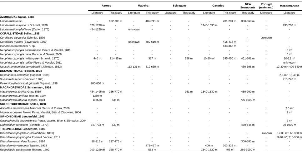
Tab. II - Records of lithistid sponges from the literature and found in this study with
an indication of their bathymetric range; (-) no record; (*) occurring inside sublitoral caves.
LIST OF ACRONYMS AND ABBREVIATIONS
AIC Akaike Information Criterion
CaCO3 Calcium carbonate
COI Mitochondrial cytochrome c oxidase subunit I
Coleta Sponges’ collection of the Department of Oceanography and
Fisheries of Azores University, Portugal
DB/Uaç Biology Department of Azores University, Ponta Delgada,
Portugal
DBUA.Por Sponges’ collection of the Biology Department of Azores
University, Portugal
DNA Deoxyribonucleic acid
DOP/Uaç Department of Oceanography and Fisheries of Azores
University, Horta, Portugal
HBOI Harbour Branch Oceanographic Institute, Florida, USA
HNO3 Nitric acid
MNHN Muséum National d’Histoire Naturelle, Paris, France
OM Optical Microscopy
PCR Polymerase Chain Reaction
PorTOL Porifera Tree of Life Project
ROV Remotely Operated Vehicle
SEM Scanning electron microscopy
SGS Sponge GeneTree Server
SiO2 Silica
SPD Sponge Barconding Project
WoRMS World Register of Marine Species
WPD World Porifera Database
1.1. PHYLUM PORIFERA
The phylum Porifera (from Latin porus= pore and ferō = bearer) comprises a diverse and ecologically important group of filter-feeding invertebrates (van Soest et al., 2012). Thought to exist since the late Neoproterozoic (635 Myr ago), they are regarded amongst the most primitive extant animal groups (Love et al., 2009). Sponges inhabit a variety of environments from the poles to the equator and from shallow to deep-waters. They are mostly marine although a group of species also occurs in freshwater. They also present a variety of shapes and sizes, from thinly incrusting to large cup-shapes as a consequence of their high adaptation capacity (van Soest et al., 2012).
Sponges are sessile metazoans and efficient filter-feeders that exhibit a simple body organization lacking true tissues or organs. The body organization is divided into three distinct layers, pinacoderm, mesohyl and choanoderm. The first one is the "skin" of the sponge and is coated by flattened cells, the pinacocytes, that also covers the surface of the internal canal system; the mesohyl possesses a matrix of collagen/spongin fibres, and inorganic skeleton (spicules) composed by silica (SiO2) or calcium carbonate (CaCO3) that
confer firmness to the sponge; other specialized cells such as amebocytes and archaeocytes are also present in this layer; lastly, the choanoderm is composed by a layer of flagellated cells, the choanocytes that are responsible for the uni-directional water flow in the interior of the sponge allowing its filtration (e.g. Hooper et al., 2002a; Hajdu et al., 2011; van Soest et al., 2012). Sponges reproduce asexually by fragmentation and budding for example (Fell, 1993). Regarding sexual condition, sponges can be either gonochoristic or hermaphroditic. With respect to reproductive condition, sponges can be either oviparous, releasing the gametes into the water column; or viviparous, by brooding the embryos until their release as larvae (Maldonado & Riesgo, 2008; Riesgo et al., 2013)
.
There are nine types of sponge larvae (reviewed in Maldonado & Bergquist, 2002), being the parenchymella the most common one within the class Demospongiae. This type of larva is lecithotrophic and its dispersal may take from a few hours to several days before attachment, depending on the species. Upon attachment the larva undergoes a metamorphosis and becomes a juvenile sponge (Maldonado et al., 2003).Sponges play key-roles in the ecosystems. As filter-feeders, they are important in the bentho-pelagic coupling by either directly removing nutrients from the environment (e.g. Yahel et al., 2003; Goeij et al., 2008) or through the symbiotic microorganisms (Radax et
al., 2011; Schläppy, et al., 2010); they are also responsible for the elimination of some viruses (Hadas et al., 2006). Furthermore, sponges are major players in substrate stabilisation, consolidation and regeneration, bioerosion, reef formation, oxygen depletion, and provide shelter for other animals (reviewed in Wulff, 2001; Bell, 2008).
Sponges have been known since Greek antiquity to bring benefits for men such as in cleansing and bathing (still used today), medicine, pharmacology and also as a household object (Voultsiadou, 2007). Nowadays, sponges have a leading role in Blue Biotechnology due to their high potential in the production of marine natural products with antiviral, antibiotic, anticancer, antitumor and fungicidal properties (e.g. Pomponi, 2006; Grasela et
al., 2012). These bioactive compounds are thought to be produced by sponges as a
chemical defense against predators, as antifouling or for spatial competition purposes (e.g. Becerro et al., 1997, 2003).
Sponges were recognized as a Phylum for the first time by Grant, in 1836 but questions regarding the evolutionary history and phylogenetic relationships of the group perdure to this very day. The taxonomy and systematics of sponges is primarily based on external (shape, colour, texture) and internal morphological characters (mainly on type and nature of skeletal structures) (Hooper et al., 2002a). However high levels of spicule homoplasy have been found (e.g. Manuel et al., 2003; Cárdenas et al., 2011), rendering sponge classification based on morphological characters alone problematic and, to some extent, unreliable. In 2002 a group of authors got together to establish and summarize the classification of sponges based on classic taxonomy – the Systema Porifera (Hooper & van Soest, 2002). An online database, the World Porifera Database (WPD, http://www.marinespecies.org/porifera), was also created with the aim of listing all sponge species and their distribution, as well as reference taxonomic literature. Information concerning the taxonomic status (e.g. valid or not valid) of sponge taxa is also given.
For the past two decades, the development of molecular tools was shown to be an important resource in the clarification of some questions concerning the classification and evolution of sponges, including: the mono- vs. paraphyletic status of the phylum (Philippe, 2009); the resurrection of the class Homoscleromorpha (Gazave et al., 2012); phylogenetic relationships within the group at various taxonomic levels (e.g. Cárdenas et al., 2011; Morrow et al., 2012; Redmond et al., 2013); and the detection of several cases of cryptic speciation (e.g. Blanquer & Uriz, 2007; Xavier et al., 2010). International initiatives such as
the Sponge Barcoding Project (SPD, http://www.spongebarcoding.org), the Sponge GeneTree Server (SGS, http://www.spongegenetrees.org) and the Porifera Tree of Life Project (PorTOL, https://www.portol.org) were created with the goals of making the identifications of sponges easier (through molecular barcodes), to improve the estimate of phylogenetic relationships at various taxonomic levels, and to improve knowledge regarding the evolutionary history of the phylum.
The phylum Porifera comprises four extant classes (Homoscleromorpha Bergquist, 1978, Demospongiae Sollas, 1885, Calcarea Bowerbank, 1862 and Hexactinellida Schmidt, 1870) and one extinct (Archaeocyatha Bornemann, 1884), 25 orders, 128 families, 680 genera and a total of 8553 currently accepted species (WPD, van Soest et al., 2012) but this number is estimated to represent just half of the true diversity of the phylum (van Soest et
al., 2012).
1.2. LITHISTID SPONGES
Within Porifera, the class Demospongiae is by far the largest and most diverse, accounting for approximately 85% (i.e. 7000+) of all described species. This class is characterized by the possession of siliceous spicules and/or a fibrous skeleton (Hooper & van Soest, 2002b). Within this class are the "Lithistid" Demospongiae sensu Pisera & Lévi, 2002a, a group of species characterized by the shared possession of a hypersilified skeleton composed of articulated spicules (desmas). These interlocking spicules often give a very firm or rock-hard consistency to the sponges, the reason for which the lithistids are also known as rock sponges (Kelly-Borges & Pomponi, 1994; Pisera & Lévi, 2002a).
Lithistid sponges were formerly considered a group (Order Lithistida Schmidt, 1870), which included all desma-bearing sponges. In 1888, Sollas established the first clear classification of lithistids based on ectosomal (surface) spicules and microscleres (small spicules) instead of shape of desmas. Two suborders were created: Hoplophora, which contained ectosomal spicules and microscleres, and Anoplia, characterized by the absence of spicules. For Sollas (1888), desmas represented a single evolutionary event, and therefore Lithistida was a monophyletic group. However a substantial body of mostly morphological and some molecular evidence has revealed the polyphyletic nature of lithistids and the astrophorid, halichondrid and spirophorid affinities of several of its families
(e.g. Kelly-Borges & Pomponi, 1994; McInerney et al., 1999; Cárdenas et al., 2011; Morrow
et al., 2012; Redmond et al., 2013). However, for practical purposes, lithistids were kept
together. Globally, 14 families, 51 genera and 198 extant species are currently accepted (van Soest et al., 2012; Tab. I), many of which are thought to be relicts of a much more diverse and abundant fauna occurring in the Mesozoic (Reid, 1967; Lévi, 1991). Given the high fossilization potential of the desmas, lithistids are, of all sponges, the best represented ones in the fossil record (Finks, 1970; Rigby, 1991; Pisera, 2002) and are thus considered a key group for the understanding of the evolutionary history of the phylum (Kelly-Borges & Pomponi, 1994).
Tab. I - Current classification and diversity account of 'Lithistid' Demospongiae (Source: Pisera & Lévi, 2002 and WPD, van Soest et al., 2012)
Family Genus Nº of
species
AZORICIDAE Sollas, 1888 Desmascula de Laubenfels, 1950 1
Jereicopsis Lévi & Lévi, 1983 1
Leiodermatium Schmidt, 1870 11
CORALLISTIDAE Sollas, 1888 Awhiowhio Kelly, 2007 3
Corallistes Schmidt, 1870 14
Herengeria Lévi & Lévi, 1988 2
Isabella Schlacher-Hoenlinger, Pisera & Hooper, 2005 1
Neophrissospongia Pisera & Lévi, 2002 6
Neoschrammeniella Pisera & Lévi, 2002 6
DESMANTHIDAE Topsent, 1894 Desmanthus Topsent, 1894 5
Paradesmanthus Pisera & Lévi, 2002 1
Petromica Topsent, 1898 8
Sulcastrella Schmidt, 1879 3
ISORAPHINIIDAE Schrammen, 1924 Costifer Wilson, 1925 2
MACANDREWIIDAE Schrammen, 1924 Macandrewia Gray, 1859 7
NEOPELTIDAE Sollas, 1888 Callipelta Sollas, 1888 6
Daedalopelta Sollas, 1888 1
Homophymia Vaclet & Vasseur, 1971 3
Neopelta Schmidt, 1880 5
PHYMARAPHINIIDAE Schrammen, 1924 Exsuperantia Özdikmen, 2009 1
Kaliapsis Bowerbank, 1869 3
Lepidothenea de Laubenfels, 1936 1
PHYMATELLIDAE Schrammen, 1910 Neoaulaxinia Pisera & Lévi, 2002 3
Neosiphonia Sollas, 1888 4
Reidispongia Lévi & Lévi, 1988 2
Pleroma Sollas, 1888 4
SCLERITODERMIDAE Sollas, 1888 Aciculites Schmidt, 1879 12
Amphibleptula Schmidt, 1879 1
Microscleroderma Kirkpatrick, 1903a 7
Pomelia Zittel, 1878 1
Scleritoderma Schmidt, 1879 4
Setidium Schmidt, 1879 1
SIPHONIDIIDAE Lendenfeld, 1903 Gastrophanella Schmidt, 1879 6
Lithobactrum Kirkpatrick, 1903 1
Siphonidium Schmidt, 1879 5
THEONELLIDAE Lendenfeld, 1903 Discodermia Du Bocage, 1869 29
Manihinea Pulitzer-Finali, 1993 2
Racodiscula Zittel, 1878 5
Siliquariaspongia Hoshino, 1981 1
Theonella Gray, 1868 14
VETULINIDAE Lendenfeld, 1903 Vetulina Schmidt, 1879 1
LITHISTIDA incertae sedis Arabescula Carter, 1873 1
Collectella Schmidt, 1870 1
Plakidium Lendenfeld, 1907 1
Lithistids are known to produce a diverse array of structurally complex natural products of great interest for biotechnology companies. In fact, to date more than 300 different compounds with antimicrobial (Matsunaga et al., 2001), antitumor (Sun & Sakemi, 1991), antifungal (Gunaskera et al., 1991), immunosuppressive (Longley et al., 1991) and anticancer (Gulavita et al., 1992; Haar et al., 1996) properties have been isolated from lithistids (reviewed in Winder et al., 2011).
Most lithistid species are found at bathyal or bathyal-like environments, such as caves, in tropical and warm temperate regions (e.g. Pomponi et al., 2001; Pisera & Vacelet, 2011). Diversity and distribution patterns of the lithistid sponge fauna are largely understudied, with the exception of some regions such as the continental shelf and slope of the tropical western Atlantic (e.g. Schmidt, 1870, 1880; van Soest & Stentoft 1988; Pomponi
et al., 2001), the southwest Pacific archipelagos of New Caledonia, New Zealand and the
seamounts of the Norfolk Ridge (e.g. Lévi & Lévi, 1983, 1988; Lévi, 1991; Schlacher-Hoenlinger et al., 2005; Kelly et al., 2007), and the Mediterranean Sea (e.g. Manconi & Serusi, 2008; Pisera & Vacelet, 2011).
In the Northeast Atlantic, knowledge of the lithistid sponge fauna is mostly restricted to the Azores archipelago for which 10 species belonging to seven genera and seven families were reported in the course of several expeditions (
e.g.
Topsent 1892a, 1904b, 1928). In fact, previous to the present study, only five lithistid species, viz.Neophrissospongia nolitangere (Schmidt, 1870), Discodermia verrucosa Topsent, 1928, Leiodermatium lynceus (Schmidt, 1870), Macandrewia azorica Gray, 1859 and Racodiscula clava sensu Topsent, 1892a were known to occur in the Canary Islands; and only three
species, Corallistes masoni (Bowerbank, 1869), Neoschrammeniella bowerbankii (Johnson, 1863) and Leiodermatium pfeifferae (Carter, 1876) were reported to Madeira.
1.3. GOALS
The main aims of this thesis were: 1) to characterize the diversity and distribution of the lithistid sponge fauna of the Macaronesian islands (Azores, Madeira, Selvagens and Canaries) and the seamounts located southern of the Azores (Great Meteor group); and 2) to investigate the phylogenetic relationships of the identified species and the affinities of their families.
2.1. SAMPLES AND STUDY AREA
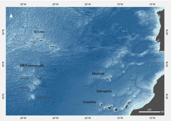
The lithistid sponges used in this work were collected in the course of several research expeditions conducted by various research institutes along the Macaronesian archipelagos of the Azores, Madeira, Selvagens and Canaries as well as on the seamounts located southern of the Azores (Fig. 1). These expeditions were conducted by: the Biology and Oceanography and Fisheries Departments of the Azores University (DB and DOP) between 2006 and 2011 to the Azores archipelago; the Harbor Branch Oceanographic Institute to Madeira, Selvagens and Canaries in 1991 aboard the RV Seward Johnson and employing the Johnson.Sea-Link I submersible; and the MNHN of Paris to the seamounts southern of the Azores (the Seamount 2 expedition) in 1993 aboard the NO Cryos. Specimens were collected by dredge, trawl, and submersible or as bycatch of longline fishing gear. Most specimens were fixed and preserved in ethanol (70%, 90%, 96%), however the specimens from the Seamount-2 expedition were fixed in formalin. Sampling information regarding the examined material (location, coordinates, sampling method and depth) is presented in Appendix I.
Fig. 1 - Study area: the Macaronesian archipelagos of the Azores, Madeira, Selvagens and Canaries, and the northeast
Atlantic seamounts located southern of the Azores.
2.2. TAXONOMY
Taxonomic and distribution data on lithistid sponges of the entire northeast Atlantic (including the Macaronesian islands) and the Mediterranean Sea were extracted from the literature.
Identifications were made from the analysis of external and internal morphological characteristics following the Systema Porifera classification system (Hooper & van Soest, 2002) and some later references (e.g. Schlacher-Hoenlinger, et al. 2005; Pisera & Vacelet, 2011). Original descriptions of all lithistid species reported for the study area were also consulted. Permanent slides of the skeleton and spicules were made for all specimens for observation under optical microscopy (OM). Skeleton slides were prepared by making thick cross and tangential tissue sections that were mounted in Canada balsam. For spicules preparations, small tissue portions were digested in bleach during 24 hours (or 48h for some
specimens), and then the bleach was removed by washing several times with distilled water and ethanol. Some drops of the spicules’ suspensions were placed on microscopy slides and then dried and fixed with Canada balsam.
Eight specimens were also prepared for observation under a scanning electron microscopy (SEM). Small portions of sponge tissue were digested in hot nitric acid (HNO3).
When all organic matter was digested, nitric acid was removed by a washing series with distilled water and ethanol. The suspension of spicules was set on the stub surface, dried and covered with gold-palladium for observations under a JEOL 5410 scanning electron microscope. A total of 162 specimens were analysed and identified to the lowest taxonomic rank possible. Small fragments of all analysed specimens, as well as spicules and skeleton preparations, are deposited in the sponge collection of the Biology Department of the University of the Azores (DBUA.Por).
2.3. PHYLOGENETIC RELATIONSHIPS
2.3.1. DNA EXTRACTION, AMPLIFICATION AND SEQUENCING
In order to investigate the phylogenetic relationships of the identified species two gene fragments were sequenced: the "Folmer fragment" of the mitochondrial cytochrome c oxidase subunit I gene (COI) (Folmer et al., 1994) and the D3-D5 domains of the nuclear ribossomal 28S gene (Morrow et al., 2012). Both genes are being used in the Porifera Tree of Life project (www. portol.org) to resolve species-level questions and in combination with other markers also allow to address higher level relationships For this purpose only specimens belonging to the COLETA, DBUA.Por and HBOI collections were used. Specimens from the MNHN Paris Seamount-2 collection were fixed in formalin and were therefore unsuitable for molecular work. A total of 70 specimens, representing all of the identified species were used.
A small fragment of choanosomal tissue (internal region of a sponge; see Glossary) was used for DNA extraction using the QIAGEN DNeasy blood and tissue kit according to the manufacturer's instructions. Since spicules are not digested by proteinase K (initial
digestion) one centrifugation step was added to remove all spicules, as suggested by Cárdenas and co-workers (2011). For the amplification of COI (612 bp) the primers LCO1490 (5'-GGTCAACAAATCATAAAGATATTGG-3') and HCO2198 (5'-TAAACTTCAGGGTGACCAAAAAATCA-3') (Folmer et al., 1994) were used; the polymerase chain reaction (PCR) conditions were the following: 4 min/94ºC; 5 cycles [30 s/93ºC, 1 min/45ºC, 30 s/72ºC]; 30 cycles [30 s/93ºC, 45 s/50ºC, 30 s/72ºC]; 7 min/72ºC. For the amplification of the 28S rRNA gene (653 bp) the primers Por28S-830F (5'-CAT CCG ACC CGT CTT GAA-3') and Por28S-1520R (5'-GCT AGT TGA TTC GGC AGG TG-3') (Morrow et al., 2011) were used and the PCR conditions were: 3 min/94ºC; 36 cycles [3 min/94ºC, 30 s/94ºC, 45 s/58ºC, 40 s/72ºC]; 7 min/72ºC. In some cases other temperatures (60-62ºC) were adopted on the annealing step to get better results on PCR products.
The PCR products were purified and sequenced (in the majority of samples one direction -forward- was sufficient; in some cases both directions were used) by Macrogen, Europe.
2.3.2. DATA ANALYSIS
The obtained sequences were checked through BLAST searches (http://blast.ncbi.nlm.nih.gov/), aligned and edited in BioEdit v 7.0.5 (Hall, 2005). Additional lithistid, Astrophorida, Spirophorida, Hadromerida, Halichondrida and Poecilosclerida sequences were extracted from GenBank and the Sponge Barcoding Project website to be used in the analyses. The analysed COI and 28S datasets included 102 and 52 sequences in total of which 36 and 29 were new, respectively (Appendix II, Tab. I and Tab. II).
Bayesian analyses were implemented using Mr. Bayes v.3.1 (Huelsenbeck & Ronquist, 2001) with parameters estimated as part of the analysis. The best-fitting models for COI and 28S datasets were derived from and jModeltest 2.1.4 (Posada, 2008) under the Akaike Information Criterion (AIC). For COI the model used was GTR+I+G and for 28S was TrN+I+G. In both cases the analyses were run for 1x10⁶ generations, saving one tree each 1000 generations. The log-likelihood values of the sample point were plotted against the generation time and all the trees prior to reaching stationary were discarded, ensuring that burn-in samples were not retained. Remaining trees were combined in a 50% majority consensus tree, in which frequency of any particular clade represents the posterior probability (Huelsenbeck & Ronquist, 2001). Members of the order Spirophorida were
designated as outgroup for the COI analysis. For the 28S analysis members of the orders Hadromerida and Halichondrida were chosen as outgroups. These outgroups were chosen on the basis of sequences being available on GenBank and the Sponge Barcoding Project, of species that have demonstrated to be phylogenetically close to the ingroup.
3.1. DIVERSITY AND DISTRIBUTION OF LITHISTIDS ON THE
NORTHEAST ATLANTIC OCEAN
Records of lithistid sponges were collected from the literature for the entire northeast Atlantic Ocean and Mediterranean Sea (e.g. Topsent 1892a, 1904b for the Macaronesian islands; Schmidt, 1870 and Du Bocage, 1869 for the Portuguese mainland; Manconi & Serusi, 2006 and Pisera & Vacelet, 2011 for the Mediterranean Sea). In total, 22 lithistid species representing 14 genera and eight families have been reported for these areas (Fig. 2; Tab. II).
In this study, 162 lithistid specimens were analysed and assigned to 11 taxa representing five families and nine genera (Fig. 2; Tab. II). Of these, the Corallistidae Sollas, 1888 is the best represented of all families with a total of four genera and four species. The remaining families are represented by one or two genera maximum.
Fig. 2 - Distribution of lithistids in Macaronesian islands and neighboring seamounts: white-filled circles represent records from
Of the 10 species previously recorded from the Azores archipelago, only seven, representing five families and six genera were found in this study. Eight species were recorded for the first time on the seamounts south of the Azores, of which seven are shared with this archipelago. Three species constitute new records for Madeira Island (Neophrissospongia nolitangere (Schmidt, 1870), Discodermia verrucosa Topsent, 1928 and Racodiscula clava sensu Topsent, 1892), two for Selvagens (N. nolitangere and Macandrewia azorica Gray, 1859), and two for the Canaries, of which one is a new species for science of the genus Isabella. Of the 14 species found to occur in the Macaronesian islands, only three viz. N. nolitangere, Leiodermatium pfeifferae (Carter, 1876) and R. clava are shared among all archipelagos, with the remainder being restricted to one or two islands’ group. Similarly, only four species seem to be shared between the Mediterranean Sea and these Atlantic archipelagos: N. nolitangere (with the Azores, Madeira, Selvagens and the Canaries), Neoschrammeniella bowerbankii (Johnson, 1863) (with Madeira), Siphonidium ramosum (Schmidt, 1870) (with the Azores) and Leiodermatium lynceus Schmidt, 1870 (with the Azores and the Canaries).
Most species were found in the upper bathyal (200-800 m) although some were found at lower depths (e.g. N. nolitangere at 91 m depth and R. clava at 168 m in the Azores; Isabella harborbranchi at 133 m in Canaries). The deepest records were those of Macandrewia robusta and R. clava found at 1000 m on the northeast Atlantic seamounts.
Tab. II - Records of lithistid sponges from the literature and found in this study with an indication of their bathymetric range; (-) no record; (*) occurring
inside sublitoral caves.
NEA Seamounts
Portugal
(mainland) Mediterranean
Literature This study Literature This study Literature This study Literature This study This study Literautre Literature
AZORICIDAE Sollas, 1888
Leiodermatium sp. - 182-706 m - 402-741 m - - - 281-291 m 330-660 m -
-Leiodermatium lynceus Schmidt, 1870 370-1730 m - - - 1340-1530 m - - - 430-760 m
Leiodermatium pfeifferae (Carter, 1876) 454-1250 m - unknown - - -
-CORALLISTIDAE Sollas, 1888
Corallistes elegantior Schmidt, 1870 - - - unknown
-Corallistes masoni (Bowerbank, 1869) - - unknown 480-610 m - - - 415-417 m - -
-Isabella harborbranchi n. sp. - - - 133-366 m - -
-Neophrissospongia endoumensis Pisera & Vacelet, 2011 - - - 5 m*
Neophrissospongia nana Manconi & Serusi, 2008 - - - 6 m*
Neophrissospongia nolitangere (Schmidt, 1870) 440 m 91-435 m - 317 m - 358 m 10-20 m* 295-450 m 461-501 m - 20-22 m*
Neophrissospongia radjae Pisera & Vacelet, 2011 - - - unknown*
Neoschrammeniella bowerbankii (Johnson, 1863) - - 113-131 m 519-669 m - - - - 480-695 m - 12-30 m*; 400-640 m
DESMANTHIDAE Topsent, 1894
Desmanthus incrustans (Topsent, 1889) - - - 2-3 m*; 10-40 m
Sulcastrella tenens (Vacelet, 1969) - - - 210-240 m
Petromica (Petromica) grimaldii Topsent, 1898 200-650 m - - -
-MACANDREWIIDAE Schrammen, 1924
Macandrewia azorica Gray, 1859 454-1495 m 256-770 m - - - 361 m 1340-1530 m - 480-900 m -
-Macandrewia ramifera Topsent, 1904 1360 m - - -
-Macandrewia robusta Topsent, 1904 1165 m 635 m - - - 705-1000 m -
-SCLERITODERMIDAE Sollas, 1888
Aciculites mediterranea Manconi, Serusi & Pisera, 2006 - - - 7.5 m*
Microscleroderma lamina Perez, Vacelet, Bitar & Zibrowius, 2004 - - - 2 m*
SIPHONIDIIDAE Lendenfeld, 1903
Gastrophanella phoeniciensis Perez, Vacelet, Bitar & Zibrowius, 2004 - - - 2 m*
Siphonidium ramosum (Schmidt, 1870) 349-793 m 530 m - - - 470-545 m - 20-1000 m
THEONELLIDAE Lendenfeld, 1903
Discodermia polydiscus (Bowerbank, 1869) - - - unknown 12-30 m*; 60-360 m
Discodermia polymorpha Pisera & Vacelet, 2011 - - - 3-20 m*; 210-360 m
Discodermia ramifera Topsent, 1892 98-318 m 237-475 m - - - 300-580 m -
-Discodermia verrucosa Topsent, 1928 - - - 476-497 m - - 400 m 303-322 m - -
-Racodiscula clava sensu Topsent, 1892 200-1229 m 168-770 m - 563 m - - 1340-1530 m 408 m 280-1000 m -
-Madeira Selvagens Canaries
3.2. SYSTEMATICS
Phylum PORIFERA Grant, 1836 Class DEMOSPONGIAE Sollas, 1885 'Lithistid' Demospongiae sensu Pisera & Lévi, 2002
DEFINITION: Polyphyletic group of demosponges related by the presence of choanosomal articulated spicules (desmas) that form a rigid skeleton in the majority of the species. Lithistids are highly polymorphic and may be encrusting, massive irregular, cup- or ear-shaped, flabellate cylindrical, branched, globular, with or without pedicel, and with or without atrial cavity. Ectosomal megascleres are discotriaenes, pseudodiscotriaenes, phyllotriaenes, pseudophyllotriaenes, dichotriaenes, rhabds, oxeas, and in some species they are absent. Several types of choanosomal megascleres such as tetraxial (tetraclones), monaxial (rhizoclones, megaclones, dicranoclones, heloclones, or various complex branched forms), polyaxial or anaxial (sphaeroclones). Microscleres may be different types of rhabds, oxeas, spirasters, amphiasters, sigmaspires and raphides, or several combinations of these spicules (Pisera & Lévi, 2002a).
Family CORALLISTIDAE Sollas, 1888
DEFINITION: Sponges similar to a cup, vase, massive ear or with a lamellar shape; Ectosomal megascleres are dichotriaenes and in some cases, simple triaenes; choanosomal desmas are dicranoclones; microscleres can be two types of spirastres, streptasters/amphiasters, microxeas, microstyles and microstrongyles (Pisera & Lévi, 2002c).
Genus Corallistes Schmidt, 1870 TYPE SPECIES: Corallistes typus Schmidt, 1870
DEFINITION: Cup- to vase-shape sponges, with simple or folded plates; ectosomal megascleres are smooth dichotriaenes and large oxeas can be present; choanosomal desmas are dicranoclones very tuberculated; microscleres are usually two types of spirasters (Pisera & Lévi, 2002c).
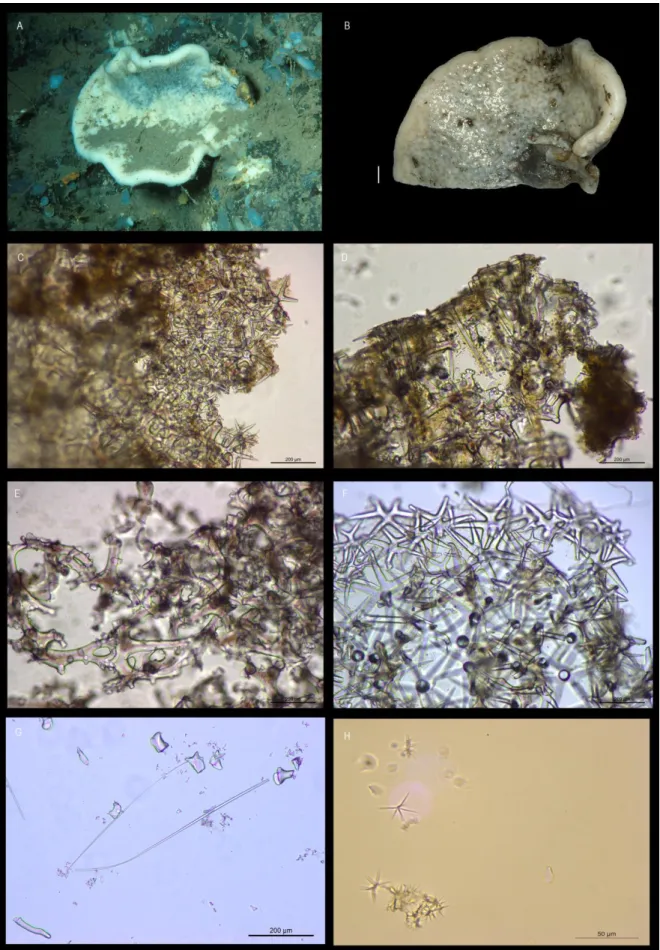
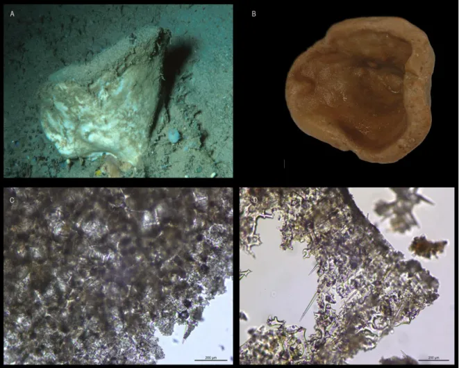
Corallistes masoni (Bowerbank, 1869)
(Figs. 3A-H)
EXAMINED MATERIAL: HBOI 27-V-91-3-006, 29-V-91-3-017, 31-V-91-1-015, 10-VI-91-1-003, 11-VI-91-1-003.
EXTERNAL MORPHOLOGY: Polymorphic sponges with cup-to vase-shape or simple to folded plates. Colour beige to light brown. Inner surface with oscules in small elevations, that may be bright blue; outer surface is smooth (Figs. 3A-B).
SKELETON: Ectosomal skeleton (Fig. 3C) contains a compact layer of smooth dichotriaenes transverse to the surface, long oxeas as megascleres, and two types of abundant spirasters with pointed arms as microscleres. Choanosomal skeleton (Fig. 3D) composed of tuberculated dicranoclone desmas that form a compact and rigid mesh (Fig. 3E); spirasters are spread throughout the choanosome but less numerous than in the ectosome.
SPICULES: Ectosomal dichotriaenes are smooth with some of the tips of the cladome directed towards the rhabdome and/or bifurcated; the cladome is very variable in length within the same specimen 80 – 202- 459 μm diameter; the rhabdome is long with pointed extremity, 124 – 425 – 950 μm length and 6 – 19 – 37 μm width (Fig. 3F). Oxeas are long, thin, and smooth and have blunt tips, 184 – 555 – 995 x 2.3 – 4.8 – 7.5 μm (Fig. 3G). Dicranoclone desmas are compact with large and rough tubercles, with brownish tone (Fig. 3E). Microscleres are two types of spirasters: type I with thin, short and numerous rays, 11 – 18 – 26 μm; type II with thin, long and few rays, 17 – 30 – 74 μm (Fig. 3H).
DISTRIBUTION: Individuals of C. masoni were sampled in Madeira and Canary islands between 415 – 610 m depth. Until the present study this species was only known from its type locality in the Madeira archipelago (unknown depth).
situ (scale 1 cm); C, ectosomal skeleton showing a layer of dichotriaenes (scale 200 µm); D, choanosomal and ectosomal
skeleton (scale 200 µm); E, dicranoclone desmas (scale 200 µm); F, dichotriaenes (scale 200µm); G, oxeas (scale 200 µm);
H, two types of spirasters: type I with thin, short and numerous rays, and type II with thin, long and few rays (scale 50 µm).
REMARKS: C. masoni was originally described by Bowerbank (1869) based on a specimen collected at an unknown depth in the Madeira archipelago. Since then the species was never reported again for the NEA but it has been often reported (misidentified) in the Mediterranean Sea as Neoschrammeniella bowerbankii (Pisera & Vacelet, 2011). With this study the known geographic range, as well as the bathymetric distribution of this species was expanded.
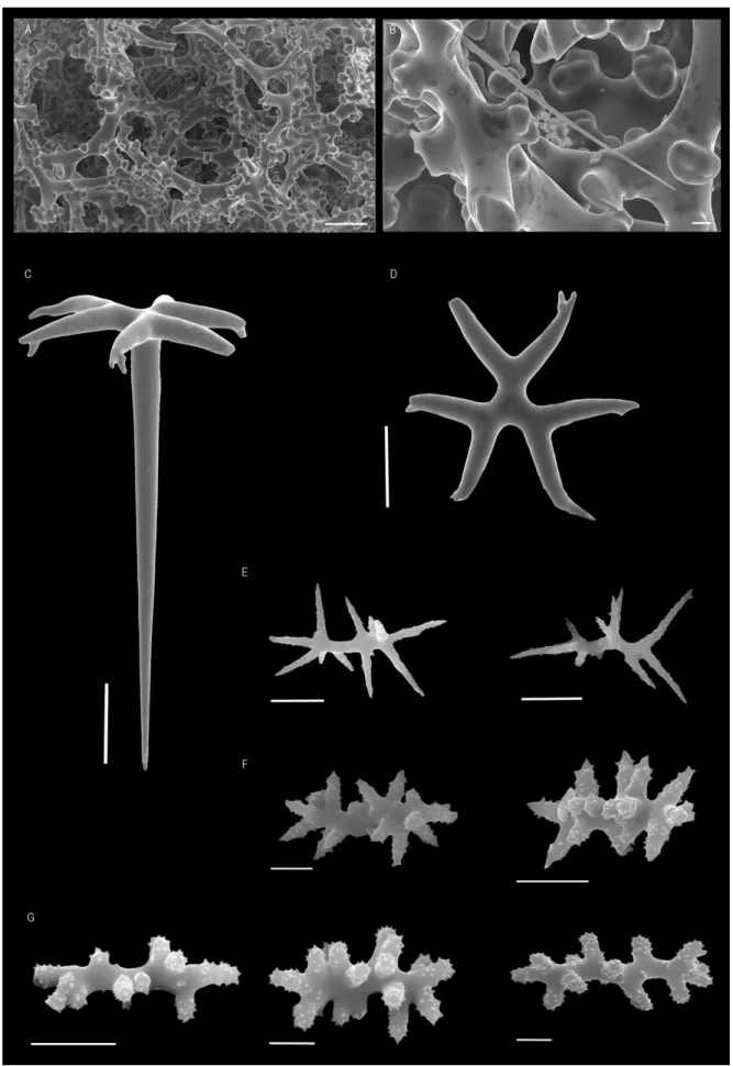
Genus Neophrissospongia Pisera & Lévi, 2002c TYPE SPECIES: Corallistes nolitangere Schmidt, 1870.
DEFINITION: Sponges with irregular cup or ear shape, contorted flabellate masses or clavate growth forms; ectosomal megascleres are dichotriaenes with strong spines and/or tubercles in the upper surface of the cladome; choanosomal desmas are dicranoclones; Microscleres are streptasters/amphiasters and spinose microtylostyles (Pisera & Vacelet, 2011).
Neophrissospongia nolitangere (Schmidt, 1870)
(Figs. 4A-D; 5A-H)
EXAMINED MATERIAL: HBOI- 28-V-91-2-001, 2-VI-91-1-005, 003, 9-VI-91-4-006, 11-VI-91-1-001, 11-VI-91-1-008, 13-VI-91-4-003, 16-VI-91-2-004; DOP- 1134, 1135, 1241, 1609, 2048.1, 2048.2, 3147, 3774, 4037, 4166, 4602, 4905, 5323, 5850, 5853, 5871, 5879, 6192, 6370, 6565, 6614, 6623, 6686; DBUA.Por- SAB2011.DR12.B1.
EXTERNAL MORPHOLOGY: Large flabellate masses in old individuals (Fig. 4A) and ear-shaped forms in young specimens (Fig. 4B); large individuals are attached to the substrate by the entire lower base whereas the youngest individuals are attached by a pedicel. Colour of the specimens varies from beige to light brown, in life and alcohol. Walls are approximately 1.3 cm thick. There are significant differences between the inner and the outer surface of the sponge: the inner surface has several oscular openings in small elevations
and is smooth on the borders; some specimens have a bright blue colour on the oscular openings; the outer surface is smooth with ostia distributed over the entire surface.
SKELETON: The ectosome (Fig. 4C) is composed of a compacted layer of dichotriaenes transverse to the surface, covered by an extremely dense and compact crust of spirasters-streptasters; abundant microtylostyles can also be observed in the ectosome. Choanosomal skeleton (Fig. 4D) is constituted by strongly tuberculated dicranoclone desmas, in a compacted net with irregular cavities (Figs. 5A-B); both microtylostyles and spirasters-streptasters are also present.
SPICULES: Ectosome megascleres are dichotriaenes with cladome 87 – 225 – 367 µm in diameter that have small pointed tubercles 3 – 12 – 22 µm in size, on their upper surface; blunt rhabdome with 276 – 610 – 1117 x 5 – 18 – 35 µm, straight or sometimes slightly curved (Figs. 5C-D); in small specimens smooth dichotriaenes can be found (Fig. 5E). Choanosomal desmas are massive dicranoclones extremely articulated, covered with mushroom-shaped tubercles (Figs. 5A-B); dicranoclones are brownish. Microscleres are microtylostyles and spirasters/streptasters; microtylostyles are pin-shaped with a spinous head and some few sparse spines all over the spicule, with 30 - 77 – 188 µm long and 0.8 – 1.8 – 4.5 µm large (Fig. 5
F)
. The spirasters/streptasters are 6 – 11 – 18 µm long, with thick and spiny arms or with long and smooth arms (Figs. 5G-H).DISTRIBUTION: Specimens of N. nolitangere were collected in all archipelagos from the Macaronesian region (Azores, Madeira, Selvagens and Canaries), between 91 and 502 m depth. This species was previously known from its type-locality in Portugal (uncertain locality), and from the archipelagos of the Azores and Cape Verde. The records for Madeira and Selvagens here reported for the first time, make therefore for a much more continuous geographical range of the species.
Fig. 4 - Neophrissospongia nolitangere (Schmidt, 1870). A, adult individual from the Canaries, in situ; B, young specimen collected in the Azores, ex situ (scale 1 cm); C, ectosomal skeleton with a compact layer of dichotriaenes and microscleres (scale 200 µm); D, choanosomal skeleton (scale 200 µm).
REMARKS: Manconi & Serusi (2008) suggested the revision of the genus
Neophrissospongia erected by Pisera & Lévi (2002c) due to the rare presence of smooth
dichotriaenes in young individuals and microstyles in some specimens, that were not described before. In their work concerning the lithistid sponges of the Mediterranean Sea Pisera & Vacelet (2011) redescribed this genus by adding the rare presence of spinose microtylostyles but they did not mention the presence of smooth dichotriaenes. In some specimens analysed in the present work smooth dichotriaenes in young ear- to cup-shaped individuals were also found. Furthermore, in the specimens here analysed, spinose microtylostyles were not at all rare, they were actually quite abundant. Accordingly, the inclusion of smooth dichotriaenes and the presence of microtylostyles in the genus definition is hereby proposed.
Fig. 5 - Neophrissospongia nolitangere (Schmidt, 1870). A, dicranoclone desmas (scale 100 µm); B, detail of dicranoclones where one can observe the circular-shape elevated tubercles (scale 100 µm); C, ectosomal dichotriaene (scale 50 µm); D, cladome of dichotriaene of an adult individual, view from below (scale 50 µm); E, smooth cladome of dichotriaene of a young
individual, view from below (scale 50 µm); F, microstyles (scale 10 µm); G, spirasters/streptasters with thick and spiny arms (scale 5 µm); H, spirasters/streptasters with long and smooth arms (scale 5 µm).
Genus Neoschrammeniella Pisera & Lévi, 2002c TYPE SPECIES: Iouea moreti Lévi & Lévi, 1988.
DEFINITION: Polymorphic sponges, cylindrical to cup- vase- shaped; ectosomal megascleres are smooth dichotriaenes; choanosomal megascleres are anisoxeas, dicronoclone desmas, and some species may have big fringing diactines; Microscleres are two or three types of acanthose spirasters, with short blunt rays, long or short pointed rays (Pisera & Vacelet, 2011).
Neoschrammeniella bowerbankii (Johnson, 1863)
(Figs. 6A-D; 7A-G)
EXAMINED MATERIAL: HBOI- 29-V-91-3-008, 30-V-91-4-007; MNHN- 2008-233, IP-2008-234.
EXTERNAL MORPHOLOGY: Polymorphic sponges, cup-shaped to undulate lamellate masses (Figs. 6A-B); colour beige to brown in life and alcohol; the walls are very thick, 1-2 cm, and both surfaces are smooth. The sponges are attached to the substrate by a small pedicel or by the entire lower base.
SKELETON: The ectosome (Fig. 6C) has a layer of smooth dichotriaenes and microscleres (two types of spirasters). In some specimens it is possible to observe some thin and long oxeas protruding the surface out from the choanosome. The choanosome (Fig. 6D) is formed by solid and brownish dicranoclone desmas (Figs. 7A-B), and dispersed spirasters of two types.
SPICULES: Ectosomal megascleres are smooth dichotriaenes and two types of spinous spirasters; dichotriaenes are smooth with 144 – 255 – 429 µm in diameter (cladome), and in some cases, with the point of the cladome directed towards the rhabdome or bifurcated; rhabdome is long and sharp 201- 470 – 755 µm long and 8 - 15 – 24 µm width (Figs. 7C-D). Two distinct types of spinous spirasters are found: type I are short, thick and have blunt
arms, 17 – 21 – 28 µm long (Fig. 7E) and type II are long, thin and have pointed arms, 13 – 25 – 39 µm long (Fig. 7F). Choanosome megascleres are dicranoclone desmas and thin oxeas; dicranoclones are dense with a lot of irregular tubercles which may be divided into small callosities (Figs. 7A-B); oxeas with blunt points measure 124 – 260 – 713 µm in length and 1.9 – 2.8 – 4.1 µm in width; most of the oxeas were broken, so this was the maximum length measured.
DISTRIBUTION: N. bowerbankii was previously known from its type-locality, Madeira Island (unknown depth), and from the Mediterranean Sea (20-22 m, occurring inside sublittoral caves). In the present study this species was found in Madeira (519-669 m) and on the Plato and Hyères seamounts (480-695 m), being the first records of this species to these seamounts.
Fig. 6 - Neoschrammeniella bowerbankii (Johnson, 1863). A, specimen collected in Madeira Island, in situ; B, specimen collected in Hyeres seamount, ex situ (scale 1 cm); C, top view of ectosomal skeleton where one can observe a layer of dichotriaenes and abundant microscleres (scale 200 µm); D, transversal view of choanosomal skeleton (scale 200 µm).
REMARKS
: The genus Neoschrammeniella was formally diagnosed by Pisera & Lévi (2002c) and the knowledge of the distribution and morphological characters of N.bowerbankii consequently increase. In their work, images of SEM were presented allowing
a better observation of microscleres, namely the two types of spirasters. In the present work SEM analysis allowed the observation of a third type of spirasters (with short, thick and pointed arms; Fig. 6G) occurring in much lower quantity. This type III seems an intermediate form of the two types previously described by Pisera & Lévi (2002c). SEM examination of further specimens will be necessary to ascertain the taxonomic significance of this finding.
dicranoclone desmas, note the tubercles divided into small tubercles and the oxea with blunt tip (scale 10 µm); C, dichotriaene (scale 100 µm); D, smooth cladome of a dichotriaene (scale 100 µm); E, spirasters of type I, with short, thick and blunt arms (scale 5 µm); F, spirasters of type II, with long, thin and pointed arms (scale 10 µm); G, spirasters of type III found in this present work, with short, thick and pointed arms (scale 5 µm).
Genus Isabella Schlacher-Hoenlinger, Pisera & Hooper 2005 TYPE SPECIES: Isabella mirabilis Schlacher-Hoenlinger, Pisera & Hooper 2005.
DEFINITION: Massive and globular lithistid; ectosomal dichotriaenes, short and long-shafted triaenes, all smooth; choanosomal dicranoclones desmas are slender, smooth and with small tubercles in some parts; two types of oxeas: type I long and thick with blunt tips, type II long, thin and curved with acerate tips; four types of microscleres: spinous fusiform microxeas, spinous centrotylotes, spirasters with small, thick and thorny arms and streptasters with long, thin and also thorny arms (Schlacher-Hoenlinger et al., 2005).
Isabella harborbranchi Carvalho, Pomponi & Xavier (in prep.)
(Figs. 8A-E; 9A-N)
EXAMINED MATERIAL: HBOI- 8-VI-91-4-005, 9-VI-91-4-013, 11-VI-91-1-002, 14-VI-91-1-003.
MORPHOLOGY: Massive and globular Corallistidae. Surface is irregular and slightly compressible, while the inner part of the sponge is rigid. Oscules/pores cannot be seen with naked eye. Colour dark brown in life and in ethanol (Fig. 8A).
SKELETON: Ectosome is rather difficult to distinguish from the choanosome due the huge quantity of microscleres spread throughout the sponge and the dark colour of its tissue. Ectosomal skeleton contains a small number of dichotriaenes and both long- and short-shafted triaenes spread throughout the sponge surface along with a large quantity of microacanthoxeas (Fig. 8B). Choanosomal skeleton (Fig. 8C) consists on dicranoclone desmas (Figs. 8D-E), two types of long oxeas and microscleres (microacanthoxeas, spirasters/streptasters).
SPICULES: Ectosomal dichotriaenes and triaenes (long and short-shafted) are irregular. The cladome of the dichotriaenes is 150 - 334 - 623 µm in diameter, 3.6 - 9.1 - 12.9 µm in width and have blunt tips; the rhabdome is 107 - 283 - 578 x 6.5 - 9.3 - 14.8 µm (Fig. 9A); some of these megascleres exhibit small arms, with blunt tips and, in some cases