Mestre em Engenharia Quimica e Quimica Molecular
Synthesis, Characterization and
Applications of Gold and Silver
Nanoparticles: From Antibacterial to
Optoelectronic Nanomaterials
Dissertação para obtenção do Grau de Doutor em Quimica
Orientador:
Prof. Dr. José Luis Capelo Martinez,
Associated Professor with habilitation
LAQV-Requimte, Faculdade de Ciências e
Tecnologia, Universidade Nova de Lisboa
Co-orientador:
Prof. Dr. Carlos Lodeiro Espiño,
Associated Professor with habilitation
LAQV-Requimte, Faculdade de Ciências e
Tecnologia, Universidade Nova de Lisboa
Dr. Javier Fernandez-Lodeiro,
Researcher
LAQV-Requimte, Faculdade de Ciências e
Tecnologia, Universidade Nova de Lisboa
Júri:
Presidente: Prof. Doutor Manuel Luís de Magalhães Nunes da Ponte Vogais: Prof. Doutor Carlos Lodeiro Espiño
Prof. Doutora Julia Lorenzo Rivera Doutor Fernando Novio Vazquez
Doutor Hugo Miguel Baptista Carreira Dos Santos Junho de 2019
Mestre em Engenharia Quimica e Quimica Molecular
Synthesis, Characterization and
Applications of Gold and Silver
Nanoparticles: From Antibacterial to
Optoelectronic Nanomaterials
Dissertação para obtenção do Grau de Doutor em Quimica
Orientador:
Prof. Dr. José Luis Capelo Martinez,
Associated Professor,
LAQV-Requimte, Faculdade de Ciências e
Tecnologia, Universidade Nova de Lisboa
Co-orientador:
Prof. Dr. Carlos Lodeiro Espiño,
Associated Professor with habilitation
LAQV-Requimte, Faculdade de Ciências e
Tecnologia, Universidade Nova de Lisboa
Dr. Javier Fernandez-Lodeiro,
Researcher
LAQV-Requimte, Faculdade de Ciências e
Tecnologia, Universidade Nova de Lisboa
Júri:
Presidente: Prof. Doutor Manuel Luís de Magalhães Nunes da Ponte Vogais: Prof. Doutor Carlos Lodeiro Espiño
Prof. Doutora Julia Lorenzo Rivera Doutor Fernando Novio Vazquez
Doutor Hugo Miguel Baptista Carreira Dos Santos
Synthesis, Characterization and Applications of Gold
and Silver Nanoparticles: From Antibacterial to
Optoelectronic Nanomaterials
Copyright © Jamila Djafari, Faculdade de Ciências e Tecnologia, Universidade NOVA de Lisboa
A Faculdade de Ciências e Tecnologia e a Universidade Nova de Lisboa têm o direito, perpétuo e sem limites geográficos, de arquivar e publicar esta dissertação através de exemplares impressos reproduzidos em papel ou de forma digital, ou por qualquer outro meio conhecido ou que venha a ser inventado, e de a divulgar através de repositórios científicos e de admitir a sua cópia e distribuição com objectivos educacionais ou de investigação, não comerciais, desde que seja dado crédito ao autor e editor.
“Nothing happens without effort.You have to have faith. And for that, you have to break down the barriers of prejudice, which requires courage.To have courage, you must conquer your fears.”
I want to thank my supervisor Prof. Dr José Luis Capelo Martinez and my co-supervisor Prof. Dr Carlos Lodeiro Espiño for giving me the opportunity to do my PhD thesis under their supervision in the BIOSCOPE Group. I also thank them for welcoming me to their laboratory and for helping me in all stages of this thesis. I thank them for their extreme kindness, their understanding, and for the beautiful people they are. I shared great moments at your side, thank you for everything because without them I could not have achieved this thesis.
I would like to thank the PROTEOMASS Scientific Association for the support for PhD inscription fees, all reagents and general funding during my thesis. Moreover, I would like to thank the FCT-MEC for my Research Contract associate with the R&D project PTDC/QEQ-MED/2118/2014 allowing me to acquire complementary skills during my thesis. To the research units, UCIBIO-REQUIMTE (2015-2107) and to the LAQV-REQUIMTE (2018-2019) for their support and help during these periods.
I sincerely thank my thesis co-supervisor Dr Javier Fernández-Lodeiro for teaching me the labor part of this thesis. I thank him for these long scientific explanations, for these late hours in the laboratory, for these moments of stress but which allowed us to live these moments of joy with each concretion of work. I thank him also for transmitting me his vision of science that is in line with its excellent values and scientific objectives. His autodidactic allowed me to realize that the most important is not only the result, but all the road travelled to reach these objectives even if it is strewn with pitfalls. Each error is learning and is necessary for scientific growth. A big thank for his patience, his foresight and for taking me under his wing to achieve all these projects. Even without having a background in nanomaterials, I was able to acquire reliable knowledge in nanoparticle synthesis that allowed me to understand theoretically but especially to precisely observe each synthesis of nanoparticles as different from each other — an enormous thanks for this.
I would like to thank all my co-workers and colleagues from the BIOSCOPE Group at the laboratory, more specially to Adrián Fernández-Lodeiro for his valuable help in the laboratory, and Susana Jorge, Eduardo Aráujo, Silvia Nuti, Gonçalo Marcelo, Dr. Elisabete Oliveira, Dr. Hugo Santos, Luis Carvalho, Gonçalo Martins, Joao Prates, and Dr. Cristian Cuerva for these shared moments, especially during the organization of the events, and during the birthdays celebrations, thank you for your good mood and your kindness. Thanks also to the colleagues working in PROTEOMASS, Ana Laço and Marta Silva.
I want to thank all the collaborators which have contributed in my work with the antibacterial, protein and SERS studies. Mainly, I would like to mention Prof. Gilberto Igrejas and Prof. Dr Patricia Poeta, and Ms. Vanessa Silva from the University of Trás-os-Montes e Alto Douro (Portugal) and Prof. Dr. Carmen Torres from the University of La Rioja (Spain) for the antibacterial studies; Prof. Dr. Emilia Bértolo Pardo, Prof. Simon C Harvey and Dr. Marie T McConnell from the Christ Church University of Canterbury (UK) for the protein and biological test on the functionalized gold@lectine nanoparticles; Dr. Benito Rodríguez-González from the University of Vigo, CACTI (Spain) for some of the HRTEM and TEM characterization, and Prof. Dr. Isabel Pastoriza, Prof. Dr. Jorge Perez-Juste and Mr. Daniel Garcia-Lojo from the University of Vigo (Spain) for the SERS studies. And for Mr. Carlos Fernández-Lodeiro for his collaboration in the synthesis of silver nanoplates.
Thanks to the Analytical Laboratory at the REQUIMTE-Chemistry Department for their support, and specially the help to Dr. Luz Fernandes, Carla Rodrigues, Nuno Costa and Ana Teresa Lopes. Thanks also to Laboratory technicians at the 4th floor of the Chemistry Department,
And finally, I would like to thank to Prof. Marco Diogo Richter Da Silva for his help as the Chemistry Doctoral Program coordinator.
D’un point de vue personnel, je souhaite remercier ma famille du plus profond de mon cœur. Tout d’abord, à mes deux modèles, aux personnes les plus importantes à mes yeux, mes parents Ivanira et Tawfik. Je vous remercie de m’avoir soutenu à chaque étape de cette expérience et d’avoir toujours été là pour moi dans les bons comme dans les mauvais moments. Je vous remercie sincèrement d’avoir toujours cru en moi et de m’avoir inculqué toutes ces belles valeurs qui font partie de votre être. Merci pour votre bienveillance, vos enseignements, et merci simplement d’être vous-même cela a permis d’être la personne que je suis aujourd’hui. Je ne vous remercierai jamais assez pour tout ce que vous avez fait pour moi car sans vous je ne serai rien. Je vous aime du fond du cœur. Un grand merci à mon petit frère adoré Yanis, mon sang, mon humain préféré, mon meilleur ami et mon confident. Merci pour ton optimisme et ta bonne humeur qui m’ont remonté le moral maintes fois. Merci pour ces good vibes, ces soirées télé, ces discussions, ces restaus et tous ces bons moments qu’on a passé ensemble. Sache que je serai toujours là pour toi et que je t’aime fort. Bien sûr, un grand merci à ma petite chienne Rubis que j’aime du fond du cœur et qui me manque terriblement.
Merci à ma tante Amel, Didier et mon petit Medhi pour tous ces moments passés ensemble lorsque je rentrais à la maison. A chaque fois, c’est un plaisir de vous voir et de pouvoir me changer les idées, merci de votre soutien.
Merci à ma famille répartie dans le monde, qui malgré la distance est toujours là pour moi notamment à mes oncles, tantes et cousines au Canada et en Algérie, merci pour leur soutien.
Obrigado tambem a minha familia brasileira com quem estou sempre em contacto, e com quem eu morro de dar risada com todas nossas discussões no cellular. Obrigado por suas belezas, vôcés são pessoas increiveis e lindas. Obrigado tambem a minha linda avo Dona Nena por ser o que ela e, uma pessoa maravilhosa, tenho muita saudades e espero verte pronto.
Une grande pensée à mes étoiles qui me manquent et qui veillent sur moi: Tata, Habibi et vovo Mario, shukraan et obrigado.
Merci à mes amies de toujours, Pauline et Camille. Merci pour ces moments partagés, et pour cette jolie amitié qui dure depuis plus de 20 ans déjà. Merci pour ces fous rires, ces discussions sans fin, pour votre bienveillance et surtout merci d’être là pour moi. Merci également à Sabrina, Yahia, Carole et Anne-Sophie.
Bien sûr, merci à Marie-Paule et Patrice pour leur aide et leur soutien, ainsi qu’à leurs familles respectives pour ces dinés passés ensemble et tous ces bons moments qui sont gravés dans ma mémoire. Un grand merci à Souheila que je considère comme ma petite sœur, et qui pour qui j’ai beaucoup d’affection. Merci d’avoir été là pour moi et merci à toute ta famille pour ces moments de joie partagés ensemble au rythme latino.
Agradezco, por supuesto, a la segunda familia que gané en los últimos años y que me recibió con los brazos abiertos en su casa. Gracias a Pilucha, por haberme considerado como su hija y por haberme dado el amor como una madre, nunca te agradeceré lo suficiente por tu sonrisa, tu bondad y por la fabulosa persona que eres. Gracias a Josecho por su sabiduría, su sentido del humor, me río mucho contigo, y gracias por aprovechar la vida como lo haces, es contagioso. Gracias a Esther, paz para su alma en el cielo, y a Avelino por su amabilidad siempre conmigo. Gracias a “Los Valentinos”, mi pequeña Valentina, Paula y Abel, y por todos estos divertidos momentos pasados juntos, siempre es un placer estar con vosotros. Gracias a Adrián, mi compañero de tesis, gracias por tu apoyo, tu ayuda y por todos tus chistes. Alguien me dijo que eras una de las personas más amables que conocía y tiene toda la verdad. ¡Gracias a mi pequeño
macroescala, correcta y brillante, especialmente mantente como eres, porque eres una persona excepcional. Gracias a Paula igualmente.
Gracias a todos mis nuevos amigos en España con quien he pasado muy buenos momentos y fiestas.
Gracias a los lindos animales españoles que compartieron buenos momentos conmigo: Luna, Athina, Piranha, Daphne, y Lila. Y a los que están en el cielo: Poncho, Hugo y Nacho.
Y, por supuesto, gracias a mi novio, que, sin él, podría haber renunciado a este reto. Gracias por apoyarme todos los días, por ser paciente conmigo y por estar siempre a mi lado. Gracias por todos estos momentos juntos que me permitieron escapar durante todos los períodos de estrés. Gracias por ser la persona más bella que conozco.
R
esumo
A investigação e o desenvolvimento de novas nanopartículas metálicas (NPs) têm crescido nas últimas décadas como resultado das propriedades ventajosas que estes nano-materiais apresentam. A sua natureza metálica associada ao seu tamanho diminuto resulta em propriedades físico-químicas que constituem uma ve ntagem real para a construção de uma nova gama de materiais nano-métricos com potenciais aplicações em campos como a biomédicina, eletrônicos, óticos ou catalíticos, entre muitos outros.
Atualmente, muitas áreas científicas e tecnológicas estão obtendo muitos benefícios pelo uso e desenvolvimento dos nano-materiais de ouro e prata. Nó entanto neste momento, é necessário desenvolver novas metodologias reproduzíveis que permitam uma maior compreensão dos materiais em escala nanométrica, favorecendo a construção das NPs de ouro e prata mais precisas e complexas, controlando seu tamanho, forma e composição, permitindo novas aplicações.
Com o objetivo de introduzir novos avanços sintéticos na área de nanotecnologia, na presente tese de doutorado temos estudado: i) A síntese e caracterização de NPs de ouro biofuncionalizadas com proteínas; ii) O desenvolvimento de uma nova ruta sintética para produzir NPs de ouro e prata usando Tetraciclina como agente sintético; iii) A síntese e funcionalização de nanoplacas triangulares de prata e seu subsequente revestimento controlado orgânico ou inorgânico (camada de sílica), e iv) Uma síntese inovadora de NPs de ouro pseudo-esféricas, multi-ponta ou tipo framboesa, utilizando Ferro(II) como agente redutor sustentável.
Além disso, as propriedades antimicrobianas e optoelectrónicas e as aplicações das NPs produzidas foram exploradas e estudadas.
The Research and development of new metallic nanoparticles (NPs) have been booming in recent decades as a result of the advantageous properties presented by these nanomaterials. Their metallic nature associated with their tiny size induce physicochemical properties that constitute a real advantage for the construction of a new range of nano-metric materials with potential applications in biomedical, electronic, optical or catalytic fields, among many others. Until now, many scientific and technological fields are obtaining great benefits by the utilization and development of gold and silver nanomaterials. However currently, it is necessary to develop new reproducible methodologies allowing a greater understanding of the nano-scale materials thus promoting the construction of most precise and more complex gold and silver NPs through the control of their size, shape and composition, allowing new reviewed applications.
With the aim to introduce new synthetic advances in the nanotechnology area, in the present doctoral thesis we have studied: i) The synthesis and characterization of bio-functionalized gold NPs with proteins; ii) The development of a nobel synthetic approach to produce gold and silver NPs using Tetracycline as synthetic agent; iii) The synthesis and functionalization of silver triangular nanoplates and their subsequent controlled organic and inorganic (silica layer) coating; and iv) the creation of a novelty synthesis of pseudo-spherical, multi-tip or raspberry like AuNPs using Iron (II) as Green reducing agent.
Furthermore, the antimicrobial and optoelectronic properties of the produced NPs and their applications have been explored and studied.
CHAPTER 1. GENERAL INTRODUCTION
... 11.1. METALLIC NANOPARTICLES BACKGROUND: RAW MATERIALS TO METAL COLLOIDS ... 1
1.2. PHYSICO-CHEMICAL PROPERTIES OF METALLIC NANOPARTICLES ... 5
1.2.1. Optical properties: origin and understanding of nanoparticles colour ... 5
1.2.2. Chemical properties of gold and silver nanoparticles ... 10
1.3. SYNTHESIS OF METALLIC NANOPARTICLES ... 11
1.4. STATE OF THE ART: APPLICATIONS OF SILVER AND GOLD NANOPARTICLES .. 16
CHAPTER 2. SYNTHESIS OF GOLD FUNCTIONALIZED NANOPARTICLES
WITH
THE
ERANTHIS
HYEMALIS
LECTIN
AND
PRELIMINARY
TOXICOLOGICAL STUDIES ON CAENORHABDITIS ELEGANS ... 23
2.1. INTRODUCTION ... 24
2.2. MATERIALS AND METHODS ... 25
2.2.1. Materials ... 25
2.2.2. Synthesis of EHL Conjugated Gold Nanoparticles ... 26
2.2.3. Nematode Assay ... 27
2.3. RESULTS AND DISCUSSION ... 27
2.3.1. Synthesis and Characterization of the Bioconjugated Gold Nanoparticles (AuNPs@EHL) ... 27
2.3.2. Biological Activity against C. elegans ... 33
2.4. CONCLUSIONS ... 36
CHAPTER 3. NEW SYNTHESIS OF GOLD AND SILVER-BASED
NANO-TETRACYCLINE COMPOSITES ... 37
3.1. INTRODUCTION ... 39
3.2.1. Chemicals Materials ... 39
3.2.2. Preparation of AgNPs@TC ... 40
3.2.3. Preparation of AgNPs@TC-2 ... 40
3.2.4. Preparation of AuNPs@TC ... 40
3.2.5. Spectrophotometric Measurements ... 41
3.2.6. Dynamic Light scattering and Z-Potential measurement ... 41
3.2.7. TEM Measurements ... 41
3.2.8. Metals screening – CLARIOstar ... 42
3.2.9. NPs Concentration and Inductively Coupled Plasma-Atomic Emission Spectrometer determination ... 42
3.2.10. Bacteria and growth conditions ... 42
3.2.11. Preparation of antibiotic and nanoparticles stock solutions ... 42
3.2.12. Determination of the minimal inhibitory concentration (MIC) of the nanoparticles ... 43
3.3. RESULTS AND DISCUSSION ... 43
3.3.1. Synthesis of AgNPs@TC and AuNPs@TC ... 44
3.3.2. Metal sensing applications ... 48
3.3.3. Exploring Antibacterial activity ... 51
3.4. CONCLUSIONS ... 53
CHAPTER 4. EXPLORING THE CONTROL IN ANTIBACTERIAL ACTIVITY
OF SILVER TRIANGULAR NANOPLATES BY SURFACE COATING
MODULATION ... 55
4.1. INTRODUCTION ... 56
4.2. MATERIALS AND METHODS ... 57
4.2.1. Materials ... 57
4.2.2. Synthesis of AgNTs in water (AgNTs@PVP) and MHA stabilization (AgNTs@MHA) ... 58
4.2.3. Silica coating of AgNTs@MHA to produce AgNTs@Si-OH ... 58
4.2.6. Characterization technics ... 59
4.2.7. Bacterial strains, culture media and growth conditions. ... 60
4.2.8. Preparation of stock solutions ... 60
4.2.9. Antibacterial susceptibility test ... 60
4.3. RESULTS AND DISCUSSION ... 61
4.3.1. Synthesis of silver triangular nanoplates and alkane-thiol functionalization ... 61
4.3.2. Silica coating of AgNTs@MHA ... 64
4.3.3. Exploring bactericidal properties ... 69
4.3.4. Proposed mechanism ... 71
4.4. CONCLUSIONS ... 72
CHAPTER 5. IRON(II) AS GREEN REDUCING AGENT IN GOLD
NANOPARTICLE SYNTHESIS ... 75
5.1. INTRODUCTION ... 76
5.2. MATERIALS AND METHODS ... 77
5.2.1. Materials ... 77
5.2.2. Iron(II) mediated synthesis of AuNPs. ... 77
5.2.3. PSS-assisted Au nanoparticles synthesis ... 77
5.2.4. (Fe2+/Citrate)-mediated synthesis of Au nanoparticles ... 77
5.2.5. Characterization techniques. ... 78
5.3. RESULTS AND DISCUSSION ... 79
5.3.1. Modulation of Fe(III)/Fe(II) redox potential. ... 87
5.3.2. Mechanism of AuNPs synthesis induce by Fe(II) ions. ... 90
5.3.3. SERS performance ... 90
5.4. CONCLUSIONS ... 94
L
ist of
F
igures
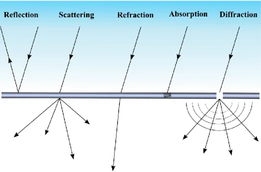
Figure 1.1. Photos of the Lycurgus Cup in (a) transmitted and (b) refracted light, photos from the British museum free service. (c) Image TEM of metallic nanoparticles composing the Lycurgus Cup, image reprinted from the reference [10]. ... 2 Figure 1.2. The different pathways resulting from the interaction of light with matter. ... 6
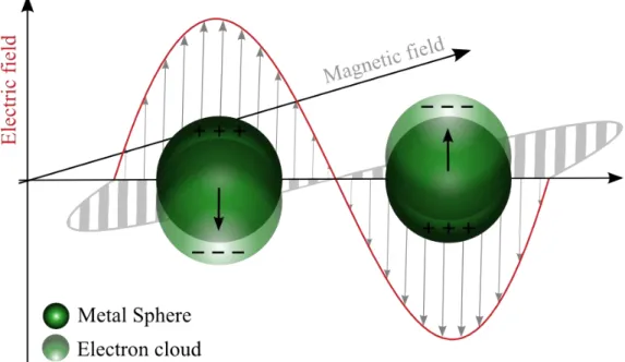
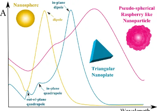
Figure 1.3. Localized Surface Plasmon Resonance of metallic nanoparticles under light illumination ... 7 Figure 1.4. UV-Vis spectra profile of metallic NPs in function of their size and composition. (Left) Silver NPs and (Right) gold NPs based on reference [28, 29]. ... 8 Figure 1.5. Different plasmon band evolution in function of the shape of nanoparticles. ... 9
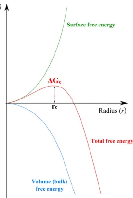
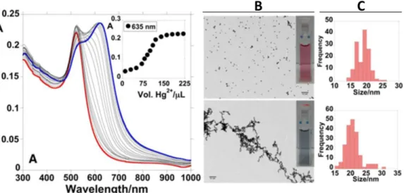
Figure 1.6. Variation of the Gibbs free energy in function of the radius of the spherical nuclei. ... 13 Figure 1.7. Schematic LaMer plot, atom concentration in function of time, representing the different stage of nucleation and growth processes ... 14 Figure 1.8. Representation of the different NPs growth evolution: Ostwald Ripening, Coalescence, Aggregation and Orientated Attachment. Crystalline planes are represented in NPs stripes. ... 15 Figure 1.9. Aggregation response of AuNPs functionalized with thiol terminated fluorescein derivative in presence of mercury ions in solution. (A) AuNPs UV-Vis spectra profile with the addition of Hg2+ in solution. (B) Images TEM of AuNPs (up) before and (down) after mercury
detection showing aggregation of nanoparticles by chain like formation. (C) Corresponding size histograms of AuNPs (up) before and (down) after mercury detection. Figure adapted from the reference [76]. ... 19 Figure 1.10. SERS response profile in function of the size and shape of AuNPs. The yellow color corresponds to the presence of hot spot in NPs. ... 21 Figure 1.11. (a) Schematic plot of a gold atom concentration versus time illustrating the growth of mesoparticles. (b) SEM images of different surface topographies, types I-V, synthesized changing the experimental conditions. Figure reprinted from the reference [96] ... 22 Figure 2.1. (a) UV-Vis spectrum, (b) histogram and (c,d) low magnification TEM images of AuNPs@Citrate. The histogram is derived from measurements of 300 nanoparticles made in ImageJ software. ... 28
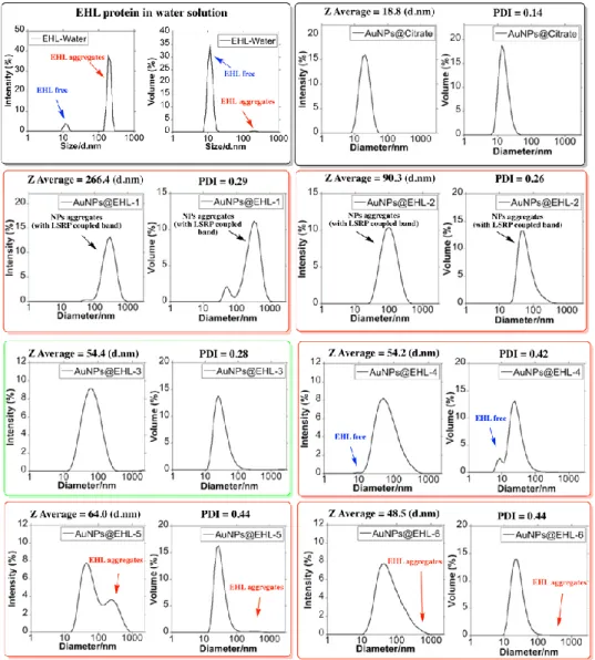
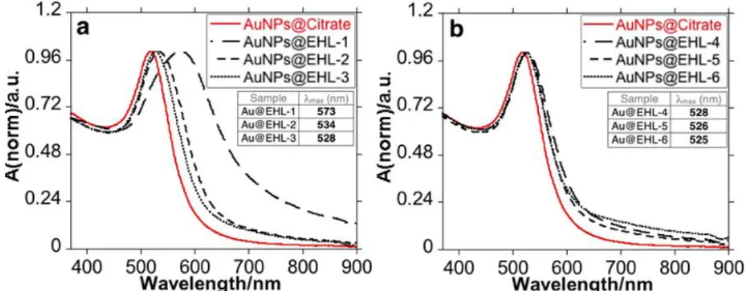
Figure 2.2. Size distribution measured by Dynamic Light Scattering. Distribution by %Intensity and %Volume ... 30 Figure 2.3. Size distribution measured by Dynamic Light Scattering of AuNPs@EHL-3 in water and PBS. Distribution by %Intensity and %Volume ... 30 Figure 2.4. UV-Vis spectra of the different AuNPs@EHL samples synthesized: (a) AuNPs@Citrate, AuNPs@EHL-1, AuNPs@EHL-2, and AuNPs@EHL-3, and (b) AuNPs@Citrate, AuNPs@EHL-4, AuNPs@EHL-5, and AuNPs@EHL-6. ... 31 Figure 2.5. (a) Z-average (red dot) and LSPR (Localized Surface Plasmon Resonance) maximum (blue dot) and (b) PDI of the AuNPs@EHL obtained as a function of EHL amounts added. .... 31 Figure 2.6. TEM images of AuNPs@EHL with different amounts of protein added: (a) AuNPs@EHL-2 (25 µL), (b) AuNPs@EHL-3 (100 µL), (c) AuNPs@EHL-4 (200 µL), and (d) AuNPs@EHL-6 (500 µL). In all cases, the nanoparticles go through two centrifugation cycles (14,000 rpm × 25 min) and are resuspended in MilliQ water. ... 32 Figure 2.7. UV-Vis study of the effects of adding 200 µL NaCl 2M to 3 mL of (a) AuNPs@Citrate and (b) AuNPs@EHL-3. Dilution factor 1:10. ... 33 Figure 2.8. EHL conjugated nanoparticles (AuNPs@EHL) affect early reproduction (early fecundity), but not total reproduction (lifetime reproductive success) of C. elegans L4 stage. .. 35 Figure 3.1. Structure of Tetracycline and its deprotonation sites with corresponding pka values. ... 44 Figure 3.2 UV-Vis absorption spectra of titration of tetracycline with aqueous solution of NaOH. ([Tetracycline]=1.10-5M in DMSO) ... 44
Figure 3.3. UV-Vis absorption spectra, color solution, histogram and TEM images of AgNPs@TC ... 45 Figure 3.4. UV-Vis absorption spectra, Z potential and color solution of (Up) AgNPs@TC and (Down) AuNPs@TC as a function of pH (pH between 1 and 12). ... 46 Figure 3.5. UV-Vis absorption spectra, color solution, histogram and TEM images of AuNPs@TC. ... 47 Figure 3.6. UV-Vis absorption spectra, color solution, histogram and TEM images of AgNPs@TC-2. ... 47 Figure 3.7. Bar diagram showing the intensity of LSRP band at 606 nm for addition of 500 nM of Na+, K+, Hg+, Mg2+, Ca2+, Mn2+, Cu2+, Zn2+, Cd2+, Hg2+, Pb2+, Cr3+, Fe2+, Fe3+, Al3+, Ga3+, In3+
Figure 3.8. Spectrophotometric titration of AgNPs@TC with the addition of increasing amounts of Al(NO3)3 and naked eye detection. ... 49
Figure 3.9. Spectrophotometric titration of AuNPs@TC with the addition of increasing amounts of Al(NO3)3 and naked eye detection. ... 49
Figure 3.10. Reversible colorimetric change upon addition of EDTA in aqueous solution and TEM images of AuNPs@TC with the addition of 500 nM of Al(NO3)3 ... 50
Figure 3.11. Spectrophotometric titration of AuNPs@TC with addition of different fixed quantities of Al(III) with A= 0.1 ... 50 Figure 3.12. Three different replics of spectrophotometric titration of AuNPs@TC with the addition of 500 nM of Al(NO3)3. ... 51
Figure 3.13. Illustrative representation of reversible naked eye sensing of AuNPs@TC towards Al3+. ... 51
Figure 4.1. Spectroscopic profile and color of AgNTs@PVP resuspended in (blue) EtOH and (purple) Water. ... 61 Figure 4.2. (a) Spectroscopic profile of AgNTs@PVP and AgNTs@MHA in EtOH solution. (b) Size histogram of lateral distance of AgNTs@MHA. (c) Color solution of AgNTs@MHA resuspended in EtOH and water. (d, e, f) TEM images obtained of AgNTs@PVP and (g, h, i) AgNTs@MHA. ... 63 Figure 4.3. FT-IR spectroscopic profile of 16-MHA and AgNTs@MHA in KBr disk. Overview between (a) 4000-2400 cm-1 and (b) 2000-400 cm-1. (c) Enlargement spectra in the S-H region
between 2700-2400 cm-1 and FT-IR peak table. ... 64
Figure 4.4. Low-resolution TEM images in different magnification of AgNTs@Si-OH obtained under [DMA] = 0.5 M and 3 hours of reaction using different TEOS concentrations. (a) 0.9mM, (b) 0.7 mM, (c) 0.6mM and (d) 0.5 mM. ... 65 Figure 4.5. Low-resolution TEM images in different magnification of AgNTs@Si-OH obtained under [TEOS] = 0.5mM, [DMA] = 0.5 M for (a and b) 180 min and (c and d) 90 min. ... 66 Figure 4.6. Low magnification TEM images at different magnifications obtained for (a to c) AgNTs@Si-OH and (d to f) AgNTs@Si-NH2 and size histogram of silica coating for (g)
AgNTs@Si-OH and (h) AgNTs@Si-NH2. ... 67
Figure 4.7. Spectroscopic profile of (a) AgNTs@MHA, AgNTs@Si-OH and AgNTs@Si-NH2 in
EtOH, and graphic representation of the Z-potential for (b) AgNTs@MHA, (c) AgNTs@Si-OH and (d) AgNTs@Si-NH2. ... 67
Figure 4.8. FT-IR spectroscopic profile of AgNTs@Si-OH in KBr disk. Overview between (a) 4000-2400 cm-1 and peak table and (b) 2000-400 cm-1. ... 68
Figure 4.9. Spectroscopic profile of (a) AgNTs@Si-COOH and (b) graphic representation of the Z-potential for AgNTs@Si-COOH. ... 69 Figure 4.10. Plausible antibacterial mechanisms of AgNTs action ... 72
Figure 5.1. (Left) Extinction spectra and (Right) corresponding photographs of AuNPs dispersions obtained with different Au(III):Fe(II) ratios, as indicated, at 30 ºC (A) and 60 ºC (B). ... 79 Figure 5.2. Representative TEM images of AuNPs obtained with an Au(III):Fe(II) molar ratio of 1:9 at 60 ºC ... 80 Figure 5.3. (A and B) FT-IR spectra of AuNPs synthesized at 60 ºC for Au(III):Fe(II) molar ratio of 1:9 (black spectra) and the oxidation product (blue spectra) obtained in the absence of Au(III) under similar conditions. (C) Table showing the vibrational assignments ... 81 Figure 5.4. (A) Normalized extinction spectra of AuNPs synthesized at 60 ºC for Au(III):Fe(II) molar ratios of 1:6 (red spectrum) and 1:9 (black spectrum). [Au(III)] was 0.25 mM and [PSS] was 0.5 mg/mL. (B-D) Corresponding TEM images of AuNPs for Au(III):Fe(II) molar ratios of 1:6 (B) and 1:9 (C-D). ... 82 Figure 5.5. (A and C) Representative TEM images and (B and D) the corresponding size distribution histograms of AuNPs synthesized at 60 ºC for Au(III):Fe(III) molar ratios of (A and B) 1:6 and (C and D) 1:9. [Au(III)] was 0.25 mM and [PSS] was 0.5 mg/mL. ... 82 Figure 5.6. (A and B) FT-IR spectra of AuNPs synthesized at 60 ºC for Au(III):Fe(II) molar ratio of 1:9 (black spectra) in the absence (black spectrum) and in the presence (blue spectra) of PSS (0.5 mg/mL). (C) Table showing the vibrational assignment. ... 83 Figure 5.7. (Left) FT-IR spectra of AuNPs synthesized at 60 ºC for Au(III):Fe(II) molar ratio of 1:9 in the presence of 0.5 mg/mL PSS (black spectra) and PSS (blue spectrum). (Right) Table showing the vibrational assignment. ... 83 Figure 5.8. (A) Representative TEM image of AuNP showing its multi-tipped morphology. (B) Fourier Transform (FT) of the NP shown in (A). (C and D) HRTEM images showing the presence of twinning planes at the tips (indicated by arrows). (E and F) FT obtained from the tip shown in (D) demonstrating the presence of twinning planes in [011] twinning axis with rotation angle of 70.53º as shown in (F). ... 84 Figure 5.9. (A) Representative TEM image of AuNP showing its multi-tipped morphology. (B-E) HRTEM images showing the presence of twinning planes at the tips (indicated by arrows). (F) FFT obtained from the tip shown in (E) showing the growing direction of the tip. ... 84
Figure 5.10. Simulation of the electron diffraction pattern using the [011] twinning axis with rotation angle of 70.43°. ... 84 Figure 5.11. (A) Normalized extinction spectra of AuNPs synthesized at Au(III):Fe(II) ratio of 1:9 in the presence of 0.5 mg/mL PSS (red spectrum) and 2 mg/mL PSS (black spectrum). [Au(III)]= 0.25 mM and T= 60 ºC. (B and D) Representative TEM images and (C and E) the corresponding size distribution histograms of AuNPs synthesized in the presence of 0.5 mg/mL PSS (B and C) and 2 mg/mL PSS (D and E)... 85 Figure 5.12. (A) Extinction spectra of Au nanoparticles synthesized at 100 °C (red) and 60 ºC (black) for Au(III):Fe(II) molar ratio of 1:6. [Au(III)] was 0.25 mM and [PSS] was 0.5 mg/mL. (B and D) Corresponding TEM and SEM images of AuNPs obtained at 100°C and 60°C respectively. ... 86 Figure 5.13. (A and C) Representative TEM images and (B and D) the corresponding size distribution histograms (right) of AuNPs synthesized with Au(III):Fe(III) ratio of 1:6 at 30 ºC (A and B) and 100 ºC (C and D). [Au(III)]= 0.25 mM and [PSS]= 0.5mg/mL. ... 86 Figure 5.14. (A) Vis-NIR extinction spectra of Au nanoparticles synthesized at 40 ºC for Au(III):Fe(II) molar ratio of 1:3 in the presence of different amounts of citrate (0.5 (red), 0.75 (black) and 1 mM (blue)). [Au(III)] was 0.5 mM, [PSS] was 0.5 mg/mL. (B and C) Corresponding TEM images of AuNPs synthesized in the presence of citrate 0.5 mM (B) and 0.75 mM (C). The scale bar is the same for both images. ... 87 Figure 5.15. (A, C, E) Representative TEM images and (B, D, F) the corresponding size distribution histograms of AuNPs obtained in the presence of 0.50 mM (A and B) and 0.75 mM (C and D) and 1.0 mM (E and F) sodium citrate at 40 ºC. [Au(III)]= 0.5 mM, [Fe(II)]= 1.5 mM and [PSS]= 0.5 mg/mL. ... 88 Figure 5.16. (A) Normalized extinction spectra of AuNPs synthesized using FeCl2.4H2O at
Au(III):Fe(II) ratio of 1:6 in the presence of 0.5 mg/mL PSS. [Au(III)]= 0.25 mM and T= 60 ºC. (B) Size distribution histograms and (C and D) representative TEM images. ... 89 Figure 5.17. (A) Normalized extinction spectrum of AuNPs synthesized using FeSO4 at
Au(III):Fe(II) ratio of 1:6 in the presence of 0.5 mg/mL PSS. [Au(III)]= 0.25 mM and T= 60 ºC. (B) Size distribution histograms and (C and D) representative TEM images. The FeSO4 solution
was contaminated with NaCl prior to the injection in the reaction (FeSO4/NaCl molar relation =
1/2). ... 89 Figure 5.18. (A) Extinction spectra of the three selected AuNPs for SERS analysis; Cit40 (black), PSS60 (red) and PSS100 (blue). The dashed lines indicate the excitation laser line used. (B) SERS spectra of 4-NTP obtained with PSS60 for the three excitation lines as indicated. (C) SERS
three excitation laser lines, as indicated. The insets show a representative TEM image of each particle. Scales bars represent 50 nm in all cases. ... 91 Figure 5.19. Limit of detection of 4-NTP determined using PSS60 (see text for details). (A) SERS spectra for different 4-NTP concentrations (as indicated in the labels) at constant concentrations of Au (0.5 mM) and 633 nm laser line. (B) Changes in the intensities of the NO2 symmetrical
stretching (1332 cm-1, open circles) and CC stretching (1568 cm-1, closed circles). ... 92
Figure 5.20. Raman and SERS spectra of PSS and Table showing the main vibrational assignment of PSS ... 92 Figure 5.21. SERS intensity of 4-NTP signals at 1332 cm-1 (open circles) and 1568 cm-1 (closed
circles) as a function of 4-NTP concentration. The lines are linear fits in the quantitative detection region. All measurements were performed at constant concentrations of Au (0.5 mM) and using 633 nm as excitation laser line. ... 93
L
ist of
T
ables
Table 2.1. AuNPs@EHL solution composition for each experiment, DLS, and Zeta Potential Values and protein amount on the nanoparticles. ... 29 Table 2.2. EHL treatment affects survival and development of treated C. elegans L1s. When scored, adult worms were only observed on 3 of the 12 EHL plates, with worms showing varied degrees of developmental delay. ... 34 Table 3.1. Bacterial strains used in this study. ... 42
Table 3.2. Antimicrobial effect of nanoparticles functionalized with tetracycline, on tetracycline-susceptible and – resistant E. coli and S. aureus strains. ... 52 Table 4.1. Different strains used in the present study ... 60
Table 4.2. The minimum inhibitory concentration (MIC) and minimum bactericidal concentration (MBC) of AgNTs@MHA, AgNTs@Si-NH2, and AgNTs@Si-COOH toward E. coli K12 ATCC
29425 and S. aureus ATCC 25923. ... 71 Table 5.1. Summary of the experimental conditions for the synthesis of the different Au nanoparticles employed in the SERS studies. ... 91
A
bbreviations
4-NTP 4-nitrothiophenol
AEFs Analytical enhancement factors
Ag Silver
AgNPs Silver Nanoparticles
AgNPs@TC Silver Nanoparticles functionalized with Tetracycline AgNTs Silver nanoprisms
AgNTs@MHA Silver nanoprisms functionalized with 16-mercaptohexadecanoic acid AgNTs@Si-COOH Silver nanoprisms functionalized with carboxylate terminated silica AgNTs@Si-NH2 Silver nanoprisms functionalized with amine terminated silica
AgNTs@Si-OH Silver nanoprisms functionalized with alcohol terminated silica APTMS (3-Aminopropyl)triethoxysilane
a-THF Anhydrous Tetrahydrofuran
Au Gold
AuNPs Gold nanoparticles
AuNPs@Citrate Gold nanoparticles functionalized with Citrate AuNPs@EHL Gold nanoparticles conjugated with EHL
AuNPs@TC Gold nanoparticles functionalized with Tetracycline BSPP bis (p-sulfonatophenyl) phenylphosphine
CFU Colony-forming unit
CTAB Cetyltrimethylammonium bromide
DLS Dynamic Light Scattering
DMA Dimethylamine
DMF Dimethylformamide
DNA Deoxyribose Nucleic Acid
EHL Eranthis hyemalis
FT-IR Fourier Transform InfraRed
HAADF High-Angle Annular Dark-Field Imaging
HRTEM High Resolution Transmission Electronic Microscopy ICP Inductively Coupled Plasma-Atomic Emission Spectrometer
LB Luria-Bertani
LOD Limit of Detection
LSPR Localized Surface Plasmon Resonance MBC Minimum bactericidal concentration
MHA 16-Mercaptohexadecanoic acid
MIC Minimum inhibitory concentration
MQ Milli-Q
NGM Nematode Growth Media
PBS Phosphate Buffered Saline
PDI Polydispersity Index
PVP Polyvinylpyrrolidone
PSS Poly(sodium 4-styrenesulfonate)
RIP Ribosome Inactivating Protein
RT Room Temperature
SAED Selected Area Electron Diffraction SERS Surface Enhanced Raman Spectroscopy
SPR Surface Plasmon Resonance
STEM Scanning Transmission Electron Microscope
TC Tetracycline
TEM Transmission Electronic Microscopy TEOS Tetraethyl orthosilicate
S
ymbols
d Particle diameter
Cext Cross-section extinction
εm Dielectric constant of the medium
R Radius of the sphere
εr Real component of the dielectric function of nanoparticles
εi Imaginary component of the dielectric function of nanoparticles
λ Wavelength of the incident radiation ΔG Total Gibbs free energy variation
r Radius of the spherical nuclei
ΔGv Free energy of the crystal
γ Surface tension energy
ΔGc Critical global free energy
rc Critical radius kb Boltzmann constant T Temperature υ Molar volume S Supersaturation C Concentration Cs Concentration at solubility
ISERS SERS intensity for a selected mode of a given analyte
IRS Non-enhanced Raman intensity
cSERS Analyte concentration in the SERS
GENERAL INTRODUCTION
1.1. METALLIC NANOPARTICLES BACKGROUND: RAW MATERIALS
TO METAL COLLOIDS
Metals such as gold, silver or copper have been widely used by humankind for millennia as a result of their availability in the native state. Already in the time of ancient civilizations, especially in Egypt and Mesopotamia, metallurgy and mining techniques were developed and exploited for the production of metal objects, pieces of jewelry or ornaments.1–3 Among the three
metals mentioned, gold was one of the most worked metal due to its bright color, beauty, malleability and resistance to corrosion. Long considered as a symbol of the sun and a “gift from the Gods” by several civilizations, gold was widely used to represent loyalty, lust and royalty. For instance, the older collections of pure gold objects dating from 4600-4200 BCE were found in Bulgaria in 1972 and represent around 6 kg of gold jewelry, vessels and decorative objects. This overwhelming collection of more than 300 objects of solid gold was found in the richest tombs of an ancient cemetery (near the Black Sea) highlighting the mystical and religious character of gold, as well as the hierarchical culture during this period.2
Regarding silver, this metal was not used as much as gold to symbolize royalty but in
1
C H A P T E R
alloy of silver and gold) but also to produce recipient to keep fresher food and liquids.4 On the
contrary, copper acquired a more important role in the manufacture of tools, weapons and dishes among others.
Over the years, the development of alchemy allowed to perform different techniques such as heating, distillation, reflux or sublimation, to produce metals-based objects. Alchemists’ motivation at that time was based on the discovery of the "philosopher's stone". It was believed that the aforementioned stone, reddish in appearance, give eternal life and allowed the transmutation of base metals into gold or silver.5 Efforts to find this "chemical treasure" improved
alchemists’ technics arising many alloys with different colors and compositions.5,6 Besides, with
the evolution of glass manufacturing, it was possible to create different colored glasses through the addition of specific amounts of transition metals such as copper, gold and silver.7
Possibly, the most famous object in history composed of a special class of colored glass dating from the 4th century BC is known as the Lycurgus Cup which is exhibited at the British
Museum in London. This drinking cage cup is a typical Roman cup composed of a carved decorative frieze representing the scene of the triumph of Dionysus (god of the wine) on Lycurgus (king of Edoni).8 The richness of this piece is not only due to the skills developed to make this
carved piece, but to the unusual optical properties compared to other pieces found at this time. Indeed, the cup exhibits strong dichroism, when the cup is illuminated from the inside, a red ruby color is transmitted while it appears green opaque when it’s illuminated from the outside. (Figure 1.1)
Several studies have been conducted to determine the composition of the cup’s colored glass. In 1965, R. Brill was able to demonstrate the origin of the unexpected dichroism. In his work, the author demonstrated the presence of gold and silver minute units in the glass by
Figure 1.1. Photos of the Lycurgus Cup in (a) transmitted and (b) refracted light, photos from the British
museum free service. (c) Image TEM of metallic nanoparticles composing the Lycurgus Cup, image reprinted from the reference [10].
composition analysis.9 Later, in 1990, studies of the Lycurgus Cup under Transmission Electronic
Microscope (TEM) and X-ray analysis were carried out by D. J. Barber and I. C. Freestone, revealing that the Roman cup is composed of polygonal (squares) metallic particles.10 The
particles observed, in average sizes between 50-100 nm, are formed of gold, silver and copper alloy in proportion 31.2/66.2/2.5. These observations using modern techniques confirmed the results of R. Brill. (Figure 1.1). The studies also reveal the presence of non-metallic polygonal (squares and hexagonal) particles ranging from 15 to 100 nm corresponding to crystallized sodium chloride.10 Therefore, the Lycurgus cup represents the oldest colored object found composed of
metallic nanoparticles (NPs) although its color is probably induced by accident/hazard.
With this important historical example, it is clear that metals at the nanoscale level have a totally different appearance and color when compared to their massive counterpart. However, to form these types of colored glasses, the simple addition of metals to the glass is not enough to generate this unusual optical effect, because the metals must be reduced in specific conditions. Note that during Roman times, the reduction of metals was not known yet. In fact, R. Brill studies about Lycurgus cup supposes that this unusual optical effect would be coincidentally obtained by the presence of redox species in the glass such as iron, antimony and manganese which can participate in the gold and silver reduction under these conditions.9 Therefore, the Lycurgus Cup
can be considered as a “nano-accidental” object produced during this period.
However, it was not until the 8th century that alchemy brought an invaluable advance
related to the metals’ manipulation in solution. The improvement of distillation followed by the discovery of mineral acids by the well-known alchemist J. I. Hayyan (Latinization Geber) allowed the solubilization of nobel metals.11 Through the manipulation of hydrochloric acid and nitric acid
derivatives J. I. Hayyan obtained for the first time the aqua-regia known at that time as the “royal solvent” which dissolve gold and silver, but also hardly soluble non-metals such as sulfur.2,5 This
discovery influenced significantly the progress in the handling of metals for the colored glasses’ preparation.
Gold was widely applied as red pigment like copper, but the latter is difficult to handle oxidizing easily and losing part of the color. One of the most famous gold-based pigments developed was known as Purple of Cassius, composed of gold nanoparticles (AuNPs) with an average size of 10-15 nm.2 It was used to dye porcelain, ceramics or glass even before the NPs
discovery. J. R. Glauber was the first researcher that informed about the preparation of Purple of Cassius using gold and tin derivatives in 1659. Twenty years later, Purple of Cassius pigment was widely used to produce red ruby glasses on a large-scale in a Postdam factory. However, in 1985 A. Cassius Junior wrote the book “De Auro” where is presented the preparation of purple colored glass developed by his father A. Cassius and the pigment take his name until now.2,12 The
medieval cathedrals windows, some of them were analyzed and revealed to be composed of metal particles, as well as the Lycurgus cup.2,3
The first famous experiments and observations in the synthesis of ruby red AuNPs were published by Michael Faraday in 1857. M. Faraday published “The Bakerian Lecture: Experimental Relations of Gold (and Other Metals) to Light” where he describes all the observations related with his important metal NPs experiments.13 Aerosols, thin films, hydrosols
composed by metals such as gold, silver, and copper but also platinum, palladium were studied in the mentioned work. For instance, M. Faraday observed the formation of ruby red AuNPs obtained by reducing tetrachloroaurate with phosphorus in a two-phase aqueous solution and their interaction with light. The conclusion about the red solutions obtained cited verbatim “The latter, when in their finest state, often remain unchanged for many months, and have all the appearance of solutions. But they never are such, containing in fact no dissolved, but only diffused gold. The particles are easily rendered evident, by gathering the rays of the sun (or a lamp) into a cone by a lens…” reveals the high understanding of the colloidal metal state.13,14 Note that M. Faraday
studied colloids even before the term colloid was first introduced by T. Graham a few years later in 1861 even if he did not considered M. Faraday solutions as colloids.15 An appropriate definition
of colloid, as a substance dispersed in a different phase medium was later revealed, proving the colloid state of M. Faraday gold solutions.16
Thanks to M. Faraday and Berzelius works among others, the colloidal nature of Purple of Cassius has been recognized by Adolf Zsigmondy who awarded the Nobel Prize in Chemistry in 1925 "for its demonstration of the heterogeneous nature of colloidal solutions and for the methods it has used”. This become fundamental in the chemistry of modern colloids.17 A. Zsigmondy
developed with H. Siedentopf, the ultra microscope which allows to observe gold-based colored solutions and raises doubt about the questions of the time about colloids saying in his Nobel lecture that “A colloidal mixture may sometimes behave like a chemical compound and has frequently simulated one”. This discovery has confirmed the colloidal nature of these solutions and can be used to determine an approximation of the particle structure including size and shape. In addition, the invention permits to observe the dynamics of colloidal solutions after disruption of the system by salts or dilution proving the formation of particles aggregates responsible for gold solutions color change from red to blue.17
All these discoveries open the door to the study of metallic colloids which possesses interesting chemical and physical behavior.
1.2. PHYSICO-CHEMICAL PROPERTIES OF METALLIC
NANOPARTICLES
1.2.1. Optical properties: origin and understanding of nanoparticles color
The color of the gold and silver colloids covers all wavelengths of the visible spectrum. These optical properties are completely different from the bright metallic color of the raw materials. Already in the studies of M. Faraday, the diffusion of sunlight in ruby red gold colloidal solutions were reported.13 A few years later, J. Tyndall described in detail this phenomenon, which
since has been called the Tyndall or Faraday-Tyndall effect.18 This optical phenomenon is
observed when the light is dispersed through a colloid or a suspension of fine particles in all directions. The Tyndall effect is a phenomenon easily observable in nature; for example, the whitish cloudy effect produced by the scattering of visible light through fog or smoke. It is also observed when a colloidal solution is illuminated with a laser.
The light is composed of an electric and magnetic field perpendicular to each other oscillating at the same frequency. When light interacts with matter, the negative charges located in atoms/molecules began to oscillate and create an electrical dipole emitting electromagnetic radiation. Therefore, the interaction of light with matter provides relevant dynamic and structural information of the matter. Several phenomena occur when light interact with matter, namely: absorption, refraction (change of direction after contact with matter), diffraction (change of density of the incident wave), reflection (after contact within the material, the wave changes of direction) or scattering (incident wave leaves in all directions) producing different optical effects (Figure 1.2). In the case of small particles, depending on their size, the interaction modes can be notably divergent. Indeed, when the particles are smaller than the wavelength, a clear colored solution is observed while the solution is opaque when the particle size is greater than the wavelength.
To explain in more detail where the color of NPs is originated, it is necessary to focus on the phenomenon of electromagnetic wave propagation in matter, which is described by fundamental physical laws: Maxwell's equations.19,20 The application of Maxwell’s equations in
the context of light interaction with small spheres has been theoretically developed by several scientists such as J. J. Thomson, L. Lorentz, F. Hasenörl and F. Ehrenhalf.21 However, the origin
of the color was understood thanks to the Maxwell equations resolution by Gustav Mie in 1908 based on the L. Rayleigh, F. Hasenörl and L. Lorenz works.21,22 The results of G. Mie provided
theoretical solutions to the experimental observations related to colloid science, and even contributed solutions to natural macroscopic phenomena. Thought application of Maxwell's equations with spherical coordinates, G. Mie exposes a theory about the scattering of light by spherical particles. Indeed, this theory makes it possible to explain in which directions the diffusion of light is the most intense and determine a pattern of emission independently of the intensity of the wave and the nature of the particle. This light pattern intensity depends on the size of the particle studied. More specifically, when the wavelength (λ) is much smaller than the size diameter (d) of the particle (λ<< d), the scattering pattern is asymmetric predominating forward explaining the clouds white color and shape for example. In the case of metallic nanoparticles, (λ >> d), the interaction light/particles induce an electric dipole generated as a consequence of the conduction electrons displacement in opposite direction to the incident wave electric field. Then, a restorative force is generated in the particle promoting the return to equilibrium, which was altered by the charge distortion. In the situation for which the induced dipole and the restorative force are coupled, a plasmon resonance occurs.23 In other words, Localized Surface Plasmon
Resonance (LSPR) is the collective oscillation of the metallic NPs conduction electrons when they are illuminated with the appropriate wavelength (Figure 1.3).24,25
In the case of gold and silver NPs, the resonance is maximum, and its frequency occurs in the visible field of the electromagnetic spectrum producing a LSPR. The NPs’ LSPR has the ability to absorb certain frequencies of the incident light and transmit the unabsorbed frequencies associated with a certain color. Note that the observed color of colloidal suspensions is associated with the part of light transmitted by the particle.26 For instance, if we consider AuNPs of size d
<30 nm, the LSPR absorbs light from the visible area of the blue-green electromagnetic spectrum and produces red reflected light, then the colloidal solution takes an intense red color. The resonance frequency, absorption bandwidth and the LSPR’s energy depend on composition, size, shape, aspect ratio as well as the surrounding medium of metallic NPs.
Based on Mie’s theory, for the case of a spherical metal particle, Equation 1.1 can be applied to determinate the cross-section extinction Cext which is the sum of absorption and scattering24,27:
𝐶𝑒𝑥𝑡 =
24𝜋2𝑅2𝜀𝑚2/3 𝜆𝜀2
(𝜀1+ 2𝜀𝑚)2+ 𝜀22 (Equation 1.1)
Where Cext is the cross-section extinction (sum of absorption and scattering), m is the
dielectric constant of the medium, R the radius of the sphere, 1 and 2 are the real and imaginary
components of the NPs dielectric function and, is the wavelength of the incident radiation. The
cross-section extinction is strongly dependent on several factors such as the dielectric constant of the solvent, the temperature, the composition, the surface charge (stabilizing agents, ligands and indirectly the dielectric charge of the NPs environment), the size and shape of the NPs.
The analysis of Ultraviolet-Visible (UV-Vis) spectroscopy profile permit to obtain precise information about the nanoparticle size and shape. The isotropic NPs, which have the same properties in all directions are defined by a well-defined UV-Vis band in the corresponding LSPR. In the case of gold and silver small size nanospheres, the resonance condition depends mainly on the metal dielectric functions and the medium. In the case of larger nanospheres a red-shift is observe according to NPs size. For instance, 20 nm diameter aqueous AgNPs have a LSPR at a wavelength of 405 nm, and when the size increases to 120 nm, the LSPR red shifts to 500 nm (Figure 1.4).28,29 As the scattering cross section increase (increasing size), radiative damping and
different higher multipoles occur in the resonance producing significantly broadened of LSPR band.
Contrary, anisotropic NPs with well-defined shape are characterized by several LSPR bands corresponding to different excitation modes linked with their shape. These excitation modes correspond to the electromagnetic dipoles present in the NPs according to their shape (Figure 1.5).
Figure 1.4. UV-Vis spectra profile of metallic NPs in function of their size and composition. (Left) Silver
For instance, silver and gold nanotriangles present both dipole and quadrupole plasmon resonances depending on their characteristics (size, shape, medium etc…).30 Furthermore,
nanotriangles also present out of plane extinction mode related with their pointed corners that create high electromagnetic energy hot spots which are detectable in UV-Vis spectroscopy.30
Another interesting geometry for gold and silver NPs is nano-stars or multi-tips NPs also known as raspberry like, urchin like NPs. These fascinating geometries bring a high number of hot spots per particle, which arouses a growing interest in the development of new synthetic methodologies allowing the production of theses advantageous geometries. Also, nano-star or multi-tip NPs plasmon resonances result from the hybridization of individual tips and NPs core plasmons.31 Furthermore, multi-tip NPs are in particular interest once the sharp tips produce an
enhancement of the local electromagnetic field making them excellent candidates for the detection of molecules in optical applications such as SERS.32,33
1.2.2. Chemical properties of gold and silver nanoparticles
It has been known for millennia that the silver metal is unstable at room temperature and generates an oxide layer at its surface. Indeed, silver is the metal that has the highest electrical and thermal conductivity being easily sensitive to oxidation in open atmosphere. The environment humidity forms a thin aqueous layer on the metal surface favoring the adsorption of atmospheric gases such as nitrates, oxides, carbonates, sulphites among others.34 These ions and molecules can
participate in the metal corrosion by promoting the dissolution of the metal and forming an metal oxide film on its surface.
Generally, the metal NPs chemical properties are directly linked to the metal redox standard potential. For example, the difference between the standard reduction potential of silver (E°Ag+/Ag
= + 0.80 V) and oxygen in normal conditions (E°O2/H2O = + 1.23 V) shows the thermodynamically
favorable character of metallic silver oxidation in an open atmosphere. This electrochemical property is present in the raw metal but also at nanometric state, which makes silver nanoparticles (AgNPs) sensitive to their environment and easily oxidizable. This feature makes them toxic to many microorganisms and may be an advantage in antibacterial and antifungal applications among others.35 Unlike silver, gold presents a higher standard reduction potential (E°
Au3+/Au = +
1.50 V) which makes this metal more resistant to corrosion and stable at ambient air.
Compared with the AgNPs’ corrosion, AuNPs being more resistant to oxidation and biocompatible, are extensively used in biological applications. For instance, spherical AuNPs are primarily used as an X-ray contrast agent for the detection of cancer.36 Due to their
photo-resistance, AuNPs can also be used in long-term analyzes with great efficiency. Moreover, the cross-section of diffusion of the AuNPs is 106 times higher than that of the fluorescent molecules’
emission.
In addition to the specific oxidation tendency, the reactivity of metal NPs are strongly linked with the metal surface affinity. According to Pearson acid/base theory, gold and silver NPs present affinity towards soft and borderline donating atoms (oxygen, nitrogen and specially sulfur).37 As consequence, thiols, amines and carboxylates containing molecules can be employed
to functionalize metallic NPs leading to new hybrid funcionalities.38 Thiolate functions are the
most reactive group to gold and silver surfaces, then amines and finally carboxylates. The NPs chemical affinities towards the functional groups allow the production of organic modifications on the NPs surface through different metal-ligand affinity which makes them excellent carriers of organic molecules, amino acids, biomolecules and polymers.39,40
1.3. SYNTHESIS OF METALLIC NANOPARTICLES
In general, there are two main paths to synthetize metallic NPs. In one hand, the top-down strategy starts with raw material which can be mechanically shocked, compressed or rubbed, causing condensation and spraying of powder leading to the production of nanomaterials.3 In the
other hand, the bottom-up method, diametrically opposite technique, can also generate NPs from precursor metal atoms used as building blocks in the solid and liquid state. Based on the fact that the metallic NPs developed in this doctoral thesis were obtained through bottom-up strategies in the liquid state, henceforth we will continue the discussion based on these techniques. Many liquid phase methods exist to synthetize NPs such as alkaline precipitation, co-precipitation, sol-gel method, or chemical reduction among others.
Bottom-up strategies generally include the reduction of metallic precursors in solution by a reducing agent (molecules, temperature… etc). In addition to reducing agents, it is mandatory to apply additional agents in order to stabilize the metallic NPs in solution, otherwise the particles, due to their high surface to volume ratio, tend to aggregate in the raw material. The different methods to stabilize metallic NPs can be classified into two large families: electrostatic and steric stabilization.41 The electrostatic stabilization comes from the electrostatic repulsion between NPs
charge when they present a double layer of electric charge. This double layer charge, which presents a total net charge, is produced by charged species interacting with the NPs surface. This total net charge provides electrostatic repulsion between the metallic NPs in dissolution.
In contrast, steric stabilization is provided by a physical barrier created through the adsorption of high molecular weight molecules for example as polymers or hydrophobic molecules. In this way, stability is given to the colloid without attending to charge terms. It should be mentioned that both stabilization methods could exist in the same colloidal system.
Furthermore, the mechanism of NPs formation is very studied but not fully understood yet by the scientific community due to its complexity and lack of observation tools.42 Several theories
have been developed to extrapolate the different phenomena involved during the formation of NPs but there are still some questions about the first stage of NPs formation which remain under discussion by the nano-technological community. The birth phase of NPs formation is composed of two main stages: nucleation and growth processes. The classical nucleation and growth theory, based on a phenomenological approach, describes the NPs formation with macroscopic parameters, and makes it possible to present a good approximation of the system.41 Considering
the case of nucleation of a solid phase in a liquid medium, the reduction of the metal precursor produces metallic clusters which form nuclei with specific size and shape composed of a few atoms.42 These nuclei then act as a specific reduction site for the NPs growth. Additionally,
have the same probability of formation in volume; and the heterogeneous nucleation where is formed from sites such as impurities, dislocations, surface defects, bubbles, grain boundaries. The nucleation can be considered as the first stage of crystallization, in others word, the creation of a new physical phase. The classical nucleation’s theory remains one of the most reproducible and representative theories of this phenomenon. This theory highlighted during the 20th century by M.
Volmer, A. Weber, R. Becker, W. Döring, J. Frenkel or J. B. Zeldovich among others is mainly based on a phenomenological and kinetic approach using macroscopic parameters such as surface tension or density to describe the system. In this theory, it is exposed that homogeneous nucleation is allowed thanks to the variation of two energies: thermodynamic energy, which is associated with a change of the free energy of crystal formation, and a kinetic energy that depends on the diffusion of atoms to nuclei. The total Gibbs free energy variation ΔG is composed of the sum of the volume energy and the surface energy and can be obtained by the following Equation 1.2:
Equation 1.2
Where r is the radius of the spherical nuclei, ΔGv the free energy of the crystal (which
depends on the energetic volume term) and γ the surface tension energy (which depends on the energetic surface term).41,43 In the crystal creation stage where the nucleation is favored, the
energetic term of the volume is negative due to the decrease of the free energy of the crystal ΔGv,
while the surface energy term is positive due to the increase of the surface energy created by the presence of the solid phase (Figure 1.6). Nucleation is favorable when the total free energy of Gibbs reaches a maximum associated with the radius nuclei sufficiently stable in solution. Typically, when nucleis reach the critical radius rc, they can survive in solution, otherwise they
are immediately redissolved. This critical radius is reached when the total free energy is at its maximum ( ).43 The critical global free energy ΔGc obtained can be defined by Equations
1.3:
The nucleation driving force is induced by the free crystal energy ΔGv, which depends on
several parameters such as Boltzmann constant kB, temperature T, molar volume υ,
supersaturation S, and where C and Cs are the precursor concentration and solubility concentration respectively (Equation 1.4).
Equation 1.4
Therefore, nucleation and growth can be understood according to the atomic concentration with time. A first mechanism was exposed in the 1950’s by V. LaMer and R. Dinegar through analysis of sulfur soils precipitation and can be presented in the so-called LaMer curve (Figure 1.7).44 In this theory, nucleation and growth are divided into three different stages: atom
production, nucleation from atoms aggregation, and nanocrystal growth. In the first stage, the metallic atoms are reduced. When the supersaturation point is exceeded (C>Cs), in the second stage the atoms begin to aggregate forming stable nuclei via auto or homogeneous nucleation. Finally, the supersaturation falls, and no further nucleation events occur.43,44 Then, nuclei grow
via continuous atoms addition.
During the last century, several theories have appeared explaining the nucleation and growth process beyond the different stages of the LaMer curve. All these proposals can be classified as atom mediated nucleation and growth models. Diffusion-limited growth,45
Finke-Watzky mechanism,46 reaction-limited growth,47 Ostwald ripening,48 digestive ripening49 among
others have been developed to explain the different growth processes.44 With the nucleation and
growth theories discussed above, it is possible to obtain solid explanations about the formation mechanism of the different morphologies (isotropic or anisotropic) of metallic NPs. In addition, thanks to the development of these theories, the introduction of experimental variations during the synthetic processes was achieved by kinetic or thermodynamic crystal growth changes. Serious and precise improvements were reached to control NPs size and shape.
However, besides to nucleation and growth processes via atoms addition, an increasing number of metal NPs synthetic processes have appeared in the last decades, which cannot be explained through atom mediated growth theories. With the improvement of electronic microscopy technics, it has been possible a more detailed visualization of crystalline structures obtained by growth processes. These growth processes are influenced by the NPs addition rather than atoms or monomers as explained by the theories mentioned above. The final products obtained by these mechanisms usually have a mesocrystalline or polycrystalline structure. In these non-classical nucleation and growth models, the nuclei reach a stable size which is used as a building block for the final nanomaterial structure. Generically, the NPs addition forming meso or polycrystal structures can occur via an oriented attachment,50 coalescence51 or both
mechanisms (Figure 1.8).
Figure 1.7. Schematic LaMer plot, atom concentration in function of time, representing the
In an experimental point of view, there are two large aqueous synthetic methods to form silver and gold NPs found in literature: seed-mediated growth and one-pot methodologies. Seed-mediated growth process involves the production of seeds (larger nuclei) and their subsequent growth to metallic NPs in two differentiated synthetic steps.52 The separation between the
nucleation and growth process allows greater control over the final product obtained, producing higher quality metallic NPs. However, on many situations, successive synthetic steps are required with tedious experimental protocols to produce the desired geometry and size. Conversely, one-pot synthesis involves the production of metallic NPs in a single step, which requires the control of nucleation and growth at the same time. Generally, the atomic control in the metallic NPs production of desired size and shape is more complicated in this way. Currently a tremendous scientific effort is being developed to allow the control in NPs synthesis by simpler methods such
Figure 1.8. Representation of the different NPs growth evolution: Ostwald Ripening, Coalescence,