Maria João Carvalho Sebastião
therapies for myocardium regeneration
Exploiting myocardial ischemia/reperfusion
models and advanced proteomic tools
Oeiras,
September 2018
Dissertation presented to obtain the Ph.D degree in Sciences of
Engineering and Technology, Biological Engineering
protein based therapies for
myocardium regeneration
Exploiting myocardial ischemia/reperfusion models
and advanced proteomic tools
Maria João Carvalho Sebastião
Dissertation presented to obtain the Ph.D degree in
Sciences of Engineering and Technology, Biological
Engineering,
MIT-Portugal
PhD
Program
in
Bioengineering Systems
Instituto de Tecnologia Química e Biológica António Xavier | Universidade Nova de Lisboa
based therapies for myocardium
regeneration
Exploiting myocardial ischemia/reperfusion models
and advanced proteomic tools
Maria João Carvalho Sebastião
The work developed in this thesis was supervised by:
- Professor Paula Alves, Instituto de Biologia Experimental e
Tecnológica (iBET) e Instituto de Tecnologia Quimica e Biológica
António Xavier, Universidade Nova de Lisboa (ITQB-NOVA)
(supervisor)
- Doctor Margarida Serra, Instituto de Biologia Experimental e
Tecnológica (iBET) e Instituto de Tecnologia Quimica e Biológica
António Xavier, Universidade Nova de Lisboa (ITQB-NOVA)
(co-supervisor)
- Doctor Patrícia Alves, Instituto de Biologia Experimental e
Tecnológica (iBET) e Instituto de Tecnologia Quimica e Biológica
António Xavier, Universidade Nova de Lisboa (ITQB-NOVA)
(co-supervisor)
Supervisors:
- Professor Paula Alves, Instituto de Biologia Experimental e Tecnológica (iBET) and
Instituto de Tecnologia Quimica e Biológica António Xavier, Universidade Nova de Lisboa (ITQB-NOVA) (supervisor)
- Doctor Margarida Serra, Instituto de Biologia Experimental e Tecnológica (iBET) and
Instituto de Tecnologia Quimica e Biológica António Xavier, Universidade Nova de Lisboa (ITQB-NOVA) (co-supervisor)
- Doctor Patrícia Alves, Instituto de Biologia Experimental e Tecnológica (iBET) and
Instituto de Tecnologia Quimica e Biológica António Xavier, Universidade Nova de Lisboa (ITQB-NOVA) (co-supervisor)
President of the Jury:
- Doctor Adriano Henriques, Associate Professor of Instituto de Tecnologia Quimica e Biológica António Xavier, Universidade Nova de Lisboa (ITQB-NOVA)
Jury:
- Doctor Joost Sluijter, Full Professor of the University Medical Center Utrecht, The Netherlands
- Doctor Rui Tostões, Vice-president of Cell Processing, FloDesign Sonics Inc, USA. - Doctor Inês Falcão Pires, Assistant Professor, Department of Surgery and Physiology, Faculty of Medicine, University of Porto.
- Doctor Ricardo Neves, Assistant Professor, Instituto de Investigação Interdisciplinar da Universidade de Coimbra.
Fundação para a Ciência e Tecnologia (FCT)
Ph.D grant SFRH/BD/52339/2013
iNOVA4Health (UID/Multi/04462/2013), financially supported by
FCT/MEC, through national funds and co-funded by FEDER under
PT2020
CARDIOSTEM (MITP-TB/ECE/0013/2013)
NETDIAMOND (SAICTPAC/0047/2015), financially supported by
FEEI under Lisboa2020
i Towards novel stem cell and protein based therapies for myocardium regeneration: Exploiting myocardial ischemia/reperfusion models and advanced proteomic tools
Copyright © 2018 by Maria João Carvalho sebastião Instituto de Tecnologia Química e Biológica António Xavier Universidade Nova de Lisboa
ii
iii
v
Acknowledgements
During my 5-year long PhD journey I had the possibility to meet and work with fantastic people. To all of those that supported and contributed directly or indirectly to the work presented in this thesis, my deepest thank you.
To my supervisor, Prof. Paula Alves for giving me the opportunity to be your student, and to work in such a rich scientific environment at iBET. Thank you for your guidance and motivation, your trust in me, and for always pushing me in the direction of becoming a better scientist and a stronger and more assertive person.
To my two co-supervisors Dr. Patrícia Gomes Alves and Dr. Margarida Serra thank you for all your patience, for sharing your scientific (and not only) knowledge with me and to always stimulate me to be a better person and a more rigorous scientist. Thank you for always having “5 minutes” to talk and discuss when I knock at your door. No matter where life takes me, I will always carry invaluable lessons that I got from you. To Dr. Itziar Palacios, Dr. Eleuterio Lombardo, Rámon Menta and all the wonderful people I had the pleasure to work with at Coretherapix, Tigenix. Thank you for the opportunity to collaborate with you and for all I learned during my internship in your lab. I felt very welcomed and I found a new meaning for the expression “nuestros hermanos”, that we use in Portugal to talk about Spain : ).
To Marcos Sousa and Marta Paiva for all the help with bioreactors. To the UniMS team, thank you for your assistance with my proteomics samples.
To Dr. Luís Almeida and Dr. Catarina Miranda from CNC, thank you for the opportunity of doing my MIT-Portugal lab rotation in your lab. To all
vi
experience. To all my dear friends at MIT-Portugal program: Although possibly one of the most intensive academic years of my life, it was also a lot of fun thanks to you!! A special hug to Catarina, Mauro, Diogo, Cátia, Dénis, Pawel and Duarte.
A special thank you to Ivo Reis, for all the help and dedication to this demanding project that made us work so many extra hours. Your help was invaluable to me! Also thank you Rute for your precious help in the first years of my PhD.
Thank you to all my Cardio/Stem colleagues: Thank you Bernardo, Marta(s), Alexey, Cláudia, João, Henrique, and all the others for your valuable help and insights in our meetings. To all my colleagues at Animal Cell Technology Unit. A special thank you to Ana Raposo for your amazing patience in dealing with all of our questions and doubts.
Para a minha família fora de casa: Ricardo, Bárbara, Fofo, Daniela, Lara, Marta, Teresa, Luísa, Sofia, Bernardo e Daniel. Nem sei o que dizer. Se não fossem os almoços, os lanches e as jantaradas polvilhados (que poético) com a vossa amizade e companheirismo não sei o que seria. Um abraçinho ainda mais apertadinho e choramingão para os meus colegas de gabinete preferidos: Sofia, Bernardo e Daniel: obrigada por tudo.
Para os meus amigos do coração João Monteiro, Mizé, Sara, Raquel, Inês, para a malta do kickboxing, para a Catarina Duarte, um muito obrigado por todo o carinho e amizade.
Fred, não consigo escrever nada que expresse o que te quero dizer. Tornas-me uma pessoa melhor todos os dias, obrigada por seres quem és e por segurares a minha mão nos momentos bons e menos bons (ao teu lado é difícil haverem momentos maus).
vii Para toda a minha família, em especial aos meus pais e irmãos que são sempre uma fonte inesgotável de apoio e amor incondicional. Pais, manos, obrigada por toda a ajuda, paciência e conselhos, são o meu ninho e porto de abrigo.
ix
Abstract
Acute Myocardial Infarction (AMI) remains a leading cause of death worldwide. After AMI, clinical restauration of blood flow aggravates tissue damage (Ischemia/Reperfusion, I/R injury), critically decreasing the number of viable cardiomyocytes (CMs). Besides CMs, other myocardial cell populations such as cardiac stem/progenitor cells (CSCs) and cardiac fibroblasts (CFs) play key roles in tissue pathology and regeneration upon AMI. Several studies have demonstrated the relevant role of endogenous CSCs in myocardial repair after I/R injury, supported by the establishment of a paracrine cross talk between CSCs and the injured tissue. Due to their regenerative properties, human CSC (hCSC) transplantation has been arising as a promising therapy for AMI patients. Clinical trials using hCSC have demonstrated some physiological improvements, but the limited cell retention and engraftment in the heart still constitutes one of the main challenges that stand in the way of meeting hCSCs full clinical potential. The low cell engraftment efficiency further supports the hypothesis that the beneficial effects of hCSCs are mainly due to paracrine modulation. In fact, novel strategies for heart regeneration involve the direct protein and small-molecule based activation of endogenous CSCs populations. A better understanding of hCSC biology upon I/R in the context of allogeneic transplantation is therefore paramount, envisioning novel cell-based and cell-free therapies in order to fully avail and potentiate hCSC regenerative properties.
Moreover, human CFs (hCFs) also have a central role in myocardium pathophysiology, namely in fibrosis, a process of tissue reorganization upon I/R injury and other ischemic heart conditions. The study of this cell population has been limited by the lack of standardized and reliable
x
characterization and isolation is specific for hCFs.
The main focus of this thesis is to characterize human cardiac cell populations in contexts of homeostasis and AMI, including hCSCs and hCFs, as well as to develop relevant I/R in vitro human cell models. In particular, advanced mass spectrometry (MS) tools were used to unveil hCSC mechanisms of action in a myocardial I/R context as well as to provide a comprehensive description of hCF membrane molecular landscape.
Envisioning at studying hCSC response to an AMI situation, the development of a two-dimensional (2D) in vitro cell model of myocardial I/R injury was explored in Chapter II. Here, a heterotypic co-culture was used, with hCSCs and human induced pluripotent stem cell derived CMs (hiPSC-CMs) , in order to better mimic the complexity of the in vivo paracrine milieu. This model was able to recapitulate important hallmarks of I/R pathophysiology, including hiPSC-CM death, the protective effect of hCSCs on hiPSC-CM viability and hCSC proliferation activation. This model also allowed us to probe hCSCs biology in response to I/R injury with a whole-proteome approach, enabling us to propose novel pathways involved in the regenerative process activated by hCSCs, including cell cycle regulation, proliferation through EGF signaling, and reactive oxygen species detoxification.
Following this work, another I/R injury in vitro model was established in
Chapter III, taking advantage of three-dimensional (3D) hiPSC-CM
spheroid cultures and stirred-tank bioreactor technology. 3D culture enables more extensive networks of cell-cell and cell-extracellular matrix (ECM) interactions, representing a step closer to the in vivo microenvironment architecture. On the other hand, bioreactor technology
xi ensures the possibility to control/ monitor environmental parameters such as pH and dissolved oxygen, critical in the context of I/R physiology. Using this setup we were able to once again recapitulate hallmarks of AMI, including hiPSC-CM death, secretion of pro-inflammatory and angiogenic factors and changes at a cell ultra-structural level, including disruption of sarcomeres and mitochondria organization.
In order to further investigate hCSC response to the factors secreted by injured hiPSC-CM, in Chapter IV, hCSCs were incubated with the conditioned medium from these bioreactor experiments, and their response was analyzed in terms of quantitative whole-proteome profiling. Besides investigating the paracrine cross-talk between hCSCs and hiPSC-CMs in an injury setup, in Chapter V we further explored the interaction of hCSCs with T-lymphocytes, aiming at better understanding the interactions of hCSCs with the host immune system in a allogeneic transplantation context. More specifically, the effect of hCSCs and hCSC conditioned media on T-lymphocyte proliferation were accessed, shedding new light on how hCSC immunomodulatory properties are not only depending on contact-dependent programmed death ligand 1 (PDL-1) mechanisms but also on tryptophan metabolism.
Chapter VI was focused on another myocardial cell population: hCFs. In
this chapter, we examined hCF proteome profile with a focus on membrane proteins. In order to define a protein signature distinctive of this cell population, we further compared the membrane-enriched proteome of these cells with membrane-enriched fractions from another stem and cardiac cell populations, yielding a subset of 30 membrane proteins exclusively identified in hCFs, constituting a valuable source of information for further studies aiming at defining a membrane molecular signature of hCFs.
xii
applying advanced MS proteomic tools, this thesis contributed to generate novel and relevant knowledge on cell populations of the human heart with key roles in AMI pathology, regarding their molecular identity and mechanisms of action upon I/R injury. Moreover, we believe that the myocardial human I/R in vitro models established herein will constitute important tools for further studies, which will enable the development of novel therapies focused on activation, recruitment and improvement of the endogenous heart regeneration capacity.
xiii
Resumo
O infarte agudo do miocárdio (IAM) constitui uma das principais causas de morte a nível mundial. Após o IAM, a restauração do fluxo sanguíneo na área afectada agrava o dano causado no miocárdio (lesão de Isquémia/Reperfusão, I/R), diminuindo criticamente o número de cardiomiócitos (CMs) viáveis. Além dos CMs, outras populações celulares tais como células estaminais cardíacas (CSCs) e fibroblastos cardíacos (CFs) desempenham papéis fundamentais na patologia e regeneração após o IAM. Diversos estudos demonstraram que as CSCs endógenas têm um papel relevante na regeneração do miocárdio após a lesão de I/R, baseada no estabelecimento de comunicação parácrina entre estas células e o tecido afectado. Devido ás suas propriedades regenerativas, o transplante de CSCs humanas (hCSCs) tem vindo a surgir como uma nova e promisora terapia para pacientes de IAM. Vários ensaios clínicos demonstraram algumas melhorias a nível funcional, mas a limitadaretenção e inclusão das células no tecido cardíaco impedem o total aproveitamento das capacidades regenerativas destas células. Além de hCSCs, os CFs humanos (hCFs) têm também um papel central na fisiopatologia do tecido cardíaco, nomeadamente na fibrose, um processo de reorganização do tecido que ocorre após a lesão de I/R e outras patologias de isquémia cardíaca. O estudo desta população celular tem sido restringido pela falta de marcadores moleculares uniformes e fidedignos, uma vez que nenhum dos antigénios utilizados actualmente para a caracterização e isolamento de hCFs é específico para os mesmos.
Esta tese teve como principal objectivo a caracterização de populações de células cardíacas humanas em contextos de homeostase e IAM, incluido hCSCs e hCFs, bem como o desenvolvimento de modelos
xiv
utilizadas ferramentas avançadas de espectrometria de massa (MS) para estudar os mecanismos de acção de hCSCs em contextos de lesão de I/R, bem como para uma caracterização abrangente das proteínas de membrana dos hCFs.
Tendo como objectivo o estudo da resposta das hCSCs a uma situação de AMI, o Capítulo II explora o desenvolvimento de um modelo bi-dimensional (2D) in vitro de lesão de I/R. Neste estudo foi utilizada uma co-cultura heterotípica com hCSCs e CMs derivados de células estaminais pluripotentes humanas (hiPSC-CMs), com o fim de mimetizar a complexa rede de interacções parácrinas do tecido cardíaco. Este modelo demonstrou capacidade para recapitular aspectos importantes da fisiopatologia de lesão de I/R, incluindo a perda de viabilidade dos hiPSC-CMs, o efeito protector das hCSCs nos hiPSC-CMs e a activação da proliferação das hCSCs. Através deste modelo foi também possível investigar a resposta das hCSCs à lesão de I/R, através de um estudo de proteoma global, desvendando assim novas vias moleculares envolvidas nas capacidades regenerativas destas células, incluindo regulação de ciclo celular, proliferação através de sinalização por EGF e detoxificação de espécies reactivas de oxigénio.
Na sequência deste trabalho, no Capítulo III foi desenvolvido um modelo
in vitro de lesão de I/R mais avançado, utilizando sistemas de cultura
tri-dimensionais (3D) e bioreactores de tanque agitado. A utilização de sistemas de cultura 3D permite a criação de redes complexas de interacção célula-célula e célula-matriz extracelular (ECM), representanto assim um microambiente mais próximo ao tecido in vivo. O uso de bioreactores de tanque agitado possibilita a monitorização/controlo de vários parâmetros de cultura, tais como pH e oxigénio dissolvido, especialmente críticos no contexto de I/R. Utilizando
xv esta estratégia, foi possível recapitular uma vez mais aspectos da fisiopatologia de IAM, incluindo a morte de hiPSC-CMs, a secreção de factores angiogénicos e disrupção da organização de sarcómeros e mitocôndrias. Com o intuito de investigar em mais detalhe a resposta das hCSCs aos factores parácrinos secretados pelos hiPSC-CMs após a lesão de I/R, no Capítulo IV, as hCSCs foram incubadas com o meio condicionado das experiências de bioreactores (Capítulo III). A resposta das hCSCs foi analizada quantitativamente em termos de proteoma total. Além do estudo da comunicação parácrina entre hCSCs e hiPSC-CMs, a interacção de hCSCs com linfócitos T foi explorada no Capítulo V, num contexto de transplante alogénico de hCSCs. Nomeadamente, foi analizado o efeito das hCSCs e de meio condicionado das mesmas na proliferação de linfócitos T, permitindo elucidar que os mecanismos imunomodulatórios das hCSCs não dependem apenas dos previamente descritos mecanismos de contacto directo através do receptor PDL-1, mas também através do metabolismo de triptofano extracelular.
O Capítulo VI centra-se noutra população de células do miocárdio: CFs humanos (hCFs). Neste capítulo, o perfil proteómico destas células é analizado, com um foco em proteínas de membrana. Com o objectivo de definir uma assinatura molecular distintiva e específica de hCFs, foram comparados os perfis proteómicos destas células com outras populações de células cardíacas e estaminais, gerando um painel de 30 proteínas de membrana exclusivamente identificadas em hCFs, constituindo assim uma valiosa fonte de informação para novos estudos envolvendo esta população cardíaca.
Em suma, esta tese contribui para o estado da arte no campo de populações celulares cardíacas e na sua resposta em contextos de IAM, através do desenvolvimento de modelos in vitro de lesão de lesão de I/R e da aplicação de técnicas avançadas de proteómica. Os modelos
xvi
posteriores que possibilitarão o desenvolvimento de novas terapias focadas em activação, recrutamento e melhoria das capacidades regenerativas endógenas do coração humano.
xvii
Thesis Publications
Sebastião, M.J., Pereira, R., Serra, M., Gomes-Alves, P., Alves, P.M.
“Human Cardiac Fibroblast Membrane Proteome Characterization.” Proteomics e1700446, 2018. doi:10.1002/pmic.201700446.
Sebastião, M.J., Pereira, R., Palacios, I., Serra, M., Gomes-Alves, P.,
Alves, P.M. “Myocardial ischemia/reperfusion injury human in vitro model to study human cardiac stem cells activation and regeneration mechanisms.” (submitted).
Sebastião, M.J.*, Menta, R.*, Serra, M., Palacios, I., Alves, P.M.,
Lombardo E.†, Gomes-Alves†, P., “Study of immunomodulatory properties of human cardiac stem cells.” * These authors contributed equally. †Co-corresponding authors. Stem Cell Research and Therapy (in press).
Sebastião, M.J., Reis I., Palacios, I., Serra, M., Gomes-Alves, P., Alves,
P.M. “Merging bioreactor technology with 3D culture: a novel myocardial I/R in vitro model.” (manuscript in preparation).
Book Chapters:
Sebastião, M.J.*, Abecasis, B.*, Carrondo M. JT., Alves, P.M.,
Gomes-Alves, P., Serra, M. (In press) 3D Strategies for expansion of human cardiac stem cells, Bioreactors For Stem Cell Expansion and Differentiation. CRC Press Taylor & Francis Group, Florida. * These authors contributed equally.
xviii
Sebastião, M.J., Gomes-Alves, P., Pereira, R., Palacios, I., Serra, M., &
Alves, P. (2017). LB41 – Unveiling human cardiac stem cells regenerative potential in ischemia/reperfusion injury. Cytotherapy (Vol. 19).
Sebastião, M. J., Paiva, M., Reis, I., Palacios, I., Serra, M.,
Gomes-Alves P., & Gomes-Alves, P. (2018). P494 – Applying proteomic tools to disclose human cardiac stem cells regenerative potential in ischemia/ reperfusion injury. Cardiovascular Research (Vol. 114, Pages S120).
Sebastião, M. J., Pereira, R., Serra, M., Gomes-Alves P., & Alves, P.
(2018). P466 – A step closer to disclose human cardiac fibroblasts molecular profile. Cardiovascular Research (Vol. 114, Pages S112-S113).
Additional Publications
Torán, J.L., López, J.A.,Gomes-Alves, P., Aguilar, S., Torroja, C., Trevisán-Herranz, M., Moscoso, I. Sebastião, M.J., Serra, M., Brito, C., Sepúlveda, J.C. Martínez, F., Borlado, LR., Vázquez, J., Alves, P.M., Bernad, A. Definition of human cardiac progenitor cells core functions; genomic and proteomics comparative analysis with human mesenchymal stem cells. (submitted).
Correia, C., Koshkin, A., Duarte, P., Hu, D., Carido, M., Sebastião, M.J., Gomes-Alves, P., Elliott, D.A., Domian, I., Teixeira, A.P., Alves, P.M., Serra, M. 3D aggregate culture improves metabolic maturation of human pluripotent stem cell derived cardiomyocytes. Biotechnology and
xix
Table of contents
Chapter I………..…..1
Chapter II – Modeling acute myocardial infarction in vitro: using a 2D
co culture system to study human cardiac stem cells activation and regeneration mechanisms…….………..………...……...51
Chapter III – Merging bioreactor technology with 3D human
cardiomyocytes culture: a novel myocardial I/R in vitro model…………97
Chapter IV – A step closer to regeneration: using advanced quantitative
proteomic tools to unveil hCSCs response to I/R injury paracrine factors………...………..125
Chapter V – Advancing the knowledge on immunomodulatory properties
of human cardiac stem cells……..……….………….151
Chapter VI – Unveiling human cardiac fibroblast membrane
proteome...173
xxi
List of Abbreviations
2D two-dimensional
2DE two-dimensional gel electrophoresis 3D three dimensional
ACE angiotensin-converting-enzyme AMI acute myocardial infarction
ASCs adipose-derived mesenchymal stem cells bFGF basic fibroblast growth factor
CDCs cardiosphere-derived cells CFs cardiac fibroblasts
CFSE carboxyfluorescein succinimidyl ester CHF chronic heart failure
c-kit tyrosine kinase CMs cardiomyocytes
CSCs cardiac stem/progenitor cells CTGF connective tissue growth factor CXCL6 granulocyte chemotactic protein 2 DDA dependent data acquisition
DDR2 tyrosine kinase discoidin domain receptor DIA data independent acquisition
EB embryoid body
xxii
ECs endothelial cells
EGF epidermal growth factor
ELISA enzyme-linked immunosorbent assay ERS endoplasmic reticulum stress
ESCs embryonic stem cells
FACS fluorescence-activated cell sorting FBS fetal bovine serum
FDA fluorescein diacetate FDR false discovery rate FSG Fish Skin Gelatin
G-CSF granulocyte colony-stimulating factor
hASCs human adipose-derived mesenchymal stem cells hCFs human cardiac fibroblasts
hCSCs human cardiac stem/progenitor cells hDFs human dermal fibroblasts
HGF hepatocyte growth factor HIF-1α hypoxia-inducible factor 1 α
hiPSC-CMs human induced pluripotent stem cell derived cardiomyocytes HLA human leukocyte antigen
hMSCs human mesenchymal stem cells
xxiii
HPLC high performance liquid chromatography HUVECs human umbilical vein endothelial cells I/R Ischemia/ Reperfusion
ICAT isotope-coded affinity tag
iCSCs induced cardiac stem/progenitor cells IDA independent data acquisition
IDO indoleamine 2,3-dioxygenase IFN-γ Interferon gamma
IGF-1 insulin-like growth factor 1 IHD ischemic heart disease IMS ischemic mimetic solution IPA ingenuity pathway analysis iPSCs induced pluripotent stem cells Isl-1 insulin gene enhancer protein Kyn kyurenine
LAD left anterior descending LC liquid chromatography
MACS magnetic –activated cell sorting
MHC major histocompatibility complex
MPTP mitochondrial permeability transition pore MS mass spectrometry
xxiv
PD-1 programmed cell death-1 PDL-1 programmed death ligand 1 Pen/Strep penicillin/streptomycin PFA paraformaldehyde
PI propidium iodide
PSCs pluripotent stem cells ROS reactive oxygen species Sca-1 stem cells antigen-1 SCF stem cell factor
SDF-1 stromal cell derived factor 1
SILAC stable isotope labeling in cell culture SMA smooth muscle actin
SMCs smooth muscle cells
SWATH sequential window acquisition of all theoretical mass spectra T3 triiodothyronine
TCR T-cell receptor
TEM transmission electron microscopy TGF-β transforming growth factor β Trp tryptophan
TW traswells
xxv
VEGF vascular endothelial growth factor WHO World Health Organization
1
Chapter I
Introduction
This Chapter was adapted from:
Sebastião, M.J.*, Abecasis, B.*, Carrondo M.J.T., Alves, P., Gomes-Alves, P., Serra, M., 3D strategies for expansion of human cardiac stem cells, Bioreactors for stem cell expansion and differentiation. CRC Press Taylor & Francis Group, Florida (in press). * These authors contributed equally.
2
Contents
Introduction ... 3 1. Acute myocardial infarction: the unmet clinical need ... 3
1.1. Pathophysiology of myocardial ischemia/reperfusion ... 4
2. Current treatments for myocardial I/R injury ... 6 3. Novel therapeutic strategies for myocardial I/R injury ... 8
3.1. Cell therapy ... 8 3.1.1. Cardiac stem cells in cell therapy ... 9 3.2. Cell-free approaches ... 11
4. The role of endogenous cardiac stem cells in cardiac repair ... 14
4.1. Identification and isolation of endogenous CSCs ... 15 4.1.1. C-kit+ CSCs ... 15 4.1.2. Sca-1+ CSCs ... 15 4.1.3. Isl-1+ CSCs ... 16 4.1.4. CSC isolation methods ... 16 4.1.5. Derivation of cardiac stem cells from other cell sources ... 18 4.2. Cardiac stem cells regenerative mechanisms ... 20
5. Cardiac Fibroblasts: key players in heart homeostasis and
disease ... 24
5.1. Cardiac Fibroblasts molecular identity ... 25
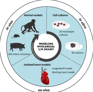
6. Modeling myocardial I/R injury ... 25
6.1. In vitro models ... 27
6.1.1. Cell sources... 28 6.1.2. Mimicking AMI physiology ... 31 6.1.3. 3D culture strategies ... 31 6.1.4. Bioreactors ... 32
7. Proteomics as powerful tool for characterization of cardiac
populations ... 33 8. Aims and scope of the thesis ... 36 9. References ... 39
3
Introduction
1. Acute myocardial infarction: the unmet clinical need
Over the last decades, better medical care and living conditions have led to a gradual increase in average life expectancy, which reached a worldwide average of 71.4 years in 2015 (WHO). According to United Nations 2015 world population ageing report, the number of people aged 60 years or over increased from 607 million in 2000 to 901 million in 2015, with projections pointing to 1.2 billion in 2030 and 2.1 billion in 2050. Such increase in life expectancy has led and will lead in the coming decades to a higher prevalence of age-related diseases including cancer, neurodegenerative disorders and cardiovascular diseases.
Ischemic Heart Disease (IHD) is one of the most common types of cardiovascular disease, and a major cause of death worldwide (Benjamin
et al., 2017). IHD consists in atherosclerosis (lipid plaque deposits) in
heart arteries inner walls, narrowing and reducing blood flow to the heart, ultimately leading to an Acute Myocardial Infarction (AMI), commonly known as heart attack. AMI consists on the cessation of blood flow to an isolated region of the heart, causing oxygen and nutrient supply depletion (ischemia), leading to myocardial tissue damage with loss of cardiomyocytes (CMs), the main cell type in the heart. Since the extent of tissue damage and cell death is influenced by both the magnitude and duration of ischemia, the revascularization and restauration of blood flow as soon as possible remains the clinical intervention of choice for AMI patients (Anderson and Morrow, 2017). However, this process, also known as reperfusion, although necessary to reestablish the delivery of oxygen and nutrients to the affected area, causes increased tissue damage (Ischemia Reperfusion I/R Injury). The elevation of molecular oxygen levels occurs at a toxic rate to cells, causing up to 50% of the
4
final damaged tissue size (Hausenloy and Yellon, 2013) and contributing to almost one fourth of AMI mortality (Yellon and Hausenloy, 2007).
Nevertheless, the current treatments are successful in reducing immediate mortality but the tissue damage is often too large (in average, about 1 billion CMs are lost during an AMI, Laflamme & Murry, 2011) to allow restauration of normal muscle function. Deposition of fibrous scar tissue leads to a decrease in myocardium tensile strength and progressive loss of cardiac output, often leading to Chronic Heart Failure (CHF), a highly fatal condition (survival rate of only 50% at 5 years, Cahill, Choudhury, & Riley, 2017) to which the only available clinical option is heart transplant. As it is known, this solution is not optimal in clinical practice due to the scarcity of available heart donors, high costs and need for immunosuppression (Lund et al., 2014).
1.1. Pathophysiology of myocardial ischemia/reperfusion
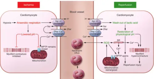
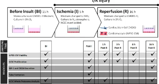
The two phases of I/R injury have distinctive physicochemical properties and therefore distinct pathophysiology mechanisms affecting myocardium tissue and CM death (figure 1.1).
During ischemia, the deprivation of oxygen and nutrients results in a series of biochemical and metabolic cellular changes including decrease of ATP and pH levels, increase in intracellular lactate and accumulation of intracellular Ca2+. Without oxygen, CMs stop oxidative phosphorylation, leading to a decrease in ATP synthesis and an increase in anaerobic respiration by glycolysis and lactic acid fermentation with accumulation of intracellular lactate. While increased glycolysis helps to compensate for the lack of aerobic ATP, this pathway binds less ATP hydrolysis-generated H+, leading to lower pH. The intracellular accumulation of H+ activates the Na+/H+ exchanger (figure 1.1). The lack
5 of ATP during ischemia also causes CM contraction arrest and inactivation of 3Na+/2K+ exchanger ATPases, leading to intracellular Na+ accumulation. In response, the activation of the 2Na+/Ca2+ pump results in intracellular Ca2+ overload (Hausenloy and Yellon, 2013; Kalogeris et
al., 2017) (figure 1.1)
Figure 1.1. Schematic illustration of the main biochemical mechanisms underlying myocardial ischemia/reperfusion induced cardiomyocyte cell injury and death. During ischemia, metabolism is switched to anaerobic respiration, resulting in production of lactate, low ATP and low pH. Ionic channels activation leads to intracellular accumulation of Na2+ and Ca2+. During reperfusion, the electron transport chain is reactivated, generating ROS that, together with higher Ca2+ accumulation, lead to MPTP opening and CM contracture. Neutrophils are recruited to the site of injury in response to the release of ROS and cytokines, contributing to tissue inflammation (Hausenloy and Yellon, 2013).
Restauration of blood flow during reperfusion provides oxygen and metabolic substrates required for aerobic ATP generation. However, the reactivation of the mitochondrial electron transport chain leads to production of cytotoxic reactive oxygen species (ROS). Acidosis is also corrected by the Na+/H+ exchanger which results in additional intracellular Na+ accumulation, resulting in the activation of 2Na+/Ca2+ exchanger. The release of extra Ca2+ from sarcoplasmic reticulum, adding to the ischemic Ca2+ accumulation culminates into a large overload of intracellular Ca2+, which in turn promotes hypercontracture
6
(sarcomeres myofibrillar contraction) (Hausenloy and Yellon, 2013; Kalogeris et al., 2017). ROS synthesis, the increase in pH and the Ca2+ overload result in the opening of the mitochondrial permeability transition pore (MPTP), a non selective channel of the inner mitochondrial membrane. MPTP opening results in mitochondrial membrane depolarization and uncoupling of the electron transport chain, leading to ATP depletion, mitochondria swelling with mitochondria membrane rupture and release of apoptotic factors culminating in activation of apoptosis and cell death (figure 1.1) (Hausenloy and Yellon, 2013; Kalogeris et al., 2017).
In response to the stress caused by reperfusion, CMs secrete several growth factors (including insulin-like growth factor 1 IGF-1, epidermal growth factor EGF, hepatocyte growth factor HGF, vascular endothelial growth factor VEGF), cytokines, chemokines and other pro-inflammatory molecules, which together with the released ROS recruit immune cells to the site of injury (figure 1.1). Neutrophils are key players in the post AMI inflammatory process: by secreting ROS, proteases, chemokines and other cytotoxic molecules, these cells further enhance inflammation and tissue damage (Frangogiannis, 2014). Phagocytic leukocytes also clear dead cells and matrix debris, setting the stage for fibrous collagen-based scar tissue deposition by activated cardiac fibroblasts (see section 5).
2. Current treatments for myocardial I/R injury
Duration of the ischemia phase is a major contributor to the extent of myocardial tissue damage. As so, the intervention of choice for a patient with AMI symptoms is the rapid reperfusion of the affected artery. The reperfusion is done mechanically by a balloon-inflated metal mesh stent
7 (percutaneous coronary intervention, PCI), by pharmacological anti-platelet and fibrinolytic (anti-thrombotic) agents, or by a combination of both (e.g., drug eluting PCI stents). Another alternative to PCI is coronary bypass surgery, in which a healthy vessel is used to divert the blood flow around the blocked artery (Anderson and Morrow, 2017). In addition, angiotensin-converting-enzyme (ACE) inhibitors and β-blockers are commonly used in the clinic to downsize myocardial scar formation (Anderson and Morrow, 2017). A substantial decrease of in-hospital AMI-related mortality rate of 7-18% was registered over the last two decades (Cahill, Choudhury and Riley, 2017). Such improvement in AMI patients prognosis is a testament to advances in PCI and pharmacological therapies as well as implementation of preventative measures. While immediate unclogging of the affected artery remains the keystone for the treatment of AMI patients, several approaches focusing in reducing the damage caused by the reperfusion are being developed and tested in clinical trials.
In contrast to classic unimpeded reperfusion, ischemic post-conditioning, also designated as slow reperfusion, consists in intermittent reperfusion, with brief repetitive interruptions of blood flow. This on/off reperfusion is thought to trigger a cascade of cell protection mechanisms that translate into a reduced myocardial injury size. Although promising results were registered in animal models, human clinical trials remain inconclusive (Giustino and Dangas, 2017). Another strategy showing more promising results in humans is remote ischemic per-conditioning, in which tissues other than the heart (usually limbs) are exposed to ischemia, resulting in the activation of systemic mechanisms of defense and myocardial protection (McLeod, Iansavichene and Cheskes, 2017).
A multitude of pharmacological approaches have also been developed to reduce the adverse impact of reperfusion injury, and several are currently
8
being evaluated in clinical trials. One such example is cyclosporine A, an inhibitor of MPTP opening. However, a recent meta-analysis has shown that the use of this drug before PCI does not translate into a better clinical outcome (Rahman et al., 2018).
Besides mechanical and pharmacological-based treatments, cell-based approaches have been emerging in the last years as a novel strategy to treat infarcted myocardium.
3. Novel therapeutic strategies for myocardial I/R injury
For many years, the adult mammalian heart has been considered an organ without regenerative potential. In 2003, Beltrami and colleagues identified cardiac stem/progenitor cells (CSCs) in the mouse heart for the first time (Beltrami et al., 2003), and in the following years CSCs were identified in other animals including humans (Bearzi et al., 2007). As most adult stem cells, CSCs have the ability to self-renew and to differentiate into tissue-specific lineages [CMs, vascular smooth muscle cells (SMCs) and endothelial cells (ECs)] (Beltrami et al., 2003).
The discovery of CSCs is an hallmark in AMI-related research, as it challenges the previous paradigm in cardiovascular research, in which the heart was seen as a post-mitotic organ without endogenous regenerative capacity. This finding opened novel avenues in regenerative medicine strategies for recovery of infarcted myocardium, including cell therapy and protein-based stimulation of endogenous heart repair.
3.1. Cell therapy
In cell therapy-based approaches several cell types were already applied in clinical trials, including “first generation” stem cell sources such as
9 bone-marrow mononuclear cells (e.g., REPAIR-ACS- NCT00711542,
BOOST-NCT00224536), bone marrow-derived mesenchymal
stem/stromal cells (e.g. BOOST trial, NCT00224536), and adipose tissue-derived mesenchymal stem/stromal cells (e.g., APOLLO trial, NCT00442806).
After “first generation” stem cell sources, more focus is now being devoted to therapies with purified and homogeneous “second generation” stem cell sources, such as CSCs.
3.1.1. Cardiac stem cells in cell therapy
CSCs are considered by several authors as the preferable candidate cell therapy for cardiac diseases, mainly due to their physiologic location and function in the heart, their potential to differentiate into myocardial lineages, and the promising regenerative effects of CSCs transplantation in myocardial infarction preclinical models. In a recent preclinical meta-analysis, an estimated improvement of 10.7% of left ejection fraction was registered, with superior effects of CSCs compared with other cell types in mice (Zwetsloot et al., 2016). However, such trend seems to be lost when moving to large animal models (Zwetsloot et al., 2016).
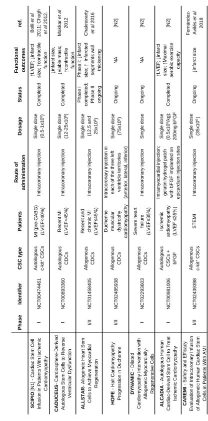
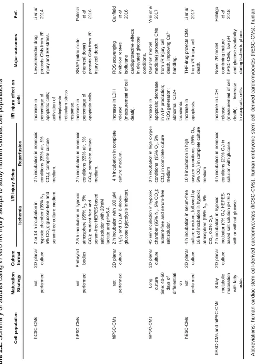
The first clinical trials using CSCs were based on autologous therapies (Chugh et al., 2012; Makkar et al., 2012; Ishigami et al., 2015) (table 1.1), which have the great advantage of not presenting immunogenicity risks to the patients. However, autologous cell therapy is associated with serious limitations that compromise widespread “off-the-shelf” clinical applications such as the difficult logistic, economic and time-constraints in patient specific tissue harvesting and expansion. Moreover, cell’s phenotype, regenerative potential, and quality will be highly variable and dependent on patient’s age, co-morbidities, and genetic background
10
(Dimmeler and Leri, 2008; Wu et al., 2016; Sharma et al., 2017). To overcome such limitations, the field has been moving towards allogeneic clinical approaches, such as the clinical study ALLSTAR and more recently, the CAREMI trial (Sanz-Ruiz et al., 2017; Fernández-Avilés et
al., 2018), which had success in demonstrating safety and lack of
rejection of transplanted cells, as well as improvements in infarct size (table 1.1). Moreover, a cross-talk of transplanted allogeneic c-kit+ hCSCs with innate natural killer cells has been shown to result in attenuation of myocardium inflammation and prevention of adverse scar tissue formation (Boukouaci et al., 2014). The same group has also identified programmed death ligand 1 (PDL-1) interaction with T regulatory cells as one of the mechanisms involved in this immunomodulatory capacity and suggested PDL-1 as a marker to identify and select low immunogenic risk allogeneic c-kit+ CSCs (Lauden
et al., 2013). Another cell therapy approach being explored is the use of
CSCs derived from pluripotent cell populations such as the ESCORT trial, in which CSCs derived from embryonic stem cells (ESC-CSCs) have been applied (table 1.1).
For all different CSC subpopulations tested, clinical trials have demonstrated some physiological improvements, namely increase in viable tissue and in heart functional outcome (table 1.1), but very limited cell retention and engraftment in the heart was observed, regardless of the route of administration and cell dosage. Within 24 hours of delivery, less than 10% of injected cells remain at the targeted location, and most of the successfully retained cells die, probably due to the inflammatory environment in the infarct and infarct border zones of the myocardium (Hong and Bolli, 2014; Mathur et al., 2017). In order to further improve the physiologic benefit of cell transplantation, several strategies have been pursued to increase cell retention, including preconditioning of cells
11 to be transplanted, for example with an hypoxia cultivation priming phase (Hosoyama et al., 2015; Hernandez et al., 2018), preconditioning the target tissue (Assmus et al., 2013), repeated cell dosage (Tokita et al., 2016), and biomaterial-based approaches (Hosseinkhani et al., 2010; Kryukov, Ruvinov and Cohen, 2014; Rajabi-Zeleti et al., 2014; Gaetani et
al., 2015; Menasché, Vanneaux, Hagège, et al., 2015).
3.2. Cell-free therapeutic approaches
The low cell engraftment efficiency in preclinical and clinical studies strongly supports the hypothesis that the beneficial physiological effect of transplanted cells is due to paracrine modulation rather than to cells engraftment and differentiation (Madonna et al., 2016).
In fact, novel strategies for heart regeneration involve the activation of endogenous CSCs populations directly with growth factors. Examples of growth factors investigated include IGF-1 and HGF (Urbanek et al., 2005; Ellison et al., 2011; Koudstaal et al., 2014; O’Neill et al., 2016; Blanco Blazquez et al., 2017), granulocyte colony-stimulating factor (G-CSF) (STEMMI-NCT00135928), erythropoietin (REVIVAL-3-NCT0039083), as well as VEGF (NORTHERN-NCT00143585). Such single growth factor therapies showed only little benefit in AMI patients, possibly due to the rapid diffusion and short half-lives of the injected molecules (Awada, Hwang and Wang, 2017). Other approaches try to overcome this by combining growth factor therapy with cell therapy, such as ALCADIA trial, in which a sustained release of basic fibroblast growth factor (bFGF) from a gelatin hydrogel sheet was used in order to augment the effect exerted by the transplanted CSCs (Takehara et al., 2012) (table 1.1).
12 Ta bl e 1 .1 . Cl in ic a l T ri a ls wi th Tra n s p la n ta ti o n o f h C S Cs . P has e Ident if ie r C S C t y pe P a ti e nt s R oute of a dm ini s tra ti on D osa ge S ta tus Funct ional outc om e s re f. SC IPI O [N 1] -C ardiac St em C ell Inf us io n in Pat ient s W it h Is c hem ic C ardiom y op at hy I N C T 004744 61 Aut olog ou s c -k it +C SC s M I (pre -C ABG) (LVEF < 40% ) Int rac or on ar y inj ec tio n Single dos e (0. 5 -1x 10 6) C om plet ed ↑LVEF ; ↓ inf arc t siz e; ↑ cont ra ct ile fun c tio n Bolli et al 2011; C hugh et al 2012. C A D U C EU S -C ardios ph e re -D e ri v e d Aut olog ou s St em C ells t o R ev e rs e Vent ric u la r D y s func tio n I N C T 008933 60 Aut olog ou s CDCs R ec ent M I (LVEF < 45% ) Int rac or on ar y inj ec tio n Single dos e (12 -25x 10 6) C om plet ed ↓inf arc t s iz e, ↓v ia bl e m as s; ↑c ont ra ct ile func tio n M ak k a r et al 2012 A L L ST A R -Allogen ei c H eart Sem C ells t o Ac hie v e M y oc ard ia l R egener at io n I/II N C T 014584 05 Allogen ou s CDCs R ec ent and c hronic M I (LVEF ≤45% ) Int rac or on ar y inj ec tio n Single dos e (12. 5 and 25x 10 6) Phas e I c om ple te d ; Phas e II on go in g Phas e I: ↓ inf arc t siz e; ↑ inf arc ted s egm e nt s w all thic k e ni ng C hak ra v ar ty et al 2016 H OPE -H alt C ardiom y o pa th y Pr og res s ion in D uc he n n e I/II N C T 024859 38 Allogen ou s CDCs D uc henn e m us c u la r dy s trophy c ardiom y o pa th y Int rac or on ar y inj ec tio n in eac h of t he three lef t v ent ri c le t errit orie s (ant erio r, lat eral , inf erior ) Single dos e (75 x 10 6) Ongoing NA [N 2] D YN A M IC -D ilat ed C ard iom y op at hy I nt erv e nt io n w it h Allogen ei c M y oc ardi al ly -R egener at iv e C ells I N C T 02 29 36 03 Allogen ou s CDCs Sev er e heart failure (LVEF ≤35% ) Int rac or on ar y inj ec tio n Single dos e Ongoing NA [N 2] A L C A D IA -Au tolo go us H um an C ardiac -D er iv ed St em C ell to T reat Is c hem ic C ardiom y op a th y I N C T 009810 06 Aut olog ou s C SC s + bF GF Is c hem ic ardiom y o pa th y (LVEF ≤ 35%) Int ram y oc a rdi a l inj ec tio n; gelat in hy drogel pat c h w it h bF GF im plant ed on epic ard iu m inj ec ti on s it es Single dos e (0. 5x 10 6/k g) ; 200m g bF GF C om plet ed ↑LVEF ; ↓ inf arc t siz e; ↑ M ax im al aerobic ex erc is e c apac it y [N 2] C A R EM I -Saf et y and Ef fic ac y Ev alu at io n of I nt rac oro n ar y I nf us ion of Allogen e ic H um an C ardiac St em C ells in Pat ient s W it h AM I I/II N C T 024393 98 Allogen ou s c -k it +C SC s ST EM I Int rac or on ar y inj ec tio n Single dos e (35x 10 6 ) Ongoing ↓inf arc t s iz e F ernández -Av ilé s et al 2018
13 A b b re v iat ion s : C a rd iac s te m / p ro g e n it o r c e lls ( C S C s ); C a rd io s p h e re -d e riv e d c e lls ( C D C s ); E mbr y o n ic s te m c e lls ( E S C ); B o n e mar ro w -d e riv e d mes e n c h y mal s te m c e lls ( B M -M S C s ); mes e n c h y mal s te m c e lls ( MS C s ); My o c a rd ial inf a rc tion ( MI ); C o ro n a ry a rt e ry b y p a s s g ra ft ing ( C A B G) ; L e ft v e n tr ic u lar e je c tion f ra c tion ( L V E F) ; S e g men t e lev a tion my o c a rd ial in fa rc tion ( S TE MI ); R igh t v e n tr ic u lar e jec tion f ra c tion ( R V E F) . N A – n o t a v a ila b le [ [N 1 ]: s u b je c t to e x p re s s ion o f c o n c e rn ; [N 2 ]: I n fo rma tion w e re o b ta ine d f ro m w w w .c lin ic a lt rial s .g o v , a c c e s s e d Mar c h 2 0 1 8 . P has e Ident if ie r C S C t y pe P a ti e nt s R oute of a dm ini s tra ti on D osa ge S ta tus Funct ional outc om e s ref. ESC OR T -T rans pla nt at io n of H um an Em bry oni c St em C ell -deriv ed Progeni to rs in Sev er e H eart F ailure I N C T 020579 00 ESC deriv ed C D 15+ Is l-1+ C SC s Sev er e heart failure (LVEF ≤ 35% ) Epic ard ia l deliv e ry of c ells em bed de d in fibrin pat c h Single dos e (4x 10 6) R ec ruit ing NA M enas c h e et al 2005 T IC A P -T rans c oro n ary Inf us ion of C ardiac Progenit or C ells in Pat ient s W it h Single Vent ric le Phy s iolo g y I N C T 012738 57 Aut olog ou s CDCs Pediat ri c pat ient s w it h hy poplas ti c lef t heart s y ndrom e Int rac or on ar y inj ec tio n Single dos e (0. 3x 10 6/k g) C om plet ed ↑R VEF ; ↑s om at ic grow th Is higam i et al 2015 PER SEU S -C ardiac Progenit o r C ell Inf us io n to T reat U niv e n tric u la r H eart D is eas e II N C T 018297 50 Aut olog ou s CDCs U niv e nt ric u la r heart dis eas e Int rac or on ar y inj ec tio n Single dos e (0. 3 x 10 6 c ells /k g ) C om plet ed ↓inf arc t s iz e; ↑s om at ic grow th; ↑ f ac tor s produc tio n Is higam i et al 2017 C ON C ER T -HF -C om bin at io n of M es en c h y m al and C -k it + C ardiac St em C ells as R egenera ti v e T herapy f or H eart F ailure II N C T 025018 11 Aut olog ou s BM _M SC s , c -k it + c C SC s alone and in c om bin a tio n Is c hem ic c ardiom y o pa th y T rans end oc a rdi al inj ec ti on Single dos e (150x 10 6 BM -M SC s ; 5x 10 6 C SC s ) Ongoing NA [N 2] T rans plan ta ti on of Aut olog o us C ardiac St em C ells in Is c hem ic H eart F ailure II N C T 017584 06 Aut olog ou s C SC s Is c hem ic heart failure Int rac or on ar y inj ec tio n Single dos e (5 -100x 10 6) R ec ruit ing NA [N 2] A POL L ON -C ardiac St em /Pro ge ni to r C ell Inf us io n in U niv e nt ric u la r Phy s iolo gy III N C T 027819 22 Aut olog ou s C SC s Pediat ri c pat ient s w it h heart f ailure Int rac or on ar y inj ec tio n Single dos e (0. 3x 10 6 c ells /k g ) R ec ruit ing NA [N 2] TAC -HFT -II -T he T rans end oc a rdi al Aut olog ou s C ells (hM SC ) or (hM SC ) and (hC SC ) in Is c hem ic H eart F ailure T rial I/II N C T 025032 80 C om bina ti on of au tolo g o us M SC s and c -k it +C SC s or M SC s alone is c hem ic lef t v ent ri c ul ar dy s func tio n and/ or heart failure s ec ond a ry t o M I T ran s en d oc a rdi al inj ec ti on Single dos e (200x 10 6BM -M SC s or 190x 10 6BM -M SC + 1x 10 6 C SC s ) R ec ruit ing NA [N 2]
14
Despite promising, several challenges still need to be addressed in order to reach the full clinical potential of CSCs. Further understanding of CSC biology is needed in order to discover and modulate molecular pathways involved in the regenerative potential of these cells. Novel findings will be pivotal for the development of improved clinical strategies, including enhanced activation of endogenous hCSCs, preconditioning of cells to be transplanted and protein-based therapies. New relevant human cell-based models of cardiac tissue damage should also be pursued to characterize CSC response to myocardial injury in vitro.
International collaborative consortiums and multicenter studies such as Translational Alliance for Regenerative Therapies in Cardiovascular Syndromes (TACTIC) (Fernández-Avilés et al., 2017), Consortium for Preclinical Assessment of Cardioprotective Therapies (CAESAR) (Jones et
al., 2015), and Cardio Repair European Multidisciplinary Initiative
(CARE-MI) (www.cordis.europa.eu) have been emerging as important platforms to bring basic researchers and clinicians together to discuss and define common goals and strategies, as well as to standardize protocols and analytical techniques in preclinical and clinical studies.
4. The role of endogenous cardiac stem cells in cardiac repair
Endogenous CSCs play important roles in cardiac homeostasis and in response to physiological stress and cardiac injury. The mammalian adult heart harbors a small percentage of endogenous CSCs (about one stem cell per 8000-20000 CMs (Anversa et al., 2006)), which are located in organized hypoxic niches within the myocardium, more abundant in lower hemodynamic stress areas such as the atrium and apex. Within these
15 niches, CSCs are typically clustered together with early committed cells and adult CMs (Leri et al., 2014).
Since their discovery in 2003 (Beltrami et al., 2003), efforts have been made to isolate, cultivate, characterize and study the regenerative mechanisms of these cells.
4.1. Identification and isolation of endogenous CSCs
Several CSCs subpopulations have been described and isolated from the adult heart according to their phenotypic profile and differential expression of several surface molecular markers. These subpopulations include c-kit+, Sca1+, and Isl-1+ CSCs (reviewed in detail in Santini et al. 2016).
4.1.1. C-kit+ CSCs
C-kit+ cells were the first population of CSCs to be identified in the adult mouse heart in 2003 (Beltrami et al., 2003). These cells are characterized by expression of the stemmness marker c-kit, expression of cardiac lineage transcription factors such as GATA4, Nkx2.5, and Mef2C, and absence of hematopoietic lineage markers such as CD34 and CD45 (Santini et al., 2016). c-kit+ CSCs are one of the most extensively studied CSCs subpopulations, already employed in several clinical trials such as SCIPIO, CAREMI, CONCERT and TAC-HFT-II (table 1.1)
4.1.2. Sca-1+ CSCs
Sca-1+ CSCs are characterized by the expression of the endothelial marker stem cells antigen-1 (Sca-1). These cells have been identified in mice and human adult heart and express the cardiac transcription factors GATA4, Mef2C and Tef1, but lack other cardiac lineage markers such as Nkx2.5, the
16
hematopoietic markers CD34 and CD45, and mature endothelial markers such as CD31 (Santini et al., 2016).
4.1.3. Isl-1+ CSCs
Another population of CSCs, characterized by the expression of the LIN-homeodomain transcription factor insulin gene enhancer protein (Isl-1) has also been described in murine and human hearts. These cells do not express c-kit, Sca-1, or CD31 while expressing the cardiac transcription factors Nkx2.5 and GATA4. The expression of Isl-1 is closely associated with age: Isl-1+ cells can be found predominantly in fetal and neonatal myocardium, yet very low levels of this population could be found in adult hearts, which limits their clinical application (Weinberger et al., 2012).
4.1.4. CSC isolation methods
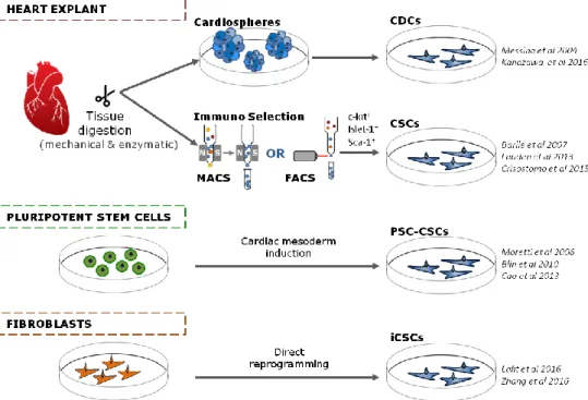
CSC subpopulations are usually isolated from the adult human heart tissue either from cadavers or from biopsies of patients undergoing heart surgery. Experimental approaches for the isolation of hCSCs from cardiac tissue can be separated into two categories: cardiosphere-based and immunoselection-based, taking advantage of the expression of specific molecular markers (figure 1.2).
The first method to isolate CSCs from human heart biopsies was reported in 2004 (Messina et al., 2004) (Patent number WO2005012510). Cardiac tissue explants were enzymatically digested and cultured as adherent explants. These explants gave rise to cellular outgrowths that spontaneously aggregate, generating three-dimensional (3D) structures, named cardiospheres, which are composed of mixed cell populations, with undifferentiated cells expressing stem cell markers in the core and differentiating cells expressing markers characteristic of cardiac, vascular
17 endothelial and stromal commitment in the periphery (Davis et al., 2010). Cardiospheres are then collected, replated in fibronectin adherent culture dishes and further expanded as monolayers to yield cardiosphere-derived cells (CDCs) (Cheng et al., 2014; Kapelios, Nanas and Malliaras, 2016).
Several preclinical studies using myocardial injury models (Kanazawa et al., 2016) have demonstrated the beneficial effects of transplantation of CDCs in improving cardiac function and decreasing scar tissue size (for detailed reviews see Marbán 2014; Kapelios, Nanas, and Malliaras 2016). These studies have already been translated into several human clinical trials such as CADUCEUS and ALLSTAR (table 1.1, page 12). However, the cell population heterogeneity inherent to the cardiosphere isolation method has been pointed as a disadvantage in terms of obtaining robust and predictable clinical outcomes.
Another method for isolating hCSCs from heart tissue is by using the enzymatic digestion of tissue explants followed by immune-selection for stem cell markers such as the surface receptors c-kit, Sca-1, Isl-1 and immunodepletion for hematopoietic and mesenchymal markers such as CD45 and Tryp, using magnetic-activated cell sorting (MACS) or fluorescence-activated cell sorting (FACS) (Barile et al., 2007; Lauden et al., 2013; Goichberg et al., 2014). In particular, CSCs immunoselected for c-kit+ represent the most extensively characterized CSC population. Although controversy exists regarding the differentiation capability of c-kit+ CSCs (Ellison et al., 2013; Nadal-Ginard, Ellison and Torella, 2014; van Berlo et
al., 2014) and the stability of c-kit marker expression during in vitro culture
(Forte et al., 2011), numerous preclinical studies (for a more detailed review consult Nigro et al. 2015) and clinical trials (table 1.1, page 12) demonstrate that c-kit+ CSCs have relevant regenerative properties, since transplantation of this cell population results in improved cardiac tissue function and reduction of scar tissue size.
18
4.1.5. Derivation of cardiac stem cells from other cell sources
Besides donated human hearts, other sources for CSC have emerged (figure 1.2). Pluripotent stem cells (PSCs), including induced pluripotent stem cells (iPSCs) and embryonic stem cells (ESCs), have unlimited self-renewal capacity and the potential to differentiate in vitro, holding great promise for the clinical translation of cell therapies. In particular, hiPSCs hold the advantage of allowing for autologous therapies without invasive isolation procedures. In fact, as development of less invasive surgical interventions techniques is further explored, the availability of tissue for isolation of autologous and allogeneic hCSCs will be scarcer in a near future. Efforts have been made throughout the years to obtain CSCs from pluripotent stem cells (PSC-CSCs).
The first study reporting PSC-CSCs (Isl-1+) was performed using murine ESCs, demonstrating the proliferative capacity of these derived CSCs on a feeder layer of mesenchymal cells (Moretti et al., 2006). Similarly, in following studies, human PSC (hPSC) were differentiated into CSCs either through an embryoid body (EB)-based spontaneous differentiation step originating Isl-1+ hCSCs (Bu et al., 2009) or a bone morphogenic protein 2 (BMP-2) directed differentiation originating stage specific embryonic antigen-1 (SSEA1+) hCSCs (Tomescot et al., 2007; Blin et al., 2010). Both types of hPSC-CSC have shown similar regenerative potential to the CSCs isolated from human fetal hearts in both rat and non-human primate models. More recently, these advances in the differentiation of hPCS-CSCs led to the translation from bench scale to a cell-based medicinal product (Menasché, Vanneaux, Fabreguettes, et al., 2015) culminating in the ongoing clinical trial with hESC-CSCs for patients with severe heart failure (ESCORT Trial, table 1.1 page 12). More recently, efforts have been made to achieve more efficient protocols for differentiation and expansion of CSCs
19 from hPSCs in a chemically defined medium under feeder- and serum-free culture conditions resulting in high purities of SSEA1+ mesoderm posterior protein 1 positive (MESP1+) cells (Cao et al., 2013).
Figure 1.2. Sources currently available for obtaining cardiac stem/progenitor cells (CSCs). Abbreviations: Magnetic activated cell sorting (MACS); Fluorescence activated cell sorting (FACS); Cardiosphere derived cells (CDCs); Pluripotent stem cell derived cardiac stem cells (PSC-CSCs); induced cardiac stem cells (iCSCs). Examples from literature are included. Adapted from Sebastião et al 2018.
Several questions still arise today on how these hPSC-CSCs compare to the CSCs isolated from the human heart. Within this context, several studies have been trying to bring light on the different cardiac progenitors generated during heart development, and which signaling pathways may be manipulated to obtain those (Birket et al., 2015). In a more recent study, global transcriptomic analysis of patient epicardium-derived CSCs and hPSC-CSCs was carried out showing that more than three thousand genes were differentially expressed between the two cell types, with hierarchical clustering analysis denoting a pronounced separation between the two
20
types of CSCs (Synnergren et al., 2016), confirming the phenotypic differences between these two populations.
Other innovative strategies for the generation of CSCs have emerged through direct reprogramming of fibroblasts (induced CSCs, iCSCs) (figure 1.2). These iCSCs have been described to i) express cardiac signature genes, ii) have extensive proliferative capacity, and iii) be able to differentiate into the three main cardiac lineages in vitro and in vivo after transplantation into infarcted mouse hearts (Lalit et al., 2016; Zhang et al., 2016). Although these iCSCs have shown promising in vivo regeneration capacity, their direct comparison to isolated endogenous CSCs or hPSC-CSCs is still lacking. In addition, these studies were performed using murine cells and further studies with human cells are still required (Chen and Wu, 2016).
4.2. Cardiac stem cells regenerative mechanisms
Endogenous CSCs play important roles in cardiac tissue cell turnover, as well as in response to physiological stress, including I/R injury. Numerous studies suggest that these cells become activated after injury, having an immuno-supressive role, proliferating, differentiating into cardiomyogenic lineages, migrating to the site of injury, and secreting important paracrine factors involved in the modulation of cell proliferation, angiogenesis, vasculogenesis and pro-survival of CMs (figure 1.3).
Although one of the hallmarks used to define CSCs is their ability to differentiate in vitro into the three major cells in the myocardium (CMs, SMCs and ECs), doubts and controversy still exist regarding the ability of endogenous and transplanted CSCs to differentiate in vivo upon injury. While some authors defend that endogenous CSCs can differentiate in vivo into functional cardiomyocytes, contributing to tissue regeneration (Ellison et
21
al., 2011, 2013; Vicinanza et al., 2017, 2018), others defend that CSCs
differentiation to cardiac lineages occurs at a very low rate, making it unlikely to be the main mechanism for CSCs-induced tissue recovery(van Berlo et al., 2014, 2018). Additionally, the origin of new CMs during tissue homeostasis and upon injury is still a matter of debate, with different authors defending different origins, ranging from CSCs to division of pre-existing CMs (Senyo et al., 2013; Sereti et al., 2018), reviewed in more detail in (Cahill, Choudhury and Riley, 2017).
Despite such doubts regarding the differentiation capacity of CSCs, there is a general consensus in the scientific community regarding CSCs-derived paracrine mechanisms. CSCs have been shown to exert potent paracrine regenerative effects both in vivo and in vitro (Li et al. 2012; Sharma et al. 2016; Miyamoto et al. 2010; Torella et al. 2007). Upon injury, CMs activate the expression and secretion of a large number of growth factors and cytokines (e.g. IGF-1, HGF, stem cell factor SCF and stromal cell derived factor 1 SDF-1) (X. Li et al., 2014a) that bind to receptors expressed by CSCs (Gomes-Alves et al. 2015; Ellison et al. 2012; Li et al. 2014; Urbanek, Rota, et al. 2005). Consequently, an auto/paracrine cross-talk is established between CMs and CSCs, leading to a continued production of growth factors by CSCs, which have been shown to contribute to the modulation of angiogenesis, vasculogenesis, pro-survival of CMs and activation of CSCs proliferation and migration as well.
Cardiac stem cell secretome studies have revealed that these cells secrete a large array of signaling molecules, including the growth factors IGF-1, EGF, VEGF, transforming growth factor beta (TGF-β), HGF and several interleukins and chemokines (Stastna et al., 2010; Albulescu et al., 2015; Sharma et al., 2015; Park et al., 2016; Torán et al., 2017). Several of these cytokines and growth factors have shown to induce CSCs proliferation in