Stimulation of GABA-Induced Ca
2+
Influx Enhances
Maturation of Human Induced Pluripotent Stem
Cell-Derived Neurons
David J. Rushton
1, Virginia B. Mattis
2, Clive N. Svendsen
2, Nicholas D. Allen
1*, Paul J. Kemp
1*1 Divisions of Pathophysiology & Repair and Neuroscience, School of Biosciences, Cardiff University, Cardiff, United Kingdom, 2 Regenerative Medicine Institute, Cedars-Sinai Medical Center, Los Angeles, California, United States of America
Abstract
Optimal use of patient-derived, induced pluripotent stem cells for modeling neuronal diseases is crucially dependent upon the proper physiological maturation of derived neurons. As a strategy to develop defined differentiation protocols that optimize electrophysiological function, we investigated the role of Caβ+ channel regulation by astrocyte
conditioned medium in neuronal maturation, using whole-cell patch clamp and Caβ+ imaging. Standard control
medium supported basic differentiation of induced pluripotent stem cell-derived neurons, as assayed by the ability to fire simple, single, induced action potentials. In contrast, treatment with astrocyte conditioned medium elicited complex and spontaneous neuronal activity, often with rhythmic and biphasic characteristics. Such augmented spontaneous activity correlated with astrocyte conditioned medium-evoked hyperpolarization and was dependent upon regulated function of L-, N- and R-type Caβ+ channels. The requirement for astrocyte conditioned medium could
be substituted by simply supplementing control differentiation medium with high Caβ+ or -amino butyric acid (GABA).
Importantly, even in the absence of GABA signalling, opening Caβ+ channels directly using Bay K8644 was able to
hyperpolarise neurons and enhance excitability, producing fully functional neurons. These data provide mechanistic insight into how secreted astrocyte factors control differentiation and, importantly, suggest that pharmacological modulation of Caβ+ channel function leads to the development of a defined protocol for improved maturation of
induced pluripotent stem cell-derived neurons.
Citation: Rushton DJ, Mattis VB, Svendsen CN, Allen ND, Kemp PJ (β01γ) Stimulation of GABA-Induced Caβ+ Influx Enhances Maturation of Human
Induced Pluripotent Stem Cell-Derived Neurons. PLoS ONE 8(11): e810γ1. doi:10.1γ71/journal.pone.00810γ1
Editor: Steven Barnes, Dalhousie University, Canada
Received June 18, β01γ; Accepted October 18, β01γ; Published November ββ, β01γ
Copyright: © β01γ Rushton et al. This is an open-access article distributed under the terms of the Creative Commons Attribution License, which permits unrestricted use, distribution, and reproduction in any medium, provided the original author and source are credited.
Funding: This work was supported by: Medical Research Council (G0900180), http://www.mrc.ac.uk; and CHDI (A-γγγ7 & A-45β8), http:// chdifoundation.org. The funders had no role in study design, data collection and analysis, decision to publish, or preparation of the manuscript.
Competing interests: The authors have declared that no competing interests exist. * E-mail: [email protected] (PJK); [email protected] (NDA)
Introduction
Induced pluripotent stem cells (iPSCs) from human patients with well-defined, and often genetically determined neuronal pathology, have huge potential both for disease modelling and for reliable high-throughput drug screening. Consistent and reproducible generation of functional neurons from iPSC is crucial to the development of cellular models of neurological disease. Although a plethora of protocols are available which might produce such neuronal models, only a few have been driven by robust functional characterisation of neuronal maturation and synaptogenesis [1-γ], which limits their utility for both disease modelling and the development of novel therapies
in vitro.
Pluripotent stem cells (PSCs) differentiate into neurons through a program of neuralisation, neuronal-subtype fate specification, cell cycle exit, post-mitotic neuronal differentiation
CHIR990β1, which together with DAPT effectively promoted post-mitotic nociceptor neural differentiation [9].
In part, the slow yet progressive nature of neuronal maturation in differentiating PSC cultures may be due to the delayed emergence and development of astrocytes [10,11]. Glia/neuron interactions have major positive effects on the functional maturation of neurons in vitro; both co-culture with astrocytes, and treatment with astrocyte conditioned medium (ACM), have been shown to enhance neuronal synaptogenesis [1β-14]. The astrocyte-secreted factors that accelerate neurogenesis, promote neurite outgrowth and enhance the synaptogenesis of cultured neurons include thrombospondins, transforming growth factor (TGF ), WNTs, glial-derived neurotrophic factor (GDNF) and the chemokine C-C motif ligand 5 (CCL5) [1β,15,16]. Similarly, the maturation of human PSC-derived neurons is promoted by glial co-culture or ACM [1,17], although the underlying physiological mechanisms have yet to be determined.
Here, the effects of ACM on the electrophysiological properties of maturing human iPSC-derived neurons have been investigated. Neuronal maturation was characterized by a hyperpolarised resting membrane potential (Vm) and the transition from a capacity to fire only evoked action potentials to spontaneous activity as a consequence of in vitro
synaptogenesis. Fundamental to these processes in other systems is the precise regulation of Caβ+ homeostasis and the developmental regulation of voltage-gated ion channels. Indeed, enhanced L-type voltage-gated Caβ+ channel activity promotes mouse progenitor cell neurogenesis [18], and changes in L-type and N-type Caβ+ channel functional expression have been implicated in mouse ESC-derived neuronal differentiation [19]. Furthermore, astrocytes increase N-type channel expression in adult hippocampal cultures [β0], where Caβ+ influx through L-type and N-type Caβ+ channels has been implicated in excitation-coupled neurogenesis [β1]. Based on these data, it was hypothesized that one mechanism by which ACM might promote neuronal maturation is through the upregulation of voltage-gated Caβ+ channel activity.
A further neuromodulatory pathway which is active in immature and differentiating neurons is excitatory -amino butyric acid (GABA) signaling [ββ-β4] We have previously reported that human PSC-derived neurons show ubiquitous Caβ+ responses to GABA, even at early stages of differentiation [7,β5], an observation similar to that reported in mouse neuroepithelial cells [β6]. Early GABAA-evoked Caβ+ responses, ahead of synaptogenesis, have also been observed in other systems, including retinal neurogenesis, where it was proposed that GABA might act as a trophic factor by activating L-type Caβ+channels [β7]. Given the established excitatory role of GABA in early fetal development [β4,β6,β8], the role of GABA in the regulation of the maturation of PSC-derived neurons was also investigated in this study.
The data presented herein suggest that functional maturation of iPSC-derived neurons is dependent upon an active GABAA receptor/Caβ+ channel pathway. Importantly, they also strongly suggest that direct manipulation of Caβ+ influx and/or Caβ+ channel activity could provide a simple and convenient strategy
to accelerate the functional maturation of immature PSC-derived neurons in vitro.
Materials and Methods
Ethics statement
Cardiff University's Biological Standards Committee performs the functions of the Animal Welfare and Ethics Body, as required by the UK's Animals (Scientific Procedures) Act 1986, in relation to its ethical oversight of the use of animals for scientific purposes. Cardiff University is authorized to carry out such work under Establishment License γ0/βγ05, granted by the UK Home Office. The humane killing of animals for scientific purposes in the UK is authorized by Schedule 1 to the Animals (Scientific Procedures) Act 1986, which specifies humane methods according to the species, size and stage of development of the animal. All animals in this study were killed humanely in accordance with this guidance by persons registered with the University as trained and competent in these methods, and thus no specific project license authority was required.
Pluripotent stem cell culture and neuronal differentiation
medium. For the first 7 days 10µM DAPT (Sigma-Aldrich, Poole, Hants., U.K.) was added to the differentiation medium, after this the cells were cultured in just differentiation medium for a further 14 days. Therefore the cells underwent differentiation for a total of γ weeks.
Differentiation medium was supplemented with ACM (1:1), additional CaClβ, GABA or specific ion channel modulators included: 1.βmM CaClβ to raise [Caβ+] from 0.6mM to 1.8mM; γ00µM GABA ( -Aminobutyric acid, Sigma-Aldrich, Poole, Hants., U.K.); 10µM bicuculline (Tocris, Bristol, Avon, U.K.); βµM nifedipine (Tocris, Bristol, Avon, U.K.); 0.1µM conotoxin Aldrich, Poole, Hants., U.K.); 0.1µM agatoxin (Sigma-Aldrich, Poole, Hants., U.K.)); 0.1µM SNX48β (Tocris, Bristol, Avon, U.K.); 1µM BayK 8644 (Tocris, Bristol, Avon, U.K.).
Astrocyte isolation, culture and medium conditioning ACM was produced from primary mouse astrocyte cultures. Striata were dissected from newborn C57Bl/6J mice. Tissue was dissected into cold Hank’s buffered saline (HBS, Peprotech, London, U.K.), fragmented by gentle trituration, washed in HBS, and then digested by incubation with accutase (PAA Labs, Yeovil, Somerset, U.K.) and DNAse1 (0.1mg.ml-1, Sigma-Aldrich, Poole, Hants., U.K.) for approximately γ0 minutes at γ7°C. Digestion was stopped by addition of astrocyte growth medium (DMEM supplemented with 1% Glutamax (Life Technologies, Paisley, Strathclyde, U.K.), 10% fetal bovine serum (Sigma-Aldrich, Poole, Hants., U.K.) and 1% of a cocktail of penicillin, streptomycin and fungizone (Anti-Anti, Life Technologies, Paisley, Strathclyde, U.K.). A cell suspension was derived by further trituration using a P1000 pipette. Dissociated, cells were washed twice with culture medium and plated onto culture flasks. Cultures were passaged at a 1:6 ratio upon reaching confluency. Confluent cultures at P1 and Pβ were used to condition neural differentiation base medium (DMEM:F1β (1:γ), β % Bβ7, 1% non-essential amino acids; all from Life Technologies, Paisley, Strathclyde, U.K.). ACM was harvested after 7β h, filter sterilised, aliquoted and stored for later use at -80°C. Different batches of ACM were compared using a mouse CCLβ (chemokine C-C motif ligand β) ELISA (Quantikine Mouse CCLβ/JE/MCP-1 immunoassay. R & D Systems, Abingdon, Oxon., U.K.) and normalised to a constant final concentration of CCLβ of 1 mg.ml-1. For differentiating neurons, ACM was mixed with differentiation base medium in a 1:1 ratio.
Electrophysiology
Unless otherwise stated, all reagents for electrophysiology and Caβ+ imaging were purchased from Sigma-Aldrich (Poole, Hants., U.K.). Coverslips with attached neurons were transferred to a perfusion bath mounted on the stage of an inverted microscope (Olympus CK-40, Olympus microscopes, Essex, UK) with phase contrast and rapid perfusion system capable of β0ms solution changes (Intracel RSC160, Intracel, Royston, UK). Conventional, whole cell patch clamp was performed on differentiating neurons at 1, β and γ weeks post plate-down using an Axopatch β00B amplifier (Molecular Devices, Sunnyvale, California, USA) interfaced with a computer using an Axon Digidata 1γβ0 DAC converter
(Molecular Devices, Sunnyvale, California, USA). In voltage-clamp mode, voltage protocols were generated with, and evoked currents were recorded by, the Axon Instruments ClampEx acquisition software in the PClamp 9.β suite (Molecular Devices, Sunnyvale, California, USA). Similarly, in current clamp mode, ClampEx was used to generate current protocols and to record membrane potentials (Vm). All analyses of currents and Vm were performed off-line using ClampFit 9. For all recordings, pipettes were manufactured using a two-stage electrode puller (PP-8γ0, Narishige International ltd., London, U.K.), were heat-polished using a Narishige Microforge (Narishige International ltd., London, U.K.) and had tip resistances of between 4 and 7 MΩ when filled with an the internal solution containing (in mM) 117 KCl, 10 NaCl, 11 EGTA, β Na.ATP and 11 HEPES. The control external solution (ESC) contained (in mM) 1γ5 NaCl, 5 KCl, 5 HEPES, 10 glucose, 1.β MgClβ and 1.β5 CaClβ. Vm and spontaneous action potentials were recorded in fast current clamp mode, where current was clamped at 0mV in ECS. Induced action potentials were recorded in fast current clamp mode during a current step protocol; current was injected to hold the membrane potential at ca. -70 mV and then 100 ms current injection was applied, starting with 0 pA and increasing to 180 pA in 10 pA increments in each successive sweep. Voltage-gated Na+ and K+ currents were measured using a voltage-step protocol; the voltage was held at -70mV and then stepped for β00 ms from -1β0 mV to 50 mV in 10 mV increments in each successive sweep. Leak currents were subtracted on-line using a P/N = 8 pre-pulse voltage-protocol. ESC was either supplemented with 10 mM tetraethyl ammonium chloride (TEA, a broad-spectrum K+ channel blocker) or NaCl was completely replaced with N-methyl-D-glutamine chloride (NMDG, a non-permeable substituent of Na+) to confirm dissect Na+ and K+ current from each other. The voltage-dependence of activation and steady-state inactivation of the Na+ currents were measured using a dual voltage-step protocol. From holding potential of -90 mV, the first β00 ms steps increased in +5mV increments to 0mV (to elicit current activation) and these were followed by a β00 ms step to 0 mV (to assess current inactivation). Following conversion to conductance (G), the G/Gmax values were plotted against the voltage to give activation and inactivation curves; the point of transection of the two curves is the voltage of peak available current, or peak Na+ window.
clamp at -β0 mV over several minutes with high analogue gain (50x).
Calcium imaging
Fura-β Caβ+ imaging was performed using a monchromator based fluorimeter system (Cairn Research, Faversham, Kent, U.K.). Cells plated on glass coverslips were incubated in β50µl of culture medium with a final concentration of 1µl of fura-β-AM (Life Technologies, Paisley, Strathclyde, U.K.) for γ0 minutes at γ7°C. Coverslips were then placed in the perfusion chamber mounted on an inverted Olympus IX70 inverted microscope (Southend-on-Sea, Essex, U.K.) and continuously perfused using an RSC, rapid perfusion system (RSC160, Intracel RSC160, Intracel, Royston, UK). Fura-β was alternately excited through a quartz, oil immersion objective with light at γ40 and γ80 nm (50-100ms) and re-emission from each wavelength was measured at 505 nm using a CCD camera (Hamamatsu Orca, Tokyo, Japan). βx binning was applied to each image to increase signal intensity but at the cost of resolution. Off-line, regions of interest were circled and all intensities were background subtracted before the γ40:γ80 emission ratio was calculated. Images were taken every γ s.
A high K+ (50 mM KCl isoosmotically replacing NaCl) solution was applied for 10 s at the beginning of each experimental protocol to determine which cells expressed functional, voltage-gated Caβ+ channels and to establish the magnitude of the control response. Caβ+ channel blocking agents were applied in the same high K+ solution. The area under the curve for each high K+ application (or high K+ with agent) was calculated using the sum of the integrals for each data point, subtracting the integral of a line starting just before the application and ending following complete recovery.
To examine GABAA responses a 10s high K+ was followed by a β00s rest and a 10s pulse of γ00µM GABA in ECS. Any significant Caβ+ influx (in terms of a change in Fura-β emission intensity ratio (iγ40/iγ80) was interpreted as an excitatory GABA response, as the cell depolarised in order to activate voltage activated Caβ+ channels. After β00s rest, a 10s pulse of γ00µM GABA in reduced Cl- solution (isosmotic replacement of NaCl with Na.isethionate in ECS, resulting in 9.9mM Cl -concentration), thus cells functionally expressing GABAA channels and will depolarise the cell and evoke a voltage activated Caβ+ influx. The background subtracted fura-β intensity ratio traces were visually inspected for each cell, only cells showing a Caβ+ response to both high K+ and GABA with low Cl- solution were included. The included cells were then sub-divided into cells with a Caβ+ response to GABA in ECS, considered to have an excitatory GABA response, and those without a Caβ+ response to GABA in ECS, considered to have an inhibitory GABA response.
Statistical analyses
For continuous data-sets where β means were compared the data were checked for an approximate fit to a Gaussian distribution. Assuming an approximately Gaussian distribution a two sample unpaired, two tailed, T-test was performed. If the data distribution was significantly distinct from a Gaussian distribution then data transformations were considered in order
to apply a T-test. However, if there was no obvious data transformation which resulted in an approximately Gaussian distribution then the medians were compared using a Mann-Whitney U test. For binomial data sets comparing two proportions we used two-tailed Chiβ tests. Statistics were reported as means or proportions followed by the sample size (number of cells) and standard error for continuous data, the probability value, test statistic and degrees of freedom (if relevant) were included for any statistical test performed. Statistics and graphical representations of the data were performed using either R (x64) β.14.0 with the default installed libraries or Graphpad Prism version 5.01 (La Jolla, Ca. USA.).
Results
ACM dramatically enhances spontaneous activity of iPSC-derived neurons
In order to establish how ACM effects the electrophysiological maturity during differentiation of iPSC-derived neurons, cells were examined over the course of three weeks for spontaneous action potentials. Whether differentiated in control medium or ACM, the proportion of cells exhibiting spontaneous action potentials (sAPs) was almost zero at week 1 (Figure 1A). By week β, this had increased significantly from 0% (n = 19) to β9 % (n = β1, P < 0.05) in the control cells and from γ % (n = γ7) to γ7 % (n = γ5, P < 0.0001) in ACM-treated cells (Figure 1A). The differences between control and ACM at both weeks were not significant. However, after γ weeks, the proportion of cells demonstrating sAPs diverged dramatically and significantly (P < 0.0001), dropping to 1γ % (n= 16) in control conditions but continuing to rise to 7γ % (n = 19) in ACM (Figure 1A). Additionally, of the small proportion of cells in control medium which did exhibit sAPs, these were often only single events or, very occasionally, short trains of single events (see, for example, Figure 1 B). In complete contrast, a large proportion of neurons (14/19) differentiated using ACM exhibited complex spontaneous activity with trains of action potentials, often with a biphasic Vm and rhythmic bursting behaviour (Figure 1C). Such behaviour suggests that ACM significantly enhanced neuronal maturation and synaptogenesis. To assay synaptogenesis more directly, miniature synaptic currents were recorded in the presence of 100 nM tetrodotoxin (TTX) at either -β0 mV or +β0 mV. At γ weeks, no control cells (n = 9), but γ0% of ACM neurons (n = 10) exhibited inward miniature synaptic currents. 10 μM of the GABAA antagonist, bicuculline, reduced the mean frequency of spontaneous inward currents from 15.5/minute to 6.5/minute, implying these were GABA-evoked synaptic currents (Figure 1D). Taken together, these data suggest a functional maturation in ACM of GABAergic neurons due to enhanced synaptogenesis.
Of the basic electrophysiological characteristics of differentiating neurons, only Vm was significantly affected by ACM
again examined over a three week time course, with and without ACM. Differentiation of iPSCs in either condition resulted in a rise in the proportion of cells able to fire induced action potentials (iAPs) upon current injection (Figure βA, left) between weeks 1 and β, from 5β% (n = β1) to 89% (n = 19) in control medium and from 41% (n = β4) to 86% (n = β9) in ACM (Figure βA, right panel). This pattern was sustained into week γ (80% in control medium (n = 15) vs. 9γ% in ACM (n = 14), Figure βA, right), where the differences between the culture conditions were not significant (P > 0.1). Thus, although ACM promoted a large increase in spontaneously active cells by week γ, this was not a direct consequence of differences in the expression of the basic cellular machinery for generation of an iAP. This idea was supported by there being no differences at
any time point (P > 0.1, by chiβ test) in the proportion of cells expressing either voltage-dependent Na+ (Figure βB, left panel) or K+ (Figure βB, right panel) currents. Furthermore, although there were modest differences in maximal current densities at weeks β and γ (i.e. control cells expressed smaller Na+ at week β (P < 0.05, t-test) and larger K+ at week γ (P < 0.01, t-test)), these small differences did not apparently affect the ability of cells to generate iAPs (Figure βB, C). These data are consistent with the notion that once minimum expression levels of these channels have been attained, cells are then able to generate iAPs. Indeed, upon analysing the entire cohort of cells (n = 818) using frequency histograms for cells able and unable to generate action potentials (Figure βD), it became clear that the average functional expression of both Na+ and K+ voltage-Figure 1. Effect of ACM on the development of spontaneous activity. A. A bar graph showing the proportion of cells (%) at each week which fired sAPs during current-clamp (I = 0 pA) at the native membrane potential (Vm). Chiβ tests were performed at each week to compare control (ACM-) and ACM-treated (ACM+) iPS-derived neurons. ***P < 0.0001, nsnot significant; n = 147 . B. Exemplar current-clamp recording (I = 0) from a neuron differentiated for γ weeks in control medium. Arrow indicates example action potential.
C. Typical current-clamp recording (I = 0) from a neuron differentiated for γ weeks in ACM. Arrow indicates first action potential in a spontaneous train.
D. Example current recording during voltage-clamp (Vh = -β0mV) showing tetrodotoxin-insensitive spontaneous miniature inwards currents in cells differentiated for γ weeks in ACM in absence (upper trace) and presence (lower trace) of GABAA receptor blockade with10 μM bicuculline.
doi: 10.1γ71/journal.pone.00810γ1.g001
activated channels merely had to exceed a minimal, and necessary level in order for the cell to fire an iAP.
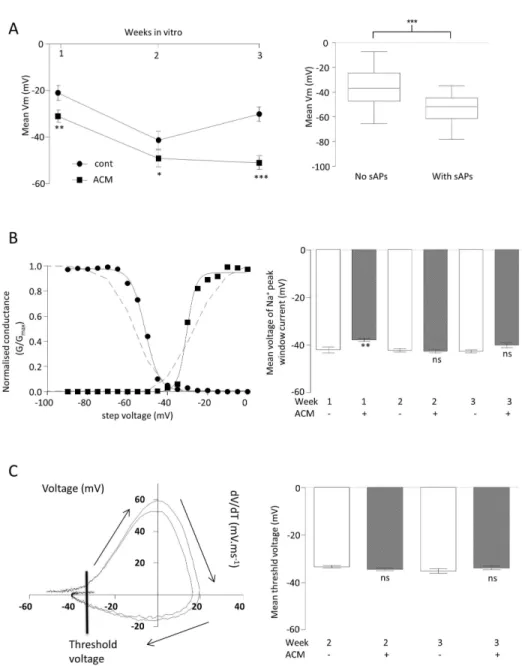
Other than the essential requirement for a minimal functional expression of Na+ and K+ channels, both Na+ current availability and resting Vm are crucial to spontaneous activity. At all weeks, but most strikingly at week γ, cells in ACM expressed significantly hyperpolarised Vm (Figure γA left, control: week 1 = -β1.0 ± γ.β mV (n=19), week β = -41.γ ± γ.8 mV (n=β1) and week γ = -γ0.5 ± γ.1 mV (n=16), ACM: week 1 = -γ1 ± β.6 mV (n=γ7), P<0.01; week β = -49.β ± β.β mV (n=γ5), P<0.05; week γ = -50.0 ± β.8 mV (n=19), P<0.0001). Furthermore, across the whole data set (independent of treatment and time), those cells which generated sAPs were significantly more hyperpolarised (P < 0.0001, Figure γA, right). Thus, ACM hyperpolarized neurons and this resulted in enhanced spontaneous activity.
That it was the differences in Vm rather than inherent properties of the Na+ currents themselves was tested by studying Na+ current availability (Figure γB). Na+ current activation/inactivation curves demonstrated that ACM evoked a very modest depolarisation of the peak Na+ current voltage (control = -4β ± 1.β (n = 8) vs. ACM = -γ7.7 ± 0.7 (n = β1), P < 0.01) at week 1. However, at later time points, where the differences in spontaneous activity were most marked, ACM evoked no significant changes in peak Na+ current voltage (week β: control = -4β.β ± 0.7 (n = 7) vs. ACM = -4β.6 ± 0.7 (n = β1); week γ: control = -4β.5 ± 0.7 (n = 10) vs. ACM = -40 ±1.1 (n = 10)). Consistent with there being few differences in peak Na+ current voltages, the rate of change of voltage (dV/dt) versus voltage plots showed that ACM evoked no significant changes in the threshold for iAPs (Figure γC, control = -γγ.γ ±
Figure 2. Effect of ACM on iAPs and voltage-gated Na+ and K+ currents of iPSC-derived neurons . A. Exemplar voltage
recording (upper left) during a current injection of 1β0 pA (lower left) from a holding current which maintained Vm at ca. -70mV. Arrow indicates iAP. Right panel shows the comparison of the proportion (%) of control (ACM-) and ACM-treated (ACM+) cells which could be induced to fire an action potential by the current-step protocol at each week of differentiation. Chiβ tests were performed at each week to compare control medium (ACM-) and ACM-treated (ACM+) iPSC-derived neurons. nsnot significant; n = 1ββ.
B. Bar graphs comparing the proportion of cells (%) demonstrating voltage-activated Na+ (left) or K+ (right) currents in response to a voltage-step protocol. Chiβ tests were performed as in A, above. nsnot significant; n = 106.
C. Mean Na+ current density (left) and K+ current density (right) versus voltage plots (V
h = -70mV) for control (circles) and ACM-treated (squares) cells during γ weeks of differentiation. T-tests were performed to compare the mean maximal current densities of cells in control medium and ACM-treated cells at each week. *P < 0.05, **P < 0.01, nsnot significant; n = 106.
D. Frequency histograms representing the number of cells within set ranges (5 pA.pF-1) of Na+ (left) and K+ (right) current densities for all neurons. Open bars represent neurons with iAPs and filled bars represent all neurons without iAPs (n = 818).
Figure 3. Effect of ACM on basic biophysical properties of iPSC-derived neruons . A. Comparison of mean resting membrane potentials (Vm) of neurons differentiated in control medium (cont, circles) or ACM (squares) for up to γ weeks. T-tests were performed at each week to compare control medium- and ACM-treated cells. *P < 0.05, **P < 0.01, ***P < 0.001,nsnot significant; n = 147 (left). Box and whisker plots comparing all cells which either did, or did not, demonstrate spontaneous activity plotted against Vm. Horizontal bar is the mean, box shows the 95% confidence and whiskers show upper and lower quartiles. T-test was performed to compare the two means. ***P < 0.001; n = β98 (right).
B. Exemplar, normalized conductance (G/Gmax) vs. voltage plots (adjusted for series resistance and junction potential). Squares represent activation (and circles represent inactivation of Na+ channels. Sigmoid Boltzmann curves was fitted to both data sets (Rβ > 0.99 for both), which intersected at the voltage at which the voltage at which the hypothetical peak Na+ current occured. The dashed lines are re-presentations of data from [γ6]showing voltage activation and inactivation curves for NaV1.β (left). Bar graphs comparing mean voltages at which the peak Na+ window current occurs for cells differentiated in control medium and ACM for up to γ weeks. T-tests were as in A, above. **P < 0.01,nsnot significant; n = 77 (right).
C. Rate of change in voltage (dV/dT) vs. voltage orbital plots from β exemplar iAPs Arrows indicate the direction of time. Line indicates voltage threshold (left). Bar graph showing the mean voltage thresholds for iAPs in ACM and control at weeks β and γ. T-tests were performed to compare control medium and ACM-treated cells. nsnot significant; n = β9 (right).
doi: 10.1γ71/journal.pone.00810γ1.g00γ
0.56 mV (n = 6) vs. ACM = -γ4.5 ± 0.41 mV (n = 10) at week β; control = -γ5 ± 0.98 mV (n=6) vs. ACM = -γγ.7 ± 0.67 mV (n=7) at week γ). Therefore, in these iPSC-derived neurons, the most important determinant of spontaneous activity is a Vm which is hyperpolarised sufficiently to release the Na+ channels from inactivation, a situation which essentially only holds true for ACM-treated cells at week γ.
Taken together, these data suggest that while ACM did not change the inherent ability of a neuron to fire an induced action potential, it increased spontaneous activity by three weeks of differentation, indicative of a more mature neuron, through an increase in the availability of Na+ channels because neurons were more hyperpolarized.
ACM enhances functional expression of voltage-gated Ca2+channels
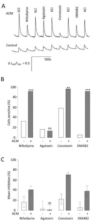
Neuronal voltage-gated Caβ+ channels, particularly N- and L-type, are associated with the pre- and post-synaptic membranes respectively [γ1]. Caβ+ channel expression has been associated with neuronal maturation [18]. Therefore, in order to investigate further the mechanism by which ACM increases functional maturity, Caβ+ channel functional expression was examined over the course of the three week differentiation. Peak inward Caβ+ current densities (carried by Baβ+) at each time point were larger in the cells treated with ACM (control week 1 = 0 ± 0 pA.pF-1 (n = 6) vs. ACM week1 = -γ.β ± 0.6 pA.pF-1 (n = 15, P < 0.001); control week β = -1.9 ± 0.4pA.pF-1 (n = 5) vs. ACM week β = -8.7 ± 1.9 pA.pF-1 (n = 1β, P < 0.05); control week γ = -5.6 ± 1.γ pA.pF-1 (n = 8) vs ACM week γ = -9.β ± 1.0 pA.pF- 1 (n = 9, P < 0.05). To dissect the channel subtypes responsible for carrying the Caβ+ current at the early time points, fura-β based Caβ+ imaging was employed. 50mM KCl (high K+) solution was used to activate all voltage-gated Caβ+ channels in the absence and presence of 10µM Nifedipine (L-type blockade), 100nM conotoxin (N-type blockade), 100nM agatoxin (P/Q-type blockade) or 100nM SNX48β (R-type blockade) (Figure 4A). Consistent with the whole-cell electrophysiology, cells differentiated in ACM not only produced larger responses, reflected in larger areas under the curve (AUC) for each high K+ application (Figure 4A), but significantly more cells responded to this stimulus. At week 1, in control medium, 89% of cells responded to high K+ with a mean AUC of β.8 ± 0.4 (n = 19), whereas 100 % of cells grown in ACM responded with a mean AUC of 6.1 ± 0.7, (n = βγ, P < 0.01). Furthermore, there were significant differences in the proportion of cells in each culture condition which were sensitive to nifedipine (γ1.γ % in control medium (n = 19) vs. 91.γ % in ACM (n = βγ, P < 0.0001), conotoxin (57.9 % in control medium versus 95.7 % in ACM) and SNX48β (5.γ % in control versus 91.γ % in ACM, Figure 4B). Agatoxin sensitivity was very small, and not significantly different under the two conditions, implying that few cells expressed P/Q- type Caβ+ channels (Figure 4B). These data show that ACM promoted increased expression of L- N- and R-type Caβ+ channels. Additionally, ACM also increased significantly the magnitude of the inhibition caused by each of the specific channel blockers (Figure 4C). Thus, nifedipine-sensitive AUC went from 16.4 ± 10.7 % (n = 19) of the high K+ response to 40.8 % ± 7.5 % (n =
βγ, P < 0.001), conotoxin-sensitive AUC went from ββ.β ± 10.1 % to 70.β ± 5.1 (P < 0.001) and SNX48β-sensitive AUC went from 5.β ± 11.7 % to γ8 ± 10.4 % (P < 0.001). In conclusion, ACM not only promoted a greater proportion of the cells to express Caβ+ channels, but those channels also made an increased functional contribution to each neuron, again indicative of a more mature neuronal phenotype.
ACM does not alter functional expression of ionotropic GABA currents
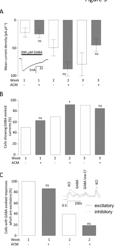
GABA can be either excitatory or inhibitory, depending upon the neuronal Cl- equilibrium potential, a parameter which is itself developmentally controlled via differential expression of several Cl- transporters [β4]. As these excitatory or inhibitory GABA-responsive phenotypes are indicative of neuronal immaturity or maturity, respectively, they were examined in order to establish whether ACM was increasing neuronal maturity regulation of the GABA-response per se. The magnitude of γ00 μM GABA-evoked inward currents increased dramatically from week 1 to β in ACM (week 1 = -β4.8 ± 6.8 pA.pF-1 (n = 8) to week β = -87.8 ± 1β.5 pA.pF-1 (n = β0)) with smaller increases observed in control medium (week 1 = -ββ.1 ± 8.β pA.pF-1 (n = 6) to week β = -49.4 ± 14.9 pA.pF-1 (n = 7)). From weeks β to γ no significant increases were observed in the median GABA current densities in either culture condition (control = -79.1 ± β0.7 (n = 11) vs. ACM = -44.9 ± 11.4 (n = 7); Figure 5A). However, ACM did not evoke a significant difference in the magnitude of GABA-evoked currents at any week, comparing either the means or medians. Although there was a modestly higher proportion of cells which expressed GABA-evoked currents in the ACM than in control medium at β weeks (control = 70 % (n = 10), ACM = 9β % (n = β5), P < 0.05), the control cultures had caught up by week γ (control = 91 % (n = 1β) vs. ACM = 85 % (n = 1γ), Figure 5B). Using conventional patch-clamp, it is not possible to ascertain whether the cellular responses to GABAA are normally excitatory or inhibitory. Therefore, Caβ+ influx in response to GABA was measured by fura-β-based Caβ+ imaging. Where the GABA response was excitatory, Caβ+ influx could be observed as a consequence of GABA-evoked depolarization to cause activation of voltage gated Caβ+ channels. If no response to GABA was observed, this could be the result of either GABA acting as an inhibitory neurotransmitter (to produce a hyperpolarization), or lack of expression of GABAA receptors and/or Caβ+ channels. To distinguish between these possibilities, GABA was also applied in a low Cl- extracellular solution; in this 9.9 mM Cl- solution, GABA
Figure 4. Effect of ACM on functional expression of voltage-activated Ca2+ channels. A. Mean traces of the ratio of intensity
(Iγ40/Iγ80) from fura-β Caβ+ imaging for control medium (solid line) and ACM-treated (dashed line) cells at week 1. Caβ+ influx was evoked using 50mM K+ solution (High K+) to depolarise the neurons. To measure the influence of different sub-types of voltage activated Caβ+channel on the depolarisation-evoked peak as a whole (the sum of the voltage activated Caβ+ influx) antagonists of specific channels were added to the High K+ solution: 10µM nifedipine (L-type Caβ+ channels), 100nM conotoxin (N-type Caβ+ channels), 100nM agatoxin (P/Q- type Caβ+ channels) and 100nM SNX48β (R-type Caβ+ channels).
B. Bar graph comparing the proportion (%) of neurons which showed significant (> 5%) inhibition by each antagonist for control medium- and ACM-treated cells at week 1, compared by chiβ tests. ***P < 0.001, **P < 0.01, nsnot significant; n = 4β.
C. Bar graph showing the reduction of Caβ+ influx (mean % inhibition) elicited by each antagonist for control medium- and ACM-treated cells at week 1, compared by t-tests. ***P < 0.001, **P < 0.01, nsnot significant; n = 4β.
doi: 10.1γ71/journal.pone.00810γ1.g004
Caβ+ channel expression, it does not aid in this maturity via altering the balance between neuronal excitatory and inhibitory responses to GABA.
Neuronal phenotype is remodeled by manipulating Ca2+ handling
The early up-regulation of voltage-gated Caβ+ channels by ACM would be predicted to result in long-lived and robust influx of Caβ+ upon stimulation. Such stimulation might well be via GABA, either at new synapses or extrasynaptically, which would then elicit an excitatory response in most cells at week 1. To investigate whether ACM might be accelerating the rate of neuronal maturation via augmentation of GABAA-dependent regulation of Caβ+ channels, cells were differentiated in ACM for γ weeks in the absence and presence of bicuculline (GABAA receptor block) or specific Caβ+ channel blockers. Remarkably, blockade of GABAA receptors, L-type, R-type or N-type Caβ+ channels all significantly depolarised the neurons (from -50.8 ± β.8 mV (n = 19) in the ACM to -β7.1 ± 1.β mV (n = 10) with 10 µM bicuculline, -β1.0 ± β.5mV (n = 9) with β µM nifedipine (L-type Caβ+ channel block), -β7.γ ± γ.9 mV (n = 9) with 100 nM conotoxin (N-type Caβ+ channel block), and -β5.5 ± β.4 mV (n = 10) with 100 nM SNX48β (R-type Caβ+ channel block), Figure 6A) and abolished the ACM-evoked increases in spontaneous activity (P < 0.0001, Figure 6B). To test the idea that Caβ+ influx was an important determinant of neuronal maturation, extracellular Caβ+ concentration was increased in the control medium from 0.6 mM to 1.8 mM and both resting Vm and spontaneous activity measured using whole cell patch-clamp. The increased Caβ+ concentration resulted in an enhancement in the proportion of cells generating spontaneous activity, which became significant at week β, from β9 % (n = β1) in 0.6 mM Caβ+ to 56 % (n = 16) in 1.8 mM Caβ+ (P < 0.05, Figure 6C). Increased Caβ+ also evoked significant hyperpolarisation of mean resting Vm at week 1 (from -β1 ± γ.β mV (n=β5) in 0.6mM Caβ+ to -4β ± 4.5 mV (n=16), P<0.0001, Figure 6D.). By week β, the resting Vm of both groups had stabilized at this hyperpolarised level (Figure 6D). These data suggested that increasing Caβ+ influx could partially mimic ACM. However, without a significant depolarizing stimulus, neither increased Caβ+ channel expression nor an increased driving force, alone or in combination, would necessarily result in increased intracellular Caβ+. Importantly, the ability of high extracellular Caβ+ to hyperpolarize the cell and augment spontaneous activity was completely ablated when GABAA receptors were blocked by 10 µM bicuculline (Figure 6C, D). Addition of bicuculline to the high extracellular Caβ+ medium abolished spontaneous activity (from 56% (n = 16), to zero (n = 19), P<0.0001) and significantly depolarised Vm (from -41.4 ± γ.β mV (n = 16) to -19.5 ± β.7 mV (n = 19), P<0.0001). Moreover, addition of γ00 µM GABA was as effective as the increased extracellular Caβ+ at week 1 in augmenting spontaneous activity (from 0% (n = β5) in control to β5 % (n = 8), P < 0.01, Figure 6C) and hyperpolarising the Vm (from -19.8 ± β.9 mV (n = β5) to -4β ± γ.γ mV (n = 8), P < 0.001, T = 4.γ, df = γ1, Figure 6D). These data show that GABA-dependent depolarisation or high extracellular Caβ+ are both capable of mimicking the ACM-evoked maturation of differentiating neurons. However, beyond
week β, the differential effects of high Caβ+ or GABA are lost and both the Vm and rates of spontaneous activity return to levels closer to those observed in control medium. This might have been attributed to a loss of excitatory GABA stimuli. Indeed, only half of the cells in the high Caβ+ cultures showed an excitatory GABA response (control = 100 % (n = 17) vs. high Caβ+ = 5γ % (n = 17), P < 0.001).
Pertinent channel blockers were employed to test whether high Caβ+ might augment the maturation of neurons via GABA
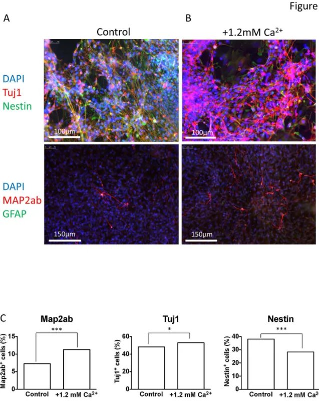
A -dependent Caβ+ influx through specific Caβ+ channels. In this separate set of experiments, β weeks of differentiation in 1.8 mM Caβ+ resulted in the proportion of cells exhibiting spontaneous activity being increased from 19 % (n = β1) to 56 % (n = 16) (P < 0.05, Figure 7A) and hyperpolarizing the Vm (Figure 7B). Importantly, in the presence of 1.8 mM Caβ+, bicuculline, nifedipine or conotoxin completely blocked spontaneous activity (P < 0.001) and depolarised the Vm (high Caβ+ = -47.1 ± 5.4 mV (n = 16); bicuculline = -γ0 ± β.6 mV (n = 19), P < 0.0001; conotoxin = -β1.1 ± 4.6 mV (n = 15), P < 0.0001, and; nifedipine = -β1.8 ± β.8 mV (n = 1γ), P < 0.0001). In contrast, agatoxin was completely without affect (Figure 7A, B), suggesting that the effect of high Caβ+ was dependant upon L- and N- type Caβ+ channels and GABA signaling. In further support of the idea that high extracellular Caβ+ promotes neuronal maturation are the data presented in Figure 8 which show that, compared to control, iPSCs differentiated in high Caβ+ for three weeks demonstrate smaller nestin-positive and larger Mapβab- & Tuj1-positive populations of cells, indicative of enhanced neuronal differentiation (Figure 8).
Since either GABAA receptor or N-type Caβ+ channel blockade diminished the positive effects of high Caβ+ on functional maturation, attempts were made to bypass the effects of each by activating alternative Caβ+ influx mechanisms. Thus, conotoxin co-treatment with high Caβ+ completely blocked the ability of high Caβ+ to augment spontaneous activity (Figure 6C) and depolarised the cells (Figure 7D). Remarkably, the effect of conotoxin was reversed by GABA (Figure 7C, D), which re-established the ability of cells to fire action potentials (from none (n = 15) with conotoxin to 50% (n = 10) with both GABA and conotoxin, P < 0.01, chiβ = 9.γ8, Figure 7C) and evoked a hyperpolarisation of the membrane (from -β0.7 ± γ.5mV (n = 15) with conotoxin to -47.6 ± γ.9 mV (n = 10) with both GABA and conotoxin, P < 0.0001, Figure 7D). Equally striking was the ability of the L-type Caβ+ channel opener, Bay K8644, to rescue the bicuculline-evoked diminution of excitability (Figure 7C, D). Thus, Bay K8644 increased the number of cells firing spontaneous action potentials (from zero with bicuculline (n = 19) to 57% (n = 7) with Bay K8644 and bicuculline, P < 0.01, Figure 7C) hyperpolarised the membrane (from -19.5 ± β.7mV (n = 19) with bicuculline to -4γ.0 ± 4.4mV (n = 1γ) with Bay K8644 and bicuculline (P < 0.0001, Figure 7D).
Figure 5. Effect of ACM on GABAA function. A. Bar graph comparing the mean GABA-evoked current densities (Vh = -70mV) for control medium- and ACM-treated neurons. Mann Whitney U tests were performed comparing the medians (due to the data showing an extremely non-normal distribution) at each week. nsnot significant. n =59.
B. Bar graph comparing the proportion of cells demonstrating a GABA-evoked current. Chiβ tests were performed to compare the proportion of cells with GABA currents at each week. *P < 0.01, nsnot significant. n =95.
C. Bar graph comparing the proportion of cells with GABA-evoked current that is excitatory. Inset shows fura-β recordings exemplifying neurons with either excitatory (solid line) or inhibitory (dotted line) GABA responses. Statistics as in B, above. *P < 0.01, nsnot significant. n =58.
doi: 10.1γ71/journal.pone.00810γ1.g005
Discussion
Although there have been many reports detailing protocols for generating neurons from human pluripotent stem cells, all rely on molecular and cellular markers as the primary measure of effectiveness; for example, III tubulin and microtubule-associated protein (MAP) β together with markers of neuronal subtype-specific fate determination (e.g. 5,6) Such markers are useful in determining how neurogenic each protocol might be, but they give very little indication as to whether the differentiated cells express the critical characteristics of functional neurons. Of particular importance are electrophysiological measures of Vm and the ability to fire action potentials, either spontaneously, or upon current injection. Using a standard, serumfree differentiation medium
-containing Bβ7 supplement, BDNF, GDNF and ascorbic acid [7] - only a small percentage of human iPSC-derived neurons were ever able to generate sAPs. However, differentiation in ACM resulted in a dramatic augmentation in the proportion of neurons which were spontaneously active, up to 74 % by week γ (Figure 1A). Action potentials are definitive characteristics of neurons and there are several factors which are absolutely required before a cell can become spontaneously excitable. Paramount is the adequate expression of voltage-gated Na+ and K+ channels. In this study, it became clear that once particular functional expression levels of these currents had been achieved (Figure βD), the cells could be induced to fire action potentials, and this ability was not affected by ACM (Figure βA). Exceeding the minimum functional level of voltage-gated channels was alone not sufficient to facilitate
Figure 6. The effects of manipulating Ca2+ and GABA signaling in the culture medium. Bar graphs comparing the proportion
of cells firing sAPs (A) and mean Vm (B) following γ weeks of differentiation in ACM alone, or ACM with 10µM bicuculline (a GABAA antagonist), with β µM nifedipine (an L-type Caβ+ channel antagonist), with 100 nM conotoxin (a N-type Caβ+ channel toxin) or with 100 nM SNX48β (an R- type Caβ+ channel toxin). Chiβ tests were performed comparing the ACM with each toxin against ACM alone. ±P < 0.05, ***P < 0.001, nsnot significant, n = 57.
Bar graphs comparing the proportion of cells firing sAPs (C) and mean Vm (D) following 1 and β weeks of differentiation in control medium (0.6 mM Caβ+), high Caβ+ medium (1.8mM Caβ+), control medium with γ00µM GABA and, high Caβ+ medium with 10µM bicuculline). T-tests were performed comparing the GABA and high Caβ+ media with control medium and the bicuculline medium with high Caβ+ medium for each week. ±P < 0.05, ***P < 0.0001, nsnot significant, n = 1γγ.
spontaneous activity. Indeed, ACM did not evoke the observed augmentation in spontaneous activity by increasing either the extent (Figure βB) or the magnitude (Figure βC) of Na+ and K+ current expression per se. Furthermore, the activation/ inactivation characteristics of the Na+ currents were not influenced by ACM (Figure γB), implying that ACM did not change the ratio of different Na+ channel sub-types which were functionally expressed during differentiation, consistent with the demonstration that iAP thresholds were also unaffected by ACM (Figure γB). Another requirement for the generation of spontaneous activity is that the Vm is sufficiently hyperpolarized to remove the voltage-dependent inactivation of Na+ channels. ACM evoked significant hyperpolarization of Vm at all weeks, most strikingly at week γ (Figure γA) where 74 % of ACM treated cells were spontaneously active and, across the whole cohort (β98 cells), there was a strong correlation between Vm and the ability of cells to generate action potentials spontaneously (Figure γA). Therefore, ACM promotes the
ability of differentiating neurons to generate action potentials spontaneously by hyperpolarising the membrane to levels sufficient to remove Na+ channel inactivation. However, it may also be necessary that such hyperpolarized neurons must receive depolarizing stimuli, especially if they are to generate the type of activity seen in vivo, in order to produce the complex and rhythmic activities observed in Figure 1. Such input would be the result of synaptogenesis in vitro, an idea completely consistent with the observation that ACM-treated neurons were unique in their expression of spontaneous miniature synaptic currents (Figure 1D). Although miniature synaptic potentials have been shown to be increased by astrocytes [14,γβ] or astrocyte-secreted factors [15,γγ] in rodent primary neuronal cultures and long-term hES cell differentiations [1], the data herein represent the first direct observation of a short-term, contact-independent augmentation of functional maturation and synaptogenesis by astrocyte-secreted factors in neurons differentiating from human PSCs
Figure 7. Manipulation of GABAA and Ca2+ currents in high Ca2+ medium: effects and functional rescue. Bar graph
comparing the proportion of cells firing sAPs (A) and mean Vm (B) at week β when treated with control medium (0.6mM Caβ+), high Caβ+ medium (1.8mM Caβ+) and high Caβ+ medium with 10µM bicuculline, with βµM nifedipine and with 100nM conotoxin and, with 100nM agatoxin (a potent P/Q- type Caβ+ channel toxin). Chiβ tests were performed to compare the high Caβ+ medium with each toxin against high Caβ+ medium alone. ±P < 0.05, ***P < 0.0001, nsnot significant, n = 90.
Bar graph comparing the proportion of cells firing sAPs (C) and mean Vm (D) at week β treated with high Caβ+ medium (1.8mM Caβ+) and high Caβ+ medium with 100nM conotoxin, conotoxin with γ00µM GABA, 10µM bicuculline and, bicuculline with 1µM BayK 8644 (an agonist of L-type Caβ+ channels). Chiβ tests were performed to compare the proportion of cells with treated with conotoxin vs. conotoxin with GABA and biciculline vs. bicuculline with BayK 8644. ***P < 0.0001, n = 65.
doi: 10.1γ71/journal.pone.00810γ1.g007
Figure 8. Immunofluorescent staining of cells differentiated in control and high Ca2+ media. Immunofluorescent staining of
iPSC-derived neurons differentiated for γ weeks in either control (A, 0.6mM) or high (B, 1.8mM) Caβ+ media; note that 1.8 mM Caβ+ was achieved by adding an extra 1.β mM CaClβ to the control medium. DAPI nuclear stain was used to show the number of cells in each field of view, in addition to primary antibodies raised against Tuj1 (neuron-specific class III -tubulin, red in the top β panels), nestin (top β panels), GFAP (glial fibrillary acidic protein, green in the lower β panels) and MAPβab (microtubule associated protein β-ab, red lower β panels). C, bar graphs showing percentage Mapβab-positive (left), Tuj1-positive (middle) and Nestin-positive (right) cells, each from 4 regions of interest over β coverslips (approximately β900 cells for each analysis) for both control and high Caβ+ differentiations at week γ, the two populations in each condition were compared by Chi squared test. ***P<0.0001, Chiβ = β8.04 (Mapβab) and β4.59 (Nestin); *P<4.97, Chiβ = 4.97 (Tuj1).
and are the first systematic determination of the electrophysiological basis of ACM-evoked enhancement of augmented spontaneous neuronal activity.
To determine the mechanism(s) which underlie ACM-enhanced neuronal maturation, the functional expression of voltage-gated Caβ+ channels and GABA
A receptors was investigated during early stages of differentiation. Strikingly, ACM evoked Caβ+ channel remodelling, with large increases in both the proportion of cells expressing L-, N- and R-type channels and the magnitude of the Caβ+ influx through each sub-type (Figure 4). In contrast, the ontogeny of GABAA receptors showed relatively little difference with ACM treatment. Thus, although the GABAA responses of most cells switched from excitatory to inhibitory during differentiation (presumably via the well-established, time-dependent modulation of Cl- transporters [β4,β8,γ4]), ACM changed neither the magnitude of the GABAA currents nor their mode of action at any time point (Figure 5).
Having established that ACM augmented functional expression of certain Caβ+ channels in the absence of significant changes in Na+, K+ and GABA
A channels, it seemed possible that ACM-evoked changes in functional maturation (hyperpolarized Vm and increased spontaneous activity) might be a result of this early Caβ+ channel remodeling. Indeed, inclusion of specific blockers of L-, N-, or R-type channels in the ACM resulted in depolarized Vm values (Figure 6B) and the complete abolition of spontaneous activity (Figure 6A), suggesting that Caβ+ influx is important in ACM-evoked maturation. In addition, although GABAA channels are unaffected by ACM per se (Figure 5), but that blocking them with bicuculline also impairs maturation (Figure 6A, B), they must be providing the depolarizing stimuli for voltage-activated Caβ+ influx early in differentiation. This implies that ACM may augment functional maturation via enhancement of the GABAA -dependent, Caβ+ influx pathway. This was investigated directly by raising the Caβ+ concentration of the control differentiation medium to 1.8 mM, or by supplementing the medium with γ00 μM GABA. In both cases, Vm values became hyperpolarised and spontaneous activity was augmented; an effect blocked by bicuculline (Figure 6C, D), nifedipine or conotoxin (Figure 7A, B). Notably, agatoxin (P/Q blockade), which is an ineffective blocker of Caβ+ influx (see Figure 4), was unable to affect the maturation (Figure 7A, B). Furthermore, at the cell biological level, high Caβ+ concentration also promoted the loss of
nestin-positive cells and an increase in Tuj1 and Mapβab-nestin-positive neurons, indicative of enhanced neuronal differentation (Figure 8).
Finally, and perhaps most importantly, the ability of conotoxin (N-type blockade) to ablate the high Caβ+-dependent increase in functional maturation was reversed upon addition of GABA, whilst the bicuculline ablation was rescued by opening L-type Caβ+ channels with BayK 8644. This implies that exogenous GABA treatment negates the requirement for N-type Caβ+ channel-dependent endogenous GABA release [γ5] and that in the absence of any endogenous GABA signaling (bicuculline), direct activation of L-type Caβ+ channels is sufficient to enhance neuronal excitability during differentiation.
Thus, ACM enhances neuronal maturation principally by amplification of an endogenous pathway which is dependent upon GABAA receptor-induced depolarization and consequent Caβ+ influx. This mechanism can be partially mimicked in vitro by modestly increasing extracellular Caβ+ or GABA concentrations. In the absence of a complete understanding of the factors which are secreted by astrocytes and their individual modes of action, these simple modifications can now be exploited to augment functional maturation in modified differentiation protocols. However, in contrast to ACM, this GABA-dependent mechanism is susceptible to loss of function when GABA becomes inhibitory. Therefore, in the absence of a full understanding of ACM we suggest application of Caβ+ channel openers offers the possibility to bypass such developmental effects and enhance maturation of PSCs as a one-step process.
Acknowledgements
The authors would like to thank Dr Amanda Redfern, Dr Alysia Battersby and Ms Hsiu-Er Tseng for maintaining the iPSC neurosphere cultures.
Author Contributions
Conceived and designed the experiments: DJR VBM CNS NDA PJK. Performed the experiments: DJR NDA PJK. Analyzed the data: DJR VBM CNS NDA PJK. Contributed reagents/materials/analysis tools: DJR VBM CNS NDA PJK. Wrote the manuscript: DJR VBM CNS NDA PJK.
References
1. Johnson MA, Weick JP, Pearce RA, Zhang SC (β007) Functional neural development from human embryonic stem cells: accelerated synaptic activity via astrocyte coculture. J Neurosci β7: γ069-γ077. doi: 10.15βγ/JNEUROSCI.456β-06.β007. PubMed: 17γ76968.
β. Miles GB, Yohn DC, Wichterle H, Jessell TM, Rafuse VF et al. (β004) Functional properties of motoneurons derived from mouse embryonic stem cells. J Neurosci β4: 7848-7858. doi:10.15βγ/JNEUROSCI. 197β-04.β004. PubMed: 15γ56197.
γ. Takazawa T, Croft GF, Amoroso MW, Studer L, Wichterle H et al. (β01β) Maturation of spinal motor neurons derived from human embryonic stem cells. PLOS ONE 7: e40154. doi:10.1γ71/journal.pone. 0040154. PubMed: ββ80β95γ.
4. Han SS, Williams LA, Eggan KC (β011) Constructing and deconstructing stem cell models of neurological disease. Neuron 70: 6β6-644. doi:10.1016/j.neuron.β011.05.00γ. PubMed: β16098β1.
5. Carri AD, Onorati M, Lelos MJ, Castiglioni V, Faedo A et al. (β01γ) Developmentally coordinated extrinsic signals drive human pluripotent stem cell differentiation toward authentic DARPP-γβ+ medium-sized spiny neurons. Development 140: γ01-γ1β. doi:10.1β4β/dev.084608. PubMed: βγβ50β04.
6. Aubry L, Bugi A, Lefort N, Rousseau F, Peschanski M et al. (β008) Striatal progenitors derived from human ES cells mature into DARPPγβ neurons in vitro and in quinolinic acid-lesioned rats. Proc Natl Acad Sci U S A 105: 16707-1671β. doi:10.107γ/pnas.0808488105. PubMed: 189ββ775.
7. Consortium THi (β01β) Induced pluripotent stem cells from patients with Huntington's disease show CAG-repeat-expansion-associated phenotypes. Cell Stem Cell 11: β64-β78. doi:10.1016/j.stem. β01β.04.0β7. PubMed: ββ748968.
8. Borghese L, Dolezalova D, Opitz T, Haupt S, Leinhaas A et al. (β010) Inhibition of notch signaling in human embryonic stem cell-derived
neural stem cells delays G1/S phase transition and accelerates neuronal differentiation in vitro and in vivo. Stem Cells β8: 955-964. doi: 10.100β/stem.408. PubMed: β0βγ5098.
9. Chambers SM, Qi Y, Mica Y, Lee G, Zhang XJ et al. (β01β) Combined small-molecule inhibition accelerates developmental timing and converts human pluripotent stem cells into nociceptors. Nat Biotechnol γ0: 715-7β0. doi:10.10γ8/nbt.ββ49. PubMed: ββ75088β.
10. Itsykson P, Ilouz N, Turetsky T, Goldstein RS, Pera MF et al. (β005) Derivation of neural precursors from human embryonic stem cells in the presence of noggin. Mol Cell Neurosci γ0: β4-γ6. doi:10.1016/j.mcn. β005.05.004. PubMed: 16081γ00.
11. Krencik R, Weick JP, Liu Y, Zhang ZJ, Zhang SC (β011) Specification of transplantable astroglial subtypes from human pluripotent stem cells. Nat Biotechnol β9: 5β8-5γ4. doi:10.10γ8/nbt.1877. PubMed: β160β806. 1β. Ullian EM, Christopherson KS, Barres BA (β004) Role for glia in synaptogenesis. Glia 47: β09-β16. doi:10.100β/glia.β008β. PubMed: 15β5β809.
1γ. Pfrieger FW, Barres BA (1997) Synaptic efficacy enhanced by glial cells in vitro. Science β77: 1684-1687. doi:10.11β6/science. β77.5γγβ.1684. PubMed: 9β87ββ5.
14. Hama H, Hara C, Yamaguchi K, Miyawaki A (β004) PKC signaling mediates global enhancement of excitatory synaptogenesis in neurons triggered by local contact with astrocytes. Neuron 41: 405-415. doi: 10.1016/S0896-6β7γ(04)00007-8. PubMed: 14766179.
15. Chou SY, Weng JY, Lai HL, Liao F, Sun SH et al. (β008) Expanded-polyglutamine huntingtin protein suppresses the secretion and production of a chemokine (CCL5/RANTES) by astrocytes. J Neurosci β8: γβ77-γβ90. doi:10.15βγ/JNEUROSCI.0116-08.β008. PubMed: 18γ67595.
16. Chang MY, Son H, Lee YS, Lee SH (β00γ) Neurons and astrocytes secrete factors that cause stem cells to differentiate into neurons and astrocytes, respectively. Mol Cell Neurosci βγ: 414-4β6. doi:10.1016/ S1044-74γ1(0γ)00068-X. PubMed: 1β8γ76β5.
17. Tang X, Zhou L, Wagner AM, Marchetto MC, Muotri AR et al. (β01γ) Astroglial cells regulate the developmental timeline of human neurons differentiated from induced pluripotent stem cells. Stem. Cell Res 11: 74γ-757.
18. D'Ascenzo M, Piacentini R, Casalbore P, Budoni M, Pallini R et al. (β006) Role of L-type Caβ+ channels in neural stem/progenitor cell differentiation. Eur J Neurosci βγ: 9γ5-944. doi:10.1111/j. 1460-9568.β006.046β8.x. PubMed: 16519658.
19. Arnhold S, Andressen C, Angelov DN, Vajna R, Volsen SG et al. (β000) Embryonic stem-cell derived neurones express a maturation dependent pattern of voltage-gated calcium channels and calcium-binding proteins. Int J Dev Neurosci 18: β01-β1β. doi:10.1016/ S07γ6-5748(99)00089-1. PubMed: 10715575.
β0. Mazzanti M, Haydon PG (β00γ) Astrocytes selectively enhance N-type calcium current in hippocampal neurons. Glia 41: 1β8-1γ6. doi:10.100β/ glia.101γ5. PubMed: 1β50980γ.
β1. Deisseroth K, Singla S, Toda H, Monje M, Palmer TD et al. (β004) Excitation-neurogenesis coupling in adult neural stem/progenitor cells. Neuron 4β: 5γ5-55β. doi:10.1016/S0896-6β7γ(04)00β66-1. PubMed: 15157417.
ββ. Tozuka Y, Fukuda S, Namba T, Seki T, Hisatsune T (β005) GABAergic excitation promotes neuronal differentiation in adult hippocampal
progenitor cells. Neuron 47: 80γ-815. doi:10.1016/j.neuron. β005.08.0βγ. PubMed: 16157β76.
βγ. Ben-Ari Y (β00β) Excitatory actions of gaba during development: the nature of the nurture. Nat Rev Neurosci γ: 7β8-7γ9. doi:10.10γ8/ nrn9β0. PubMed: 1ββ091β1.
β4. Ganguly K, Schinder AF, Wong ST, Poo M (β001) GABA itself promotes the developmental switch of neuronal GABAergic responses from excitation to inhibition. Cell 105: 5β1-5γβ. doi:10.1016/ S009β-8674(01)00γ41-5. PubMed: 11γ71γ48.
β5. Joannides AJ, Webber DJ, Raineteau O, Kelly C, Irvine KA et al. (β007) Environmental signals regulate lineage choice and temporal maturation of neural stem cells from human embryonic stem cells. Brain 1γ0: 1β6γ-1β75. doi:10.109γ/brain/awm070. PubMed: 1747β984.
β6. Jelitai M, Anderová M, Markó K, Kékesi K, Koncz P et al. (β004) Role of gamma-aminobutyric acid in early neuronal development: studies with an embryonic neuroectodermal stem cell clone. J Neurosci Res 76: 801-811. doi:10.100β/jnr.β0106. PubMed: 15160γ9β.
β7. Yamashita M, Fukuda Y (199γ) Calcium channels and GABA receptors in the early embryonic chick retina. J Neurobiol β4: 1600-1614. doi: 10.100β/neu.480β41β05. PubMed: 8γ01β68.
β8. Toyoda H, Yamada J, Ueno S, Okabe A, Kato H et al. (β005) Differential functional expression of cation-Cl- cotransporter mRNAs (KCC1, KCCβ, and NKCC1) in rat trigeminal nervous system. Brain. Resour - Mol Brain Res 1γγ: 1β-18. doi:10.1016/j.molbrainres. β004.09.015.
β9. Yu J, Vodyanik MA, Smuga-Otto K, Antosiewicz-Bourget J, Frane JL et al. (β007) Induced pluripotent stem cell lines derived from human somatic cells. Science γ18: 1917-19β0. doi:10.11β6/science.11515β6. PubMed: 180β945β.
γ0. Ebert AD, Shelley BC, Hurley AM, Onorati M, Castiglioni V et al. (β01γ) EZ spheres: A stable and expandable culture system for the generation of pre-rosette multipotent stem cells from human ESCs and iPSCs. Stem. Cell Res 10: 417-4β7.
γ1. Vigers AJ, Pfenninger KH (1991) N-type and L-type calcium channels are present in nerve growth cones. Numbers increase on synaptogenesis. Brain Res. Dev Brain Res 60: 197-β0γ. doi: 10.1016/0165-γ806(91)90048-N.
γβ. Ullian EM, Sapperstein SK, Christopherson KS, Barres BA (β001) Control of synapse number by glia. Science β91: 657-661. doi:10.11β6/ science.β91.5504.657. PubMed: 11158678.
γγ. Christopherson KS, Ullian EM, Stokes CC, Mullowney CE, Hell JW et al. (β005) Thrombospondins are astrocyte-secreted proteins that promote CNS synaptogenesis. Cell 1β0: 4β1-4γγ. doi:10.1016/j.cell. β004.1β.0β0. PubMed: 15707899.
γ4. Ge S, Goh EL, Sailor KA, Kitabatake Y, Ming GL et al. (β006) GABA regulates synaptic integration of newly generated neurons in the adult brain. Nature 4γ9: 589-59γ. doi:10.10γ8/nature04404. PubMed: 16γ41β0γ.
γ5. Poncer JC, McKinney RA, Gähwiler BH, Thompson SM (1997) Either N- or P-type calcium channels mediate GABA release at distinct hippocampal inhibitory synapses. Neuron 18: 46γ-47β. doi:10.1016/ S0896-6β7γ(00)81β46-5. PubMed: 91157γ9.