DOI: http://dx.doi.org/10.18363/rbo.v77.2020.e1801 Literature Review / Esthetic Dentistry
Esthetics with a Minimal Intervention Aided
by an Accessible Technology
Sayene Garcia Batista,1 Jayzon Stephan Brooks,1 Inger Teixeira de Campos Tuñas1
1Department of Social and Preventive Dentistry, School of Dentistry, Federal University of Rio de Janeiro (UFRJ), Rio de Janeiro, RJ, Brazil • Conflicts of interest: none declared.
AbstrAct
Objective: the purpose of this paper, based on a literature review, is to relate a clinical case in which the correct diagnosis, planning and conservative, restorative and
esthetic treatment in the anterior maxillary region was executed. In addition, this paper recognizes the value of the use of photographs obtained with a smartphone in the planning and completion of this case. Materials and Methods: a bibliographic search was done in PUBMed and Scielo databases, with the terms (dental esthetics) AND (composite resin OR composite resins) AND (anterior teeth) AND (anterior teeth) AND (Microdontia) AND (Dental Esthetics) AND (Composite resin) OR (Composite Resins) AND (Anterior Teeth) AND (Digital Photography or Digital Photographs). A total of 44 papers relevant to the topic were chosen, in their complete versions. Results: in respecting biological principles along with the philosophy of a minimally invasive approach, rehabilitation was successful in restoring both function and esthetics, in order to promote a more harmonious smile. Conclusion: the possibility of reproducing natural teeth artistically with composite resin, the chosen direct restorative material, represents a simple, satisfactory and reversible alternative as a solution to anomalies and deviations from dental forms. Photographs aid in making the patient more receptive to treatment and the disclosure of the final results.
Keywords: Composite resin; Dental esthetics; Conservative rehabilitation
Introduction
T
he culture of valuing the smile is reflected in the great demand for dental procedures that harmonize and beautify the facial expression. As a result, more confident individuals, with better self-esteem, are able to establish personal and professional relationships responsible for improving their quality of life.1,2 As a resultof its frank evolution, Esthetic Dentistry has achieved astonishing results in reestablishing dental harmony and, principally, in the wider context of health, has been capable of reintegrating individuals socially.3,4,5
Esthetics, in its essence, involves subjective components to create the perception of harmony and beauty.5 Therefore,
an esthetically pleasing smile involves a series of factors such as shade of the teeth, its size, form, position and alignment.6,7 Within the factors that can interfere with the
esthetic of a smile, one can find dental anomalies of which can be classified according to number, size, structure and shape.
Conoid teeth represent an alteration of the natural dental form such as an isolated microdontia in the permanent dentition. The crown is conical and the root is within its normality.8 Conoid teeth affect approximately
8.4% of the population with its greatest prevalence in women, more frequently in the permanent dentition and in the superior arch.7 This dental anomaly is a result of a
dominant autosomal inheritance that leads to an atypical mineralization and given its frequent occurrence in the anterior region, it represents one of the factors that negatively affect the harmony of the smile.5,7,8
The advances in the composition of restorative materials and development of more refined techniques have made
it easier for the production of more resistant, esthetic and long-lasting composite resin restorations.7,9 Under the
principle of minimal invasive Dentistry, the direct adhesive restorative treatment, with adequate composite resins for enamel and dentin, is a valid and an interesting option to correct disharmony of dental shape and size.5 This
procedure seeks the preservation of the dental structure, apart from presenting reversibility through the addition and wear of material, if necessary.2,10 The direct restorative
treatment presents itself as a simple, predictable, effective and minimally invasive method.2
Given the increasing demand for esthetic treatment in contemporary Dentistry, it has become fundamental to incorporate tools that can refine and optimize the predictability for treatment.11,12 Over time, more dentists
have been using more digital photography as an integral part of their day-to-day in the dental clinic.13 Photography
is a valuable resource in that it turns important images into tools for diagnostic and esthetic planning. The advancement of smartphone cameras has resulted in digital photographs of excellent quality. The photographs aid in conquering the trust of the patient and the approval of the proposed treatment besides having an important role in the diagnosis and planning.14
The objective of this paper, based on a literature review, is to relate a clinical case of the esthetic and functional resolution of anterior superior teeth through the technique of using direct veneers from nanofilled composite resin, in a patient that presented conoid lateral incisors and superior canines. Additionally, the importance of the photographic record will be made explicit to optimize the treatment with the use of a smartphone in this process.
Materials and Methods
A bibliographic search was done in PUBMed and Scielo databases, with the terms (dental esthetics) AND (composite resin OR composite resins) AND (anterior teeth) AND (anterior teeth) AND (Microdontia) AND (Dental Esthetics) AND (Composite resin) OR Composite Resins) AND (Anterior Teeth) AND (Digital Photography or Digital Photographs). The scientific articles found were analyzed based on their title and abstract in order to exclude those that were not relevant for the review. Inclusion criteria were articles published between 2001 and 2020, in their complete versions, in English and Portuguese. After applying the inclusion and exclusion criteria, 44 papers were chosen.
This paper also presents in a detailed, organized and sequenced description of the solution of the clinical case in which involves the rehabilitation of teeth in the anterior maxillary region with direct veneers of composite resin based on a minimal intervention philosophy.
Results
Literature Review
Conservative Esthetic Rehabilitation
The desire for an esthetic and harmonic smile increases the level of demand for patients since it becomes a relevant factor for their acceptance in society and self-esteem. Thus, any change in the normal pattern of the dental structure directly influences the need for correction. White teeth, well contoured and aligned are patterns of a harmonious smile that have been asserting themselves every day in the population.2,5,7,10
There are different alternatives for the esthetic and functional solution of teeth with shape changes. The making of single unit crowns or ceramic veneers are options. However, the need for greater dental wear and cost weigh when choosing the treatment plan.9 The favorable
esthetic result in a single session, lower cost for the patient - when compared to ceramic veneers - minimal or no wear of the dental structure are configured as attractions of this restorative technique, which has gained more and more followers among professionals and patients.11,12
The composite resin was developed in the 60s by Bowen, which still prevails today, a traditional material and essential for the dental practice.15 Composite resins are
basically obtained through the association of an organic matrix (BIS-GMA - bisphenol glyceryl methacrylate) with inorganic particles (colloidal silica quartz) bonded by a coupling agent - vinyl silane.16 Throughout history, various
types of resins have been developed. The classification, in regards to the filler size used, allows grouping the composite resins into three essential types: macrofilled, microfilled and hybrid (or microhybrid). Currently, the
variation in the filler size of inorganic particles has brought changes in many physical properties of composite resins, mainly due to the improvement of coupling agents between the resin matrix from Bowen and the available size.15,16,17
The most important changes were those that involved reducing the particle size to produce materials that are more effective. The application of nanotechnology in direct dental restorative materials is one of the dental advances that has stood out in the market. Thus, nanofilled composite resins were introduced to the market, with a particle size of approximately 0.02 μm, half the particle size of microfilled resins. Its composition is a combination of resin and resin / pre-polymerized particles with two types of particles: nanometric and nano-agglomerates. Its characteristics provide properties superior to those of hybrid composites, such as better polishing, easy handling, ability to maintain anatomy for long periods, adequate resistance to be indicated in posterior teeth, due to its size, and optical and smoothness properties similar to microfilled composite resins.16,17
The innovation of nanofilled resins has made spherical filler particles of small dimensions favor clinical handling, polishing and brightness that have gained much similarity to that achieved with microfilled. The mechanical properties of compressive strength, flexural strength and modulus of elasticity, surface hardness remain unchanged compared to micro-hybrids. The surface smoothness was improved by the very structural characteristics of its small inorganic particle size.16,18
Therefore, clinically, composite resins such as the nanofilled type have excellent esthetic properties, good polishing and greater maintenance capacity. Other characteristics such as opalescence and fluorescence make this restorative material capable of returning the restored tooth to its natural properties. The possibility of etching the dental structure and providing adherence to it in a lasting and efficient manner has guaranteed much indication of this material in different dental procedures.2,10,15,18
In addition, this restorative material is highlighted by its ability to mimic the natural appearance of teeth, restore the morphological function and restore characteristics such as shade, translucency, hue, chroma and value. This dental reconstruction is considered effective when it obeys the mechanical, biological, esthetic and functional principles. The esthetic recovery of the smile requires technical and scientific knowledge and manual skills in order to reproduce the fundamental anatomical details for the reproduction of the anatomical shape of the teeth as natural as possible.5,6,7,8
Planning is fundamental in the search for the best esthetic and functional results of restorative procedures. The use of some techniques combined with this planning
allows the work to be performed with greater predictability of results. This can be exemplified with the diagnostic waxing, which allows greater predictability of the clinical result, in addition to redefining the dental morphology individually for each patient and performing a restorative test with less chance of mistakes.11,12
Digital Photographs with Smartphones
The routine use of photography in the dental clinic has become a resource of great importance for the elaboration of the diagnosis, planning and documentation over the years. The images contribute to teaching, communication with the prosthesis laboratory, forensic expertise and encouragement for the patient during treatment. The establishment of a standard protocol with digital photographs optimizes the exchange of information between the team present or not during the treatment.13,14
Digital photography is the progress of the conventional method of taking photographs. Digital cameras have been used since the mid-90s, however these presented disadvantages such as high cost and the need for specific technical knowledge. Thus, the use of this resource for photographic shots in the clinic had become more restricted mainly because of the financial cost. However, the evolution of smartphones along with the development of applications allowed that the images captured with this equipment could be used to obtain quality dental photographs.39,42,44
The cell phone is easy to carry around and simple in handling as it is part of people’s daily use, without the need for manuals on how to use the camera and its functions. Current cell phone models, the smartphones, achieve satisfactory results with extraoral and intraoral photos. Another practicality regarding the use of the smartphone in the dental clinic is the presence of components of connections such as Wi-Fi, which allow the rapid forwarding of images, in order to provide an exchange of knowledge and ideas with other dentists, technicians and patients. Today’s smartphones have in their configurations advanced technology systems that allow quality photographs to be obtained with high levels of detail - compared to that of professional camera photographs.42,44
Clinical Case Report
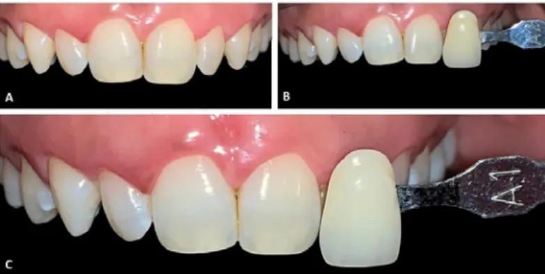
A 32-year-old female patient appeared at the dental clinic of the Federal University of Rio de Janeiro, describing that her anterior superior teeth were very small and that they negatively affected the esthetic of her smile. After a thorough anamnesis, a clinical exam along with an evaluation of the radiographic exams were done. Clinically, the patient presented reduced lateral incisors and superior canines (12,13,21,22), characteristics of conoid teeth (Figure 1A). Subsequently, for the planning of this
case, intra-oral photographs were taken with an iPhone X (iPhone - Apple (BR)), inicial dental impression with Alginate (Jeltrate®, Dentsply) and the making of plaster
dental cast (Herodent®, Coltene). The diagnostic waxing
from canine to canine was done with the intent to plan the form and final contour of the restoration.
Figure 1. A: Anterior teeth before rehabilitative treatment. B: Selection of the initial shade A2. C: Final shade A1 after teeth bleaching treatment in the clinic (1 session) and at home (14 days).
With the diagnostic waxing done, the following treatment plan was proposed to the patient: to do a tooth bleaching treatment associated with techniques from the clinic and at home to brighten the smile, besides re-anatomizing the anterior superior teeth with direct composite resin, in other words, direct resin veneers from canine to canine to return the esthetic equilibrium of the smile.
Following the explanation and approval of the treatment plan, the protocol for tooth bleaching was proceeded with in the dental clinic. Prophylaxis was done and the initial
shade according to the Vitapan Classical scale (Vitapan
Classical. Code: 005391) was recorded, the shade A2 was selected and photographs as a comparison method after treatment were taken (Figure 1B). For the protection of the soft tissues and to avoid the contact between the bleaching gel and the oral mucus, a labial retractor
was used (Arc Flex®, FGM), protecting the tongue with
gauzes and continual suction. Subsequently, a gingival
barrier was applied (Top Dam®, FGM) with the aim to
protect the gingival tissue. The chosen bleaching gel
was 35% hydrogen peroxide (Whiteness® HP, FGM) and
manipulated, according to the manufacturer’s instructions, and applied in a single session of 45 minutes.
At-home bleaching was done after the clinic for 3 weeks, during the night. For this purpose, an impression of the
superior arch was made using Alginate (Jeltrate®, Dentsply)
and afterwards the plaster dental cast (Herodent®, Coltene).
A silicone tray was made from the dental plaster cast to be used as a deposit for the bleaching gel. The patient was instructed to apply a drop of 4% hydrogen peroxide
around the region corresponding to the vestibular face of the teeth to be bleached.
Fourteen days passed after at-home bleaching and the patient returned to the clinic to do the re-anatomization. To mark the contour of the dental anatomy, a silicone
condensation guide (Putty Denso Profile®, Coltene) was
made from the diagnostic waxing – technique of silicone wall. Initially, prophylaxis was done and the selection of the resin’s shade. The selection should be done with clean and humid teeth, with natural light and without prior acid etching, with small increments added resin on the vestibular surface of the teeth to search for a shade as similar as possible to the structure of teeth. For this dental
re-anatomization case, enamel resins A1 (Vittra APS®,
FGM) and TRANS N (Vittra® APS, FGM) were selected
for the incisal edges of teeth to be treated.
The final shade after bleaching was selected (Figure 1C). The restorative incremental technique started with use of the modified rubber dam isolation of the operative field and was aided by a silicone barrier. After etching with
37% phosphoric aid (Condac®, FGM) in the vestibular
and incisal surfaces of central incisors and canines for 30 seconds, rinses with water/air spray was done in double the time for acid etching. Removing the excess of the humidity with a spray of air, the adhesive system (Ambar
APS®, FGM) was applied on the substrate with the aid
of an apllicator brush (Technobrush®, Coltene). The first
drop of adhesive was applied with active movements for 10 followed by a second application for 10 more seconds to construct a new layer on the same surface. Light sprays of air for the evaporation of the solvent for 10 seconds was applied then followed by photoactivation of the adhesive for 10 more seconds.
The correct adaptation of the silicone guide was noted followed by an increment of resin shade A1 inserted in the silicone guide to reconstruct the palatine enamel. The silicone guide was positioned and photoactivated for 20 seconds. Increments of resin TRANS N were incorporated
on the incisal areas to mimic the incisal transparency
present in natural teeth. The restoration was concluded with a final layer of A1 resin, corresponding to the vestibular enamel, smooth with the help of a brush, with the objective to reproduce the surface of the vestibular enamel and smoothness of the surface, with respect to the natural anatomy. The technique used was the stratification of layers with increments of, at maximum, 2 mm and photoactivation of each increment for 20 seconds. This technique made it possible to mimic the angles of reflection of light on the vestibular surface of all of the restored teeth,
which resulted in a natural aspect (Figure 2).
It is important to note that there was no removal of the dental surface, in that there was space for the insertion
of resin in all the vestibular surface and that the steps of acid etching and application of the adhesive system were done on the central and lateral incisors along with canines.
After the removal of the rubber dam isolation, latero-protrusive movements were executed to adjust the contact points. The occlusal contacts were verified, using a strip
of carbon (Accufilm II®). The initial dental finish was
done in the same session with a scalpel blade number 15,
fine-grained, ultra-fine finishing drills (KG Sorensen®),
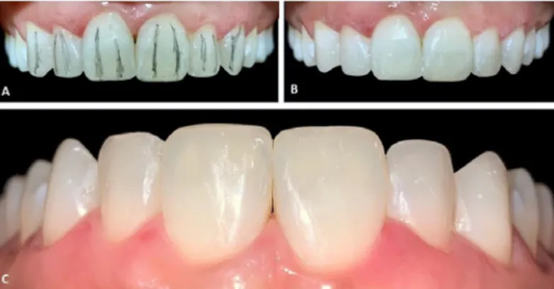
sandpaper strips and sandpaper discs (TDV) and, in subsequent consultation, a more refined finish was carried out. In the following session, the final finish and polish were done. The polyester sandpaper (TDV) was used in the proximal regions. The final finish was done mimicking the superficial texture, for this reason, a graphite point,
areas of reflection of light were noticed (Figure 3A) and
the finishing procedure with multi-laminated tips (Kerr Corporation) and fine-grained and ultra-fine diamonds
(KG Sorensen®) (Figure 3B). The polishing was done
using rubber polishers (TDV) and sandpaper discs for
finishing and polishing (Superfix®, TDV) according to
the sequence recommended by the manufacturer, finishing
system Enhance (Dentsply®) and polishing paste Enamelize
(Cosmedent) with Diamond felt disc (FMG®), obtaining a
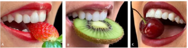
smooth and shiny final surface (Figure 3C). At the end of the treatment, an artistic photoshoot was come done with
an Iphone X (Figure 4).
Figure 2. A: Modified rubber dam isolation. B: Enamel surface etching with 37% phosphoric acid from canine to canine. C: Rinsing of phosphoric acid with air / water spray was done double the etching time. D: Application of the adhesive system. E: Photoactivation of the adhesive. F: Correct adaptation of the silicone guide.
Figure 3. A. Reflection lines demarcated with graphite. B: Appearance after finishing. C: Final appearance after finishing and polishing.
Figure 4. A, B, C: Artistic photos taken with Iphone X (iPhone - Apple (BR)) and natural light.
Discussion
Esthetic rehabilitations have become a clinical routine due to the growing interest of patients for procedures that harmonize the smile and, consequently, influence self-image, beauty and social acceptance.15,16 The increased
demand of patients for esthetics has resulted in the development of several techniques to restore, mainly, the anterior teeth that highlights the esthetic zone.9 Dental
re-anatomization can be performed with different approaches ranging from composite resins to dental ceramics, with or without dental preparation.17,18 Achieving esthetic
excellence in the construction of imperceptible restorations is a challenge in the dental clinic, as the dentist needs to be able to apply restorative materials to the tooth, harmonizing shade, shape and texture.5,19
Dental re-anatomization from the use of direct composites, currently, represents the first alternative of a minimally invasive approach, restoring form and function with preservation of healthy tissue.7,20,21,22,23
Maximum tissue preservation and the possibility of further adjustments are factors that allow the rehabilitation by the technique with direct composite resins to be the first option in the treatment plan and with better cost-benefit for the patient in the rehabilitation of the smile due to changes in shape.24 With the stimulus for the development
of composite resins and adhesive systems, the approaches with direct composite resin procedures have become standards of excellence, within an ultraconservative philosophy, with the reduction - or elimination - of the need for wear and cavity preparations.7,15,24,25
Knowledge of the physical-chemical properties of dental materials is essential for dentists to achieve results with esthetic excellence.4,26 Restorations made of composite resin
have the advantages of esthetics, adhesion, preservation of healthy dental structure and the ability to reproduce the natural, by mimicking the complexity of shades and shapes of teeth. However, depending on the composition and the improper use of resinous composites, these can result in restorations with unsatisfactory surface roughness, absence of gloss and shade instability.4,27
To resolve the case presented in this paper, a composite resin of the nanofilled type was used with nano spheroidal zirconia fillers that allowed to obtain a composite with high mechanical properties and excellent polishing and gloss capacity.28 The nanofilled composite resin has some
advantages in comparison to microfilled and hybrid resins, highlighting their mechanical properties, since they have a large amount of reduced size particles and excellent esthetic result, in order to enable the achievement of a satisfactory surface polishing, as well as long-lasting.3,15,29
The ideal relationship between the tooth-restoration interface, that is, the union of composite resins with dental structures, is due to the correct association of dental etching and adhesive systems.34 Currently, adhesive systems
can be divided into: conventional and self-etching.35 In
this context, the options of adhesive approaches used on dental structures can be varied. Conventional adhesive systems are available in three steps or two clinical steps.6 In
the three-step adhesive systems, the primer and adhesive are applied, they are not applied to the two-step systems, primer and adhesive are displayed in the single solution as used in the case of this paper - Ambar APS, FGM. As for self-etching adhesive systems, there is no step prior to acid etching. Self-etching adhesive systems can be found in two steps or in one clinical step (universal).6 In two-step
adhesive systems, acid primer and adhesive are applied separately, while in one-step systems, acid, primer and adhesive are applied at the same clinical time. Respecting the adhesive technique, following the protocol meticulously in accordance with the chosen adhesive system, will reflect in the clinical success of restorations.6,34,35
In the restoration and reconstruction of anterior teeth, the technique with the use of diagnostic waxing, associated with the use of a matrix or silicone barrier brings predictability in the resolution of the treatment plan.37 The
use of silicone barrier obtained from the impression of the arcade or waxed model can provide greater ease in the reconstruction of elements, while providing the operator security to complete the restorative treatment.3
Finishing and polishing are essential for the success and longevity of composite resin restorations. These steps are intended to enhance anatomical characteristics, reduce roughness and promote surface smoothness and shine. Thus, from the execution of these procedures, areas of accumulation of biofilm and staining of the composite will be reduced, collaborating with the maintenance of the health of the tissues, marginal integrity and esthetics of the restorations.4,31 However, over time, shade instability
and alteration of the texture of the composite resin may occur, making periodic controls necessary.34 Durability
is related to factors ranging from the technical quality of the operator to the patient’s behavioral aspects. For greater longevity of direct restorations in composite resin, the technical quality of the operator is essential, respecting each clinical step and following a meticulous restorative protocol. Behavioral aspects of patients, such as healthy habits, should be considered. It is important that patients
20. Hirata R, Ampessan RL, Lui J. Reconstrução de dentes anteriores com resinas compostas – uma sequência de escolha e aplicação de resinas. JBC. 2001;5(25):15-25.
21. Albino LGB, Rodrigues JA. Solução conservadora para o restabelecimento do equilíbrio estético e funcional de dentes anteriores. Rev Dental Press de Estét. 2012;9(1):96-105.
22. Boselli G, Pascotto RC. Incisivos laterais conóides: diagnóstico, planejamento e tratamento restaurador direto. Rev Dental Press Estét 2007;4(2):111-117. 23. Pini NIP, Khoury EMDA, Pascotto RC. Tratamento interdisciplinar para reabilitação estética do sorriso. Rev Dental Press Estét. 2010;7(2):40-50. 24. Coelho LGC, Machado WC, Soares MRPS, Melo KA. Reanatomização estética em paciente com hipodontia, dente conoide e permanência de elemento decíduo. Rev Pós-Grad. 2010;17(4):204-208.
25. Malhotra N, Mala K, Acharya S. Resin-based composite as a direct esthetic restorative material. Compend Contin Educ Dent. 2011;32(5):14-23.
26. Souza-Junior EJ, Borges BCD, Bertoldo CES, Lovadino JR, Aguiar FHB, Paulillo LAMS. Restauração estética direta de dente anterior fraturado: relato de caso clínico. Rev Dental Press Estét. 2010;7(4):42-51.
27. Veronezi MC, Brianezzi LFF, Modena K, Lima MS, Bernardi SE. Remodelação estética de dentes conóides: tratamento multidisciplinar. RDAPO. 2017;1(1):35-40.
28. Cabral L, Lindolm RN, Cunha VM, Junior CLG, Mello AMD, Mello FAS. Fechamento de diastemas em incisivos laterais conóides: relato de caso. Rev Gest Saúde. 2016;14(2):28-32.
29. Demarco F, Collares K, Coelho-de-Souza F, Correa M, Cenci M, Moraes R et
al. Anterior composite restorations: a systematic review on long-term survival
and reasons for failure. Dent Mater J. 2015;31(10):1214-1224.
30. Yanikian C, Yanikian F, Sundfeld D, Lins R, Martins L. Direct composite resin veneers in nonvital teeth: a still viable alternative to mask dark substrates. Oper Dent. 2019;44(4):E159-E166.
31. Cardoso PC, Decurcio RA, Pacheco FR, Monteiro LJE, Ferreira MG, Lima PLA, et al. Facetas diretas de resina composta e clareamento dental: estratégias para dentes escurecidos. ROBRAC. 2011;20(55):341-347.
32. Costa SXS, Becker AB, Rastelli ANS, Andrade MF. Contorno cosmético com resina composta nanoparticulada: relato de caso clínico. Rev Dental Press Estét. 2007;4(3):24-33.
33. Costa PX, Pudente HT, Almeida IMA, Lima GS, Moi GP. Otimização estética em dentes conoides: relato de caso clínico. Connection Line Rev Eletrôn UNIVAG. 2012;1(7):46-55.
34. Wagner A, Wendler M, Petschelt A, Belli R, Lohbauer U. Bonding performance of universal adhesives in different etching modes. J Dent. 2014;42(7):800-807.
35. Rosa W, Piva E, Silva A. Bond strength of universal adhesives: a systematic review and meta-analysis. J Dent. 2015;43(7):765-776.
36. Kulkarni V, Deshmukh J, Duddu M, Ragavendra T, Vanka A, Patil A. endodontic treatment and esthetic management of a primary double tooth with direct composite using silicone buildup guide. Contemp Clin Dent. 2012;3(5):92.
37. Demarco F, Collares K, Correa M, Cenci M, Moraes R, Opdam N. Should my composite restorations last forever? Why are they failing? Braz Oral Res. 2017;31(1):92-99.
References
1. Chaves LP, Schmitt VL, Consolgmano E, Frenken RP, Mondelli RFL, Wang L. Resin composite build-ups for complementing multidisciplinary esthetic and functional dental treatments: a case report. Braz Dent Sci. 2015;18(1):28-33. 2. Romero M. Esthetic anterior composite resin restorations using a single shade: Step-by-step technique. J Prosthet Dent. 2015;114(1):9-12.
3. Azevedo N, Galvão G, Nihi VSC, Hoeppner MG, Nihi FM. Otimização do sorriso com restaurações diretas de compósito resinoso nanoparticulado. UNOPAR Cient Ciênc Biol Saúde. 2015;17(1):43-49.
4. Menezes MS, Vilela ALR, Silva FP, Reis GR, Borges MG. Acabamento e polimento em resina composta: reprodução do natural. ROBRAC. 2014;23(66):124-129.
5. Lobato MF, Isaac SZ, Hilgert LA, Sbardelotto C. Reanatomização de dente conóide com resina composta por meio de técnica de aplicação simplificada. Rev Nav Odontol. 2019;46(1):31-36.
6. Van Meerbeek B, Yoshihara K, Yoshida Y, Mine A, De Munck J, Van Landuyt KL. State of the art of self-etch adhesives. Dent Mater J. 2011;27(1):17-28. 7. Cunha CTM, Torres LMS, Chaves LVF, Borges BCD, Farias-Neto A. Incisivos laterais conóides: otimização estética através do uso de resina composta direta. UNOPAR Cient Ciênc Biol Saúde. 2013;15(4):307-310.
8. Figueiredo RJA, Andrade AKM, Duarte RM, Medeiros FDFC. Otimizando a estética por meio de reanatomizações em dentes conóides. Rev Gauch Odontol. 2008;56(3):333-336.
9. Sowmya K, Dwijendra K, Pranitha V, Roy K. Esthetic rehabilitation with direct composite veneering: a report of 2 cases. Case Rep Dent. 2017; 2017:1-3. 10. Mathias P, Silva EVF, Aguiar TR, Andrade AS, Azevedo J. A conservative esthetic approach using enamel recontouring and composite resin restorations. Case Rep Dent. 2016; 2016:1-5.
11. Heintze S, Rousson V, Hickel R. Clinical effectiveness of direct anterior restorations – a meta-analysis. Dent Mater J. 2015;31(5):481-495.
12. Korkut B. Smile makeover with direct composite veneers: a two-year follow-up report. Dent Res Dent Clin Dent Prospects. 2018;12(2):146-151.
13. Xia J, Li Y, Cai D, Shi X, Zhao S, Jiang Q et al. Direct resin composite restoration of maxillary central incisors using a 3D-printed template: two clinical cases. BMC Oral Health. 2018;18(1):1-8.
14. De Oliveira A, Domingos P, Palma-Dibb R, Garcia P. Chemical and morphological features of nanofilled composite resin: influence of finishing and polishing procedures and fluoride solutions. Microsc Res Techniq. 2011;75(2):212-219.
15. Fernandes H, Silva R, Marinho M, Oliveira P, Ribeiro J, Moysés M. Evolução da resina composta: revisão de literatura. Rev Uni Vale do Rio Verde. 2014;12(2):401-411.
16. Michelon C, Hwas A, Borges MF, Marchiori JC, Susin AH. Direct posterior composites resins restoration – current considerations and clinical application. RFO UPF. 2010;14(3):256-261.
17. Silva J, Rocha D, Kimpara E, Uemura E. Composite resins: current aspects and perspectives. Rev Odonto. 2008;16(32):98-104.
18. Bispo LB. Nanoparticle Composite: Is there superiority in its use? Rev Dent online. 2010;9(19):21-24.
19. Pereira DA, Borges MG, Silva FP, Menezes MS. Reabilitação estética do sorriso por meio de procedimento restaurador direto com resina composta nanoparticulada: relato de caso. ROBRAC. 2016;25(72):54-58.
are instructed by dentists, so that they know how to take care of themselves and preserve their restorations.29,37,38
In regards to the predictability of treatment, digital photographs have been introduced in dental activities and, increasingly, clinics have used this tool as an integral part of their daily lives.39,40,41 Associated with clinical and
radiographic examinations and study cast, the photographs are auxiliary in the diagnosis and essential in creating an individualized esthetic treatment plan. In addition to being used as professional and legal documentation, photography has become an indispensable tool in Esthetic Dentistry, offering images that capture the patient’s instant emotion.39,42,43,44
Conclusion
From the literature review done, illustrated by the clinical case report, it was concluded that esthetic rehabilitation with direct composite resins represents a safe, predictable, esthetic and minimally invasive approach, as long as professionals comply with the established protocols with dedication and details. In addition, the implementation of digital technology in the treatment, through photographs obtained with a smartphone, proves to be a practical, accessible and effective tool in the detailed documentation of the execution of the case, as well as in the quality of presentation of the results achieved.
Submitted: 04/16/2020 / Accepted for publication: 04/23/2020
Corresponding author
Inger Teixeira de Campos Tuñas
E-mail: ingertunas@gmail.com
Mini Curriculum and Author’s Contribution
1. Sayene Garcia Batista - Undergraduate student in Dentistry. Contribution: Bibliographic search, preparation and writing of the manuscript, execution of the clinical case. ORCID: 0000-0003-3080-9866
2. Jayzon Stephan Brooks - Undergraduate student in Dentistry. Contribution: Writing of the manuscript. ORCID: 0000-0002-6245-6260
3. Inger Teixeira de Campos Tuñas - DDS, PhD. Contribution: Guidance of the whole process, bibliographic search, preparation, writing and revision of the manuscript, participation in the clinical case. ORCID: 0000-0001-7070-1900
38. Baldissera R, Corrêa M, Schuch H, Collares K, Nascimento G, Jardim P et
al. Are there universal restorative composites for anterior and posterior teeth?
J Dent. 2013;41(11):1027-1035.
39. Masioli M, Cunha DL, Damasio WQ,. Fotografia digital na clínica diária. In Macedo MCS, Baldacci RF, Coordenadores. E-book Jubileu de Ouro: procedimentos odontológicos. São Paulo: APCD; 2007.
40. Oliveira JP. Fotografia e video digital: a nova fronteira da odontologia. Rev Dental Press Estétic. 2005; 2(1):117-132.
41. Volpato C, Garbelotto L, Zani I, Vasconcelos D. Próteses odontológicas -uma visão contemporânea - fundamentos e procedimentos. 1nd ed. São Paulo: Grupo Gen - Livraria Santos Editora; 2011.
42. Cardoso P, Decurcio R. Facetas: lentes de contato e fragmentos cerâmicos. 2nd ed. Florianópolis: Ponto; 2015.
43. Masioli M. Fotografia odontológica. 2nd ed. Porto Alegre: Artmed; 2010. 44. Medeiros D. Click Dudu - Fotografia odontológica & marketing unindo dentistas laboratórios & clientes. 1nd ed. Florianópolis: Ponto; 2013.