ZOONOSES AND COMMUNICABLE DISEASES
COMMON TO MAN AND ANIMALS
Third Edition
Volume III
Parasitoses
Scientific and Technical Publication No. 580
PAN AMERICAN HEALTH ORGANIZATION Pan American Sanitary Bureau, Regional Office of the
WORLD HEALTH ORGANIZATION 525 Twenty-third Street, N.W. Washington, D.C. 20037 U.S.A.
Zoonosis y enfermedades transmisibles comunes al hombre y a los animales: parasitosis
ISBN 92 75 31991 X (3 volume set) ISBN 92 75 31992 8 (Vol. 3)
PAHO HQ Library Cataloguing-in-Publication Pan American Health Organization
Zoonoses and communicable diseases common to man and animals: parasitoses
3rd ed. Washington, D.C.: PAHO, © 2003.
3 vol.—(Scientific and Technical Publication No. 580)
ISBN 92 75 11991 0—3 volume set ISBN 92 75 11993 7—Vol. 3 I. Title II. (Series)
1. ZOONOSES
2. PARASITIC DISEASES 3. DISEASE RESERVOIRS
4. COMMUNICABLE DISEASE CONTROL 5. FOOD CONTAMINATION
6. PUBLIC HEALTH VETERINARY NLM WC950.P187 2003 v.3 En
The Pan American Health Organization welcomes requests for permission to reproduce or translate its publications, in part or in full. Applications and inquiries should be addressed to Publications, Pan American Health Organization, Washington, D.C., U.S.A., which will be glad to provide the latest information on any changes made to the text, plans for new editions, and reprints and translations already available.
© Pan American Health Organization, 2003
Publications of the Pan American Health Organization enjoy copyright protection in accor-dance with the provisions of Protocol 2 of the Universal Copyright Convention. All rights are reserved.
The designations employed and the presentation of the material in this publication do not imply the expression of any opinion whatsoever on the part of the Secretariat of the Pan American Health Organization concerning the status of any country, territory, city or area or of its authorities, or concerning the delimitation of its frontiers or boundaries.
The mention of specific companies or of certain manufacturers’ products does not imply that they are endorsed or recommended by the Pan American Health Organization in prefer-ence to others of a similar nature that are not mentioned. Errors and omissions excepted, the names of proprietary products are distinguished by initial capital letters.
CONTENTS
Prologue . . . vii
Preface to the First Edition . . . ix
Preface to the Second Edition . . . xi
Introduction . . . xv
Section A: PROTOZOOSES
African Trypanosomiasis . . . 3Amebiasis . . . 11
Babesiosis . . . 15
Balantidiasis. . . 20
Chagas’ Disease . . . 23
Cryptosporidiosis . . . 34
Cutaneous Leishmaniasis . . . 38
Cyclosporiasis . . . 49
Giardiasis. . . 52
Infections Caused by Free-living Amebae . . . 58
Malaria in Nonhuman Primates . . . 62
Microsporidiosis . . . 68
Sarcocystosis . . . 72
Toxoplasmosis . . . 76
Visceral Leishmaniasis. . . 86
Section B: HELMINTHIASES
1. Trematodiases
Cercarial Dermatitis . . . 99Clonorchiasis . . . 103
Dicroceliasis . . . 108
Echinostomiasis . . . 112
Fascioliasis. . . 115
Fasciolopsiasis . . . 124
Gastrodiscoidiasis . . . 127
Heterophyiasis . . . 128
Nanophyetiasis. . . 132
Opisthorchiasis. . . 135
Paragonimiasis . . . 140
Schistosomiasis . . . 146
2. Cestodiases
Bertielliasis . . . 160
Coenurosis . . . 162
Cysticercosis . . . 166
Diphyllobothriasis . . . 176
Dipylidiasis . . . 181
Hydatidosis . . . 184
Hymenolepiasis . . . 199
Inermicapsiferiasis . . . 205
Mesocestoidiasis . . . 206
Raillientiniasis . . . 208
Sparganosis . . . 210
Taeniasis . . . 214
3. Acanthocephaliases and Nematodiases
Acanthocephaliasis. . . 222Angiostrongyliasis . . . 225
Anisakiasis. . . 231
Ascariasis. . . 236
Baylisascariasis . . . 241
Capillariasis . . . 244
Cutaneous Larva Migrans. . . 249
Dioctophymosis . . . 252
Dracunculiasis . . . 254
Esophagostomiasis and Ternidensiasis . . . 259
Gnathostomiasis. . . 262
Gongylonemiasis . . . 267
Lagochilascariasis . . . 269
Mammomonogamiasis . . . 271
Micronemiasis . . . 273
Strongyloidiasis . . . 275
Thelaziasis . . . 283
Trichinosis . . . 285
Trichostrongyliasis . . . 299
Trichuriasis of Animal Origin. . . 302
Visceral Larva Migrans and Toxocariasis . . . 305
Zoonotic Ancylostomiasis . . . 312
Zoonotic Filariases. . . 317
Section C: ARTHROPODS
Dermatitis Caused by Mites of Animal Origin . . . 327Myiases . . . 331
Pentastomiases . . . 345
Tungiasis . . . 357 Zoonotic Scabies . . . 361
LIST OF TABLES
Protozooses
Tables
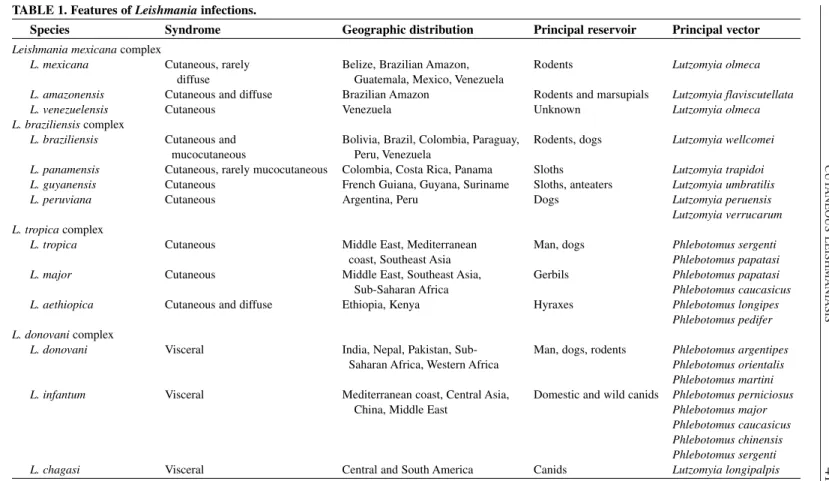
1. Features of Leishmaniainfections. . . 41 2. Plasmodiumspecies that infect primates. . . 63
Helminthiases
3. Intermediate hosts and geographic distribution of the
main zoonotic echinostomes.. . . 113
Arthropods
4. Ticks that infect man, and organisms and infections they transmit. . . 351
vii
In recent years, zoonoses and communicable diseases common to man and ani-mals have gained increasing attention worldwide. Human diseases that have their origins in infected animals, such as AIDS or Creutzfeldt-Jakob, have highlighted the need for a better understanding of animal diseases in terms of their epidemiology, mechanism of transmission to man, diagnosis, prevention, and control. Social and demographic changes have also contributed to the importance of gaining and dis-seminating knowledge about zoonoses. For instance, as people encroach further and further on ecological areas with which they had little contact and whose fauna may not be well known, their exposure to animals—and the infections they transmit— has increased. New knowledge is also being developed in the area of urban ecology. The ease and speed of modern travel also facilitates the spread of diseases once con-fined to specific geographic areas, as recently occurred with severe acute respiratory syndrome (SARS). Animal migration and trade pose a similar threat, as was shown by the outbreaks in the United States of West Nile fever, and most recently, mon-keypox—two diseases not previously known in the Western Hemisphere. Each of these examples highlights the need for improved knowledge and surveillance of and response to zoonoses.
The negative effects of zoonoses are far reaching. High incidence rates continue to cause significant morbidity and mortality in both humans and animals. Their eco-nomic impact is seen in lost labor productivity due to illness; reduced travel and tourism to affected areas; reduced livestock and food production; death and destruc-tion of affected animals; and restricdestruc-tions on and reducdestruc-tions in internadestruc-tional trade. Zoonoses can be a serious drain on a country’s economy, which in turn can have wide repercussions for a society’s health.
To help solve these problems, the Pan American Health Organization (PAHO)— an international public health organization that has devoted itself to improving the health and living conditions of the people of the Americas for over one hundred years—established the Veterinary Public Health Unit. The Unit’s overall objective is to collaborate with PAHO’s Member Governments in the development, implemen-tation, and evaluation of policies and programs that lead to food safety and protec-tion and to the prevenprotec-tion, control, or eradicaprotec-tion of zoonoses, among them foot-and-mouth disease.
To this end, PAHO’s Veterinary Public Health Unit has two specialized regional centers: the Pan American Foot-and-Mouth Disease Center (PANAFTOSA), created in 1951 in Rio de Janeiro, Brazil, and the Pan American Institute for Food Protection and Zoonoses (INPPAZ), established on November 15, 1991, in Buenos Aires, Argentina. INPPAZ’s precursor was the Pan American Zoonoses Center (CEPANZO), which was created through an agreement with the Government of Argentina to help the countries of the Americas combat zoonoses, and which oper-ated from 1956 until 1990.
medi-viii PROLOGUE
cine and public health through the promotion of veterinary health education in learn-ing, research, and health care centers. An example of this work is the preparation of several publications, among which the two previous Spanish and English editions of Zoonoses and Communicable Diseases Common to Man and Animalsstand out.
Scientific knowledge has progressed since the last edition was published in 1986. Also, the countries of the Americas have modified their livestock production strate-gies in recent years, which has affected the transmission of zoonotic infections and their distribution. The publication of this third edition is an attempt to address these changes. The third edition is presented in three volumes: the first contains bacte-rioses and mycoses; the second, chlamydioses, rickettsioses, and viroses; and the third, parasitoses.
We believe that this new edition will continue to be useful for professors and stu-dents of public health, medicine, veterinary medicine, and rural development; work-ers in public health and animal health institutions; and veterinarians, researchwork-ers, and others interested in the subject. We also hope that this publication is a useful tool in the elaboration of national zoonosis control or eradication policies and programs, as well as in risk evaluation and in the design of epidemiological surveillance sys-tems for the prevention and timely control of emerging and reemerging zoonoses. In summary, we are confident that this book will contribute to the application of the knowledge and resources of the veterinary sciences for the protection and improve-ment of public health.
ix
This book considers two groups of communicable diseases: those transmitted from vertebrate animals to man, which are—strictly speaking—zoonoses; and those common to man and animals. In the first group, animals play an essential role in maintaining the infection in nature, and man is only an accidental host. In the sec-ond group, both animals and man generally contract the infection from the same sources, such as soil, water, invertebrate animals, and plants; as a rule, however, animals do not play an essential role in the life cycle of the etiologic agent, but may contribute in varying degrees to the distribution and actual transmission of infections.
No attempt has been made to include all infections and diseases comprised in these two groups. A selection has been made of some 150 that are of principal inter-est, for various reasons, in the field of public health. The number of listed zoonoses is increasing as new biomedical knowledge is acquired. Moreover, as human activ-ity extends into unexplored territories containing natural foci of infection, new zoonotic diseases are continually being recognized. In addition, improved health services and better differential diagnostic methods have distinguished zoonoses pre-viously confused with other, more common diseases. A number of diseases described in this book have only recently been recognized, examples of which include the Argentine and Bolivian hemorrhagic fevers, angiostrongyliasis, rotaviral enteritis, Lassa fever, Marburg disease, and babesiosis.
The principal objective in writing this book was to provide the medical profes-sions a source of information on the zoonoses and communicable diseases common to man and animals. Toward that end, both medical and veterinary aspects, which have traditionally been dealt with separately in different texts, have been combined in a single, comprehensive volume. As a result, physicians, veterinarians, epidemi-ologists, and biologists can all gain an overview of these diseases from one source. This book, like most scientific works, is the product of many books, texts, mono-graphs, and journal articles. Many sources of literature in medicine, veterinary med-icine, virology, bacteriology, mycology, and parasitology were consulted, as were a large number of reports from different biomedical disciplines, in order to provide up-to-date and concise information on each disease. It is expected that any errors or omissions that may have been committed can, with the collaboration of the readers, be corrected in a future edition.
Where possible, explanations were attempted with special emphasis on the Americas, particularly Latin America. An effort was made, one which was not always successful, to collect available information on diseases in this Region. Data on the incidence of many zoonoses are fragmentary and frequently not reliable. It is hoped that the establishment of control programs in various countries will lead to improved epidemiologic surveillance and disease reporting.
More space has been devoted to those zoonoses having greatest impact on public health and on the economy of the countries of the Americas, but information is also included on those regionally less important or exotic diseases.
x PREFACE TO THE FIRST EDITION
public health and animal health administrators, physicians, and veterinarians must be familiar with the geographic distribution and pathologic manifestations of the various infectious agents so that they can recognize and prevent the introduction of exotic diseases.
We, the authors, would like to give special recognition to Dr. Joe R. Held, Assistant Surgeon-General of the United States Public Health Service and Director of the Division of Research Services of the U.S. National Institutes of Health, who gave impetus to the English translation and reviewed the bacterioses sections.
We would also like to express our utmost appreciation to the experts who reviewed various portions of this book and offered their suggestions for improving the text. These include: Dr. Jeffrey F. Williams, Professor in the Department of Microbiology and Public Health, Michigan State University, who reviewed the chapters dealing with parasitic zoonoses; Dr. James Bond, PAHO/WHO Regional Adviser in Viral Diseases, who read the viroses; Dr. Antonio Pío, formerly PAHO/WHO Regional Adviser in Tuberculosis and presently with WHO in Geneva, and Dr. James H. Rust, PAHO/WHO Regional Adviser in Enteric Diseases, both of whom reviewed the bacterioses; and Dr. F. J. López Antuñano, PAHO/WHO Regional Adviser in Parasitic Diseases, who read the metazooses.
We would like to thank Dr. James Cocozza, PAHO/WHO Veterinary Adviser, for his review of the translation and Dr. Judith Navarro, Editor in the Office of Publications of PAHO, for her valuable collaboration in the editorial revision and composition of the book.
xi
The fine reception accorded the Spanish, English, and French versions of this book has motivated us to revise it in order that it still may serve the purpose for which it was written: to provide an up-to-date source of information to the medical profession and allied fields. This book has undoubtedly filled a void, judging by its wide use in schools of public health, medicine, and veterinary medicine, as well as by bureaus of public and animal health.
The present edition has been considerably enlarged. In the seven years since the first edition was published, our knowledge of zoonoses has increased broadly and rapidly, and new zoonotic diseases have emerged. Consequently, most of the dis-cussions have been largely rewritten, and 28 new diseases have been added to the original 148. Some of these new diseases are emerging zoonoses; others are patho-logic entities that have been known for a long time, but for which the epidemiopatho-logic connection between man and animal has been unclear until recently.
The use this book has had outside the Western Hemisphere has caused us to aban-don the previous emphasis on the Americas in favor of a wider scope and geomed-ical view. Moreover, wars and other conflicts have given rise to the migration of populations from one country or continent to another. A patient with a disease heretofore known only in Asia may now turn up in Amsterdam, London, or New York. The physician must be aware of these diseases in order to diagnose and treat them. “Exotic” animal diseases have been introduced from Africa to Europe, the Caribbean, and South America, causing great damage. The veterinary physician must learn to recognize them to be able to prevent and eradicate them before they become entrenched. It must be remembered that parasites, viruses, bacteria, and other agents of zoonotic infection can take up residence in any territory where they find suitable ecologic conditions. Ignorance, economic or personal interests, and human customs and needs also favor the spread of these diseases.
xii PREFACE TO THE SECOND EDITION
noted. Another topic deeply interesting to researchers is the mystery of the radical antigenic changes of type A influenza virus, a cause of explosive pandemics that affect millions of persons around the world. Evidence is mounting that these changes result from recombination with a virus of animal origin (see Influenza). That this should occur is not surprising, given the constant interaction between man and animals. As a rule, zoonoses are transmitted from animal to man, but the reverse may also occur, as is pointed out in the chapters on hepatitis, herpes simplex, and measles. The victims in these cases are nonhuman primates, which may in turn retransmit the infection to man under certain circumstances.
Among emerging zoonoses we cite Lyme disease, which was defined as a clinical entity in 1977; the etiologic agent was found to be a spirochete (isolated in 1982), for which the name Borrelia burgdorferi was recently proposed. Emerging viral zoonoses of note in Latin America are Rocio encephalitis and Oropouche fever; the latter has caused multiple epidemics with thousands of victims in northeast Brazil. Outstanding among new viral disease problems in Africa are the emergence of Ebola disease and the spread of Rift Valley fever virus, which has caused tens of thousands of human cases along with great havoc in the cattle industry of Egypt and has evoked alarm around the world. Similarly, the protozoan Cryptosporidiumis emerging as one of the numerous agents of diarrheal diseases among man and animals, and prob-ably has a worldwide distribution.
As the English edition was being prepared, reports came to light of two animal diseases not previously confirmed in humans. Three cases of human pseudorabies virus infection were recognized between 1983 and 1986 in two men and one woman who had all had close contact with cats and other domestic animals. In 1986, sero-logic testing confirmed infection by Ehrlichia canisin a 51-year-old man who had been suspected of having Rocky Mountain spotted fever. This is the first known occurrence of E. canisinfection in a human. These two diseases bear watching as possible emerging zoonoses.
The space given to each zoonosis is in proportion to its importance. Some diseases that deserve their own monographs were given more detailed treatment, but no attempt was made to cover the topic exhaustively.
We, the authors, would like to give special recognition to Dr. Donald C. Blenden, Professor in the Department of Medicine and Infectious Diseases, School of Medicine, and Head of the Department of Veterinary Microbiology, College of Veterinary Medicine, University of Missouri; and to Dr. Manuel J. Torres, Professor of Epidemiology and Public Health, Department of Veterinary Microbiology, College of Veterinary Medicine, University of Missouri, for their thorough review of and valuable contributions to the English translation of this book.
We would also like to recognize the support received from the Pan American Health Organization (PAHO/WHO), the Pan American Health and Education Foundation (PAHEF), and the Pan American Zoonoses Center in Buenos Aires, Argentina, which enabled us to update this book.
We are most grateful to Dr. F. L. Bryan for his generous permission to adapt his monograph “Diseases Transmitted by Foods” as an Appendix to this book.
like to express our most sincere gratitude and recognition to Ms. Donna J. Reynolds, editor in the PAHO Editorial Service, for her valuable collaboration in the scientific editorial revision of the book.
xv
INTRODUCTION
This new edition of Zoonoses and Communicable Diseases Common to Man and Animals is published in three volumes: I. Bacterioses and mycoses; II. Chlamydioses and rickettsioses, and viroses; and III. Parasitoses. Each of the five parts corresponds to the location of the etiologic agents in the biological classifica-tion; for practical purposes, chlamydias and rickettsias are grouped together.
In each part, the diseases are listed in alphabetical order to facilitate reader searches. There is also an alphabetical index, which includes synonyms of the dis-eases and the etiologic agents’ names.
In this edition, the numbers and names of the diseases according to the International Statistical Classification of Diseases and Related Health Problems, Tenth Revision (ICD-10), are listed below the disease title. However, some zoonoses are not included in ICD-10 and are difficult to classify within the current scheme.
In addition, for each disease or infection, elements such as synonyms; etiology; geographical distribution; occurrence in man and animals; the disease in man and animals; source of infection and mode of transmission; role of animals in the epi-demiology; diagnosis; and control are addressed. Patient treatment (for man or other species) is beyond the scope of this work; however, recommended medicines are indicated for many diseases, especially where they are applicable to prophylaxis. Special attention is paid to the epidemiological and ecological aspects so that the reader can begin to understand the determining factors of the infection or disease. Some topics include simple illustrations of the etiologic agent’s mode of transmis-sion, showing the animals that maintain the cycle of infection in nature. Similarly, other graphics and tables are included to provide additional information on the geo-graphical distribution or prevalence of certain zoonoses.
The data on the occurrence of the infection in man and animals, along with data on the geographical distribution, may help the reader judge the relative impact that each disease has on public health and the livestock economy in the different regions of the world, given that the importance of different zoonoses varies greatly. For example, foot-and-mouth disease is extremely important from an economic stand-point, but of little importance in terms of public health, if animal protein losses are not considered. In contrast, Argentine and Machupo hemorrhagic fevers are impor-tant human diseases, but their economic impact is minimal, if treatment costs and loss of man-hours are not taken into account. Many other diseases, such as brucel-losis, leptospirosis, salmonelbrucel-losis, and equine encephalitis, are important from both a public health and an economic standpoint.
Section A
AFRICAN TRYPANOSOMIASIS
ICD-10 B56 African trypanosomiasis; B56.0 Gambiense trypanosomiasis; B56.1 Rhodesiense trypanosomiasis;
B56.9 African trypanosomiasis, unspecified
Synonyms: Sleeping sickness, trypanosomiasis; gambiense trypanosomiasis:
infection due to Trypanosoma brucei gambiense, West African sleeping sickness; rhodesiense trypanosomiasis: infection due to Trypanosoma brucei rhodesiense, East African sleeping sickness.
Etiology: African trypanosomiasis in man is caused by two subspecies of Trypanosoma (Trypanozoon) brucei:T. brucei gambiense andT. brucei rhodesiense, both of which are transmitted by the bite of tsetse flies (genus Glossina) (Bales, 1991; Dumas and Bouteille, 1996; Chimelli and Scaravilli, 1997). These try-panosomes are considered to belong to the salivarian group because of the way in which they are transmitted through the vector’s bite. Infection caused directly by a bite is considered inoculative, or via the anterior station, as opposed to contamina-tive, or via the posterior station, when the infection is transmitted by means of the fly’s excrement (see the chapter on Chagas’ Disease).
The two subspecies that affect man,T. b. gambiense and T. b. rhodesiense, as well as T. b. brucei, are morphologically indistinguishable. The latter species, while it does not affect man, is pathogenic for domestic animals in Africa, such as donkeys, horses, goats, camels, mules, sheep, dogs, and cattle. The forms that are present in blood, cerebrospinal fluid, and lymph are pleomorphic trypomastigotes (see Chagas’ Disease). The forms range from long, thin parasites (measuring 30 µm by 1.5 µm on average, with a subterminal kinetoplast, a long flagellum extending from
the anterior tip of the body, and an undulating membrane between the flagellum and the body) to short, fat parasites (averaging 15 µm by 3.5 µm, with a near-terminal
kinetoplast and no external flagellum). The long forms multiply in the fluids of the definitive host by binary longitudinal division. The short forms are the infective ele-ments for the vector and do not divide in the human host.
For a long time, the three subspecies T. b. brucei, T. b. gambiense, andT. b. rhode-siense were distinguished on the basis of their infectivity and pathogenicity for rats, their sensitivity to the drug tryparsamide, and their pathogenicity for man. To make a definitive distinction between T. b. brucei and the human trypanosomes, human volunteers were employed, with the consequent risks. In practice, differentiation
between the two human pathogens is still fundamentally based on the course and geographic distribution of the infection. Now more precise techniques are available for identifying the parasites. The blood incubation infectivity test (BIIT) consists of incubating the trypanosomes in human serum or plasma and then inoculating them in rats. In this procedure,T. b. bruceiloses its infectivity for rats whereas the sub-species that affect humans maintain it. Nevertheless, studies have revealed wide variation in the susceptibility of these trypanosomes to the effects of human serum, and some evidence exists that T. b. brucei can become resistant to the action of the serum. This would mean that T. b. brucei could become infective for man when flies infected by animals feed on human blood (Minter, 1982). Another important and increasingly used method is characterization of the trypanosomes according to the electrophoretic movement of their isozymes, which makes it possible to distinguish the different zymodemes (see definition under Chagas’ Disease). Truc and Tybayrenc (1993) have described 23 zymodemes in Central Africa, which can be divided into two groups, one corresponding to T. b. gambienseand the other to T. b. brucei. Recent findings suggest that the different zymodemes are related not only to the species but also to the geographic distribution and clinical characteristics of the infection (Smith and Bailey, 1997). Also, the use of polymerase chain reaction has made it possible to identify T. b. gambiense(Schares and Mehlitz, 1996).
In man, the trypanosomes of African trypanosomiasis multiply in the blood, lymph, cerebrospinal fluid, and intercellular spaces, but they do not penetrate cells. In the vector, the short, fat trypanosomes consumed in the process of ingesting a blood meal multiply in the lumen of the mid and hindgut for about 10 days, after which they turn into thin forms and migrate toward the proventriculus, where they multiply for another 10 days; from there they travel to the salivary glands, where they attach themselves to the epithelial cells and turn into epimastigotes (see Chagas’ Disease). The epimastigotes continue to multiply and are rapidly transformed into short, fat, metacyclic trypomastigotes, sometimes without a flagellum, which are the forms that are infective for man. Although the complete cycle of the trypanosome inside the tsetse fly can range from 15 to 35 days (average 21 days), the infection cycle up to the formation of metacyclic trypomastigotes is completed in only about 10% of the flies that ingest the parasite. The infected flies remain so for the rest of their lives and inoculate trypanosomes every time they take a blood meal.
Geographic Distribution:African trypanosomiasis in man occurs between 15°
N and 20° S Latitude in Africa, which is the vector’s area of distribution. T. b. rhode-siense is found in multiple foci in eastern Africa over an area stretching from Ethiopia to Botswana, while T. b. gambiense is found in central and western Africa, from northwestern Senegal to northeastern Sudan in the north to Angola in the south. Outside the endemic area, there are occasional cases in tourists and immi-grants from endemic countries.
Occurrence in Man:In the past, there were devastating epidemics of gambiense
estimated at less than 10,000 cases. In 1972, there were 4,126 new cases in the Democratic Republic of Congo (formerly Zaire) and 3,000 in the rest of Africa (De Raadt, 1976). However, starting in the 1970s, the disease flared up again alarmingly in some of its old foci (Kusoe, 1993; Cattand, 1994; Jusot et al., 1995). This increase was a reflection of the massive new movements of people both within and outside the endemic areas as a result of wars and social and political instability in many African countries. The situation was aggravated by shortages of human and material resources for surveillance and medical care programs in the affected countries (Mhlanga, 1996). In 1982, the World Health Organization (WHO) reported that gambiensetrypanosomiasis was endemic in 23 African countries, 45 million inhab-itants were at risk, and that every year there were nearly 10,000 new infections (Bull World Health Organ, 1982). Currently, African trypanosomiasis in man is endemic in 36 African countries south of the Sahara; the two forms of the disease together pose a risk for approximately 50 million people; and about 25,000 new cases are being reported annually, with the likelihood that not all cases were being notified (Bales, 1991; Kusoe, 1993).
Gambiense trypanosomiasis, which is chronic, tends to occur in epidemics, whereas rhodesiensetrypanosomiasis, which has a more acute course, occurs spo-radically, and gives rise to far fewer epidemics. The latter infection is endemic among livestock-raising tribes in eastern Africa and frequently affects hunters, fish-ermen, and travelers. Overall incidence is rather low because the people avoid areas infested by the vector.
The Disease in Man:The human disease usually has three phases: the primary
lesion, parasitemia, and invasion of the central nervous system. Two or three days after the bite of an infected fly, a painful inflammation (chancre) appears at the inoc-ulation site, and it disappears after two to three weeks (McGovern et al., 1995). The primary lesion is observed more frequently in infections caused by T. b. rhodesiense than in those produced by T. b. gambiense. From the chancre site, the trypanosomes invade the bloodstream, and the patient suffers from irregular and intermittent fever, mirroring the waves of parasitemia. Other signs during this acute period are painless adenopathies, especially in the posterior cervical lymph nodes, as well as edema of the eyelids and joints. The most common symptoms of the acute phase are cepha-lalgia, insomnia, arthralgia, weight loss, and generalized erythema and pruritus, par-ticularly in the sternal region. In later stages of the disease, the symptomatology is related to the affected organ. Invasion of the central nervous system is common, and a large variety of psychological, motor, and sensory perturbations may be seen. Following the meningitis that develops early in the course of the infection, a rupture occurs in the choroid plexus which allows the parasites to invade sites in the brain. The result is encephalitis, consisting of generalized inflammation with perivascular infiltrations of B and T lymphocytes, plasmocytes, and macrophages. The blood-brain barrier becomes permeable, and this condition may give rise to vasogenic cere-bral edema. Astrocytes and microglia are activated, and, together with immune cells, they begin to produce cytokines, which also contribute to progression of the disease (Pentreath et al., 1994). There is irritability, paresthesia, and insomnia, and later on, cerebral edema can cause severe headaches and edema of the optic papillae. There can also be neurologic manifestations such as epileptic seizures, chorea, psychotic episodes, euphoria, somnolence, lethargy, and coma.
As it was noted earlier,gambiensetrypanosomiasis usually follows a slow and chronic course. Weeks or months may elapse between the first and second phase, and months or years may elapse between the second and third phase. Rhodesiense trypanosomiasis has a more acute course and its phases are less marked; death may come within a few months, in contrast to patients with T. b. gambiense infection, who can live for many years. Cardiac complications are more common in rhode-siensetrypanosomiasis, and some patients die before reaching the neurologic phase (Greenwood and Whittle, 1980; WHO, 1979).
Both forms of African trypanosomiasis severely alter the patient’s immune sys-tem. The main characteristics are synthesis of large amounts of gamma globulin, autoantibody formation, and immunodeficiency (Vincendeau et al., 1996). The par-asites in the bloodstream are covered with variable glycoprotein surface antigen (VGSA), which generates powerful immune responses that rapidly suppress para-sitemia. These responses include antibody production and activation of macrophages that produce tumor necrosis factor alpha (TNF-α) and nitric acid
(NO). Some parasites, however, manage to express another of the more than 1,000 genes coded for this antigen and are covered with a different glycoprotein, thereby initiating a new wave of parasitemia. These waves recur every 7 to 15 days until the patient, if left untreated, dies. The succession of new antigens is a powerful stimu-lus for the immune response, which participates in both the defense and the pathol-ogy of the disease. Although there is epidemiologic evidence of protective immunity in gambiensetrypanosomiasis (Khonde et al., 1995), individual antigenic variation is effective protection for the parasite against the immunity of the host. In terms of immunopathology, there is no evidence that high gamma globulin levels or an abun-dance of immune complexes play an important role in pathology of the human dis-ease. Nevertheless, there is experimental evidence suggesting that autoantibodies to components of the central nervous system, such as anti-galactocerebrosides and tryptophan anti-analogous antibodies, may play a part in the development of encephalitis (Hunter et al., 1992). Although T lymphocytes diminish the parasite’s capacity to proliferate, they continue to produce gamma interferon (IFN-γ). The
macrophages and astrocytes, for their part, produce TNF-α. Although IFN-γand
TNF-α, together with their specific antibodies and the NO of the macrophages, have powerful properties to fight the trypanosomes, it has been demonstrated that TNF-α
levels are directly related to the severity of the disease (Okomo-Assoumou et al., 1995).
The Disease in Animals:Infections caused by African trypanosomes in animals
for domestic animals, but none of them infects man (Levine, 1985):T. congolense affects carnivores, swine, equines, and ruminants; T. vivaxaffects equines and rumi-nants; and T. simiaeaffects camels and swine. The primary symptoms in animals are lymphadenopathy, intermittent fever, anemia, and progressive emaciation (Urquhart, 1980). Depending on the species, the age of the host, and the parasite load, the dis-ease may be acute or chronic.
Trypanosomiasis in animals has played a role in configuring African societies: awareness of the parasite’s fatal effect on horses protected the original inhabitants from foreign invasions, while its effect on cattle has prevented ranchers from taking advantage of 7 million km2 of pastureland to raise high-yield European cattle. Another form of trypanosomiasis that occurs both in Africa and outside the conti-nent is caused by T. evansi. It is transmitted by tabanid flies and is especially path-ogenic for camels, equines, and dogs.
Source of Infection and Mode of Transmission:Man is the main reservoir of T.
b. gambienseand the source of infection for the vector. Because the infection is pro-longed and includes intervals between febrile attacks during which the patient feels relatively well, affected individuals may move about and propagate the infection in new areas where the vectors exist. There is no evidence that lower animals play a role in human T. b. gambiense infection, even though animal-to-animal transmission has been demonstrated in the laboratory (Molyneux, 1983) and parasites from swine, sheep, and dogs have been shown to be identical to human parasites in their sensitivity to human sera or their isoenzymatic profile (Scott et al., 1983; Schares and Mehlitz, 1996).The success of control programs aimed exclusively at eliminat-ing the human parasite would indicate that animal reservoirs are not important in gambiense trypanosomiasis. Nevertheless, the presence of animal reservoirs could account for maintenance of the T. b. gambienseinfection in areas where isolated human cases have occurred with long intervals between them.
The main vectors of T. b. gambiense infection are the tsetse flies Glossina fuscipes, G. palpalis, andG. tachinoide. These species belong to the palpalis, or riverine, group of flies, which inhabit dense vegetation along the shores of rivers and lakes. Human infection occurs almost always in the vicinity of watercourses or places where water pools in rural settings; tourists are rarely affected. The male and female tsetse flies are biological vectors, but they can transmit the infection mechan-ically during epidemics, when there are many patients with parasitemia. In general, the infection rate in the vectors is low. In addition, according to some reports, con-genital transmission can occur in man.
By contrast, in the case of rhodesiensetrypanosomiasis, lower animals, especially cattle, play an important role as reservoirs. T. b. rhodesiensehas been isolated from a number of wild and domestic animals; but only antelopes, hyenas, lions, sheep, and cattle develop sufficiently high and prolonged parasitemia to serve as effective reservoirs. These animals are responsible for persistence of the parasite in areas that have not been inhabited by humans for years.
The main vectors in eastern Africa are Glossina morsitans,G. pallidipes, andG. swynnertoni. These species belong to the morsitans group of flies, which inhabit savannahs and forested areas and prefer to feed on cattle and wild animals. The more acute nature of the human infection, coupled with the fact that the habitat of the vec-tors is not near homes, makes rhodesiensetrypanosomiasis more sporadic than the
gambienseform and less capable of causing epidemics. The main victims of the rhodesienseform are hunters, tourists, and persons who have contact with wild ani-mal habitats where the infection is enzootic.
Diagnosis:The disease may be suspected when its main symptoms and signs are
present, in particular intermittent fever, enlarged posterior cervical lymph glands, and cutaneous erythema. Biochemical tests do not reveal any remarkable alterations except higher cell counts and increased IgM in cerebrospinal fluid, which are con-sidered pathognomonic of invasion of the central nervous system (Bisser et al., 1997). The infection is confirmed by demonstrating the presence of the parasite in aspirate from the chancre or the lymph glands, in bone marrow, or in blood taken during the acute phase, or cerebrospinal fluid during the chronic phase. The sample to be observed may be either fresh or fixed and stained. In acute-phase patients, aspi-ration of the lymph glands is more effective for detecting T. b. gambiensethan T. b. rhodesiense. On the other hand, peripheral parasitemia is higher in rhodesiensethan in gambiensetrypanosomiasis, and it is therefore easier to demonstrate the presence of T. b. rhodesienseby examining thick blood films. In both cases, however, the lev-els of parasitemia fluctuate and are higher during febrile attacks. It is easier to find parasites in blood by centrifugation in hematocrit tubes and examination of the leukocyte layer, or by minifiltration in DEAE-cellulose, centrifugation, and exami-nation of the exudate (Bailey and Smith, 1994). To demonstrate the presence of T. b. rhodesiense,samples of blood or cerebrospinal fluid can be inoculated intraperi-toneally in mice, which develop detectable parasitemia within the second week. It is difficult to infect rodents with T. b. gambiense. When the foregoing methods have been unsuccessful, an attempt may be made to examine bone marrow or culture it in special media such as glucose, lacto-albumin, serum, hemoglobin, or GLSH. Sediment from cerebrospinal fluid should be examined immediately after it is col-lected. Serologic reactions such as the card agglutination test, indirect hemaggluti-nation, enzyme-linked immunosorbent assay (ELISA), and indirect immunofluores-cence are useful for epidemiologic studies, but they are of limited value for individual diagnosis: healthy individuals may have developed antibodies to animal trypanosomes inoculated by tsetse flies which did not produce infection, and these antibodies can cross-react with the antigens of T. b. gambienseand T. b. rhodesiense.
Control: The two main approaches to controlling the African trypanosomiases
would be to saturate the natural environment with male flies sterilized in the labo-ratory, which was successful in eradicating the fly Cochliomyia hominivorax in Libya in 1991. Empirical observations and mathematical models suggest that reduc-ing the vector population is most efficient durreduc-ing epidemics, while reducreduc-ing the human reservoir is more effective in endemic situations (Gouteux and Artzrouni, 1996). Other appropriate measures include preventing host-vector contact by the use of protective clothing, netting that keeps out flies, repellants, or simply not going into areas where there are high densities of tsetse flies. In highly endemic areas, the indiscriminate donation of blood should be prohibited. Chemoprophylaxis for visi-tors to endemic areas is not recommended because pentamidine and suramin are only effective against T. b. gambiense, they are somewhat toxic, their use can mask symptoms of the disease until it invades the central nervous system, and generalized application promotes parasite resistance to the drugs. Moreover, most tourists are more exposed to T. b. rhodesiensethan to T. b. gambiense.Wery (1990) considers that the most important advances in the control of gambiensetrypanosomiasis have been the improvements in serologic diagnosis, the demonstration of parasitemia, and the introduction of low-cost, efficient traps for tsetse flies.
The problem of antigenic variation in the African trypanosomes has impeded the production of a vaccine, but there is epidemiologic evidence that the disease gener-ates protective immunity: while 30% of the uninfected population in the Democratic Republic of Congo is at risk of contracting the infection, only 15% of those previ-ously infected run a similar risk (Khonde et al., 1995). These facts suggest that a vaccination is possible.
Bibliography
Bailey, J.W., D.H. Smith. The quantitative buffy coat for the diagnosis of trypanosomes.
Trop Doct24:54–56, 1994.
Bales, J.D. African trypanosomiasis. In: Strickland, G.T., ed. Hunter’s Tropical Medicine,
7th ed. Philadelphia: Saunders; 1991:617–628.
Bisser, S., B. Bouteille, J. Sarda,et al. Apport des examens biochimiques dans le
diagnos-tic de la phase nerveuse de la trypanosomose humaine africaine. Bull Soc Pathol Exot
90:321–326, 1997.
Cattand, P.P. Trypanosomiase humaine africaine. Situation epidemiologique actuelle, une recrudescence alarmante de la maladie. Bull Soc Pathol Exot87:307–310, 1994.
Chimelli, L., S. Scaravilli. Trypanosomiasis. Brain Pathol7:599–611, 1997.
Control of sleeping sickness due to Trypanosoma brucei gambiense. Bull World Health Organ60:821–825, 1982.
De Raadt, P. African sleeping sickness today. Trans R Soc Trop Med Hyg 70:114–116, 1976.
Dumas, M., B. Bouteille. Trypanosomose humaine africaine. C R Seances Soc Biol Fil
190:395–408, 1996.
Goodwin, L.G. The pathology of African trypanosomiases. Trans R Soc Trop Med Hyg
64:797–817, 1970.
Gouteux, P.J., M. Artzrouni. Aut-il ou non un controle des vecteurs dans la lutte contre la maladie du sommeil? Une approche bio-mathématique du problème. Bull Soc Pathol Exot
89:299–305, 1996.
Greenwood, B.M., H.C. Whittle. The pathogenesis of sleeping sickness. Trans R Soc Trop Med Hyg 74:716–725, 1980.
Hunter, C.A., F.W. Jennings, J.F. Tierney, M. Murray, P.G. Kennedy. Correlation of autoan-tibody titres with central nervous system pathology in experimental African trypanosomiasis.
J Neuroimmunol41:143–148, 1992.
Jusot, J.F., S.J. de Vlas, G.J. van Oortmarssen, A. De Muynck. Apport d’un modèle math-ématique dans le controle d’une parasitose: cas de la trypanosomiase humaine africaine à
Trypanosoma brucei gambiense. Ann Soc Belg Med Trop75:257–272, 1995.
Khonde, N., J. Pepin, T. Niyonsenga, F. Milord, P. De Wals. Epidemiological evidence for immunity following Trypanosoma brucei gambiensesleeping sickness. Trans R Soc Trop Med Hyg89:607–611, 1995.
Kuzoe, F.A. Current situation of African trypanosomiasis. Acta Trop54:153–162, 1993. Langley, P.A. Understanding tsetse flies. Onderstepoort J Vet Res61:361–367, 1994. Levine, N.D. Veterinary Protozoology. Ames: Iowa State University Press; 1985. McGovern, T.W., W. Williams, J.E. Fitzpatrick, M.S. Cetron, B.C. Hepburn, R.H. Gentry. Cutaneous manifestations of African trypanosomiasis. Arch Dermatol131:1178–1182, 1995. Mhlanga, J.D. Sleeping sickness: Perspectives in African trypanosomiasis. Sci Prog79 (Pt 3):183–214, 1996.
Minter, D.M. Trypanosomes. In: Manson, P., F.I.C. Apted. Manson’s Tropical Diseases, 18th ed. London: Ballière Tindall; 1982.
Molyneux, D.M. Selective primary health care: Strategies for control of disease in devel-oping world. VIII African trypanosomiasis. Rev Infect Dis 5:945–956, 1983.
Okomo-Assoumou, M.C., S. Daulouede, J.L. Lemesre, A. N’Zila-Mouanda, P. Vincendeau. Correlation of high serum levels of tumor necrosis factor-alpha with disease severity in human African trypanosomiasis. Am J Trop Med Hyg53:539–543, 1995.
Pentreath, V.W., P.J. Baugh, D.R. Lavin. Sleeping sickness and the central nervous system.
Onderstepoort J Vet Res61:369–377, 1994.
Schares, G., D. Mehlitz. Sleeping sickness in Zaire: A nested polymerase chain reaction improves the identification of Trypanosoma (Trypanozoon) brucei gambiense by specific kinetoplast DNA probes. Trop Med Int Health 1:59–70, 1996.
Scott, C.M., J.L. Frezil, A. Toudic, D.G. Godfrey. The sheep as a potential reservoir of human trypanosomiasis in the Republic of the Congo. Trans R Soc Trop Med Hyg 77:397–401, 1983.
Smith, D.H., J.W. Bailey. Human African trypanosomiasis in south-eastern Uganda: Clinical diversity and isoenzyme profiles. Ann Trop Med Parasitol91:851–856, 1997.
Truc, P., M. Tibayrenc. Population genetics of Trypanosoma brucei in central Africa: Taxonomic and epidemiological significance. Parasitology106(Pt 2):137–149, 1993.
Urquhart, G.M. The pathogenesis and immunology of African trypanosomiasis in domes-tic animals. Trans R Soc Trop Med Hyg 74:726–729, 1980.
Vincendeau, P., M.C. Okomo-Assoumou, S. Semballa, C. Fouquet, S. Daulouede. Immunologie et immunopathologie de la trypanosomose Africaine. Med Trop (Mars) 56:73–78, 1996.
Wery, M. Les lents progrès du controle de la maladie du sommeil. Ann Parasitol Hum Comp65(Suppl 1):89–93, 1990.
AMEBIASIS
ICD-10 A06
Synonyms:Amebiosis, amebic dysentery, entamebiasis.
Etiology:Of the numerous species of the genus Entamoeba found in mammals,
only E. histolyticaand E. poleckiare of zoonotic interest. E. disparwas recently iden-tified as a separate species, but knowledge about it is still quite limited. E. histolytica is essentially a human parasite which is also capable of infecting a number of non-human primates. In addition, it has been occasionally isolated from dogs, cats, swine, and rats, and it has produced experimental infection in rabbits and other rodents (Tsutsumi, 1994). E. poleckiwas isolated from swine and goats in 1912 and has also been identified in humans (Giboda et al., 1988), although Levine (1985) contends that the original description was inadequate to distinguish it from E. histolytica. Another human species,E. dispar, was thought for many years to be a “small race” of E. histolyticabecause it is very similar in appearance but does not have the same pathogenic power; it has now been identified as a separate species (Jackson, 1998).
Amebas have two developmental stages: the trophic (or vegetative), during which the trophozoite is formed, and the cystic (or resistant) stage, when the cyst appears. The trophozoites live in the large intestine of the host, moving around by means of pseudopodia and multiplying by binary fission. As they progress through the host intestine toward the outside, they divide into smaller forms, cease taking in nour-ishment, and develop a thin, resistant wall around themselves in preparation for turn-ing into cysts. At first the cysts are mononuclear; they then subdivide by two con-secutive mitoses, producing two and ultimately four nuclei. At that point the cysts are eliminated in the feces of the host. If they are ingested by another host via con-taminated food or water, upon reaching the small intestine they break up into four new trophozoites which then migrate to the large intestine, where the multiplication process resumes.
Geographic Distribution:Worldwide.
Occurrence in Man:E. histolyticainfection is especially prevalent in tropical and subtropical areas, and it is more frequent in developing countries than industri-alized ones. An estimated 400 to 500 million people in the world are infected, and between 5% and 10% of them present symptoms (García and Bruckner, 1997). In recent decades, prevalence of the infection has declined notably in the industrialized countries. In the US, for example, the rate in the general population fell from 7% in 1961 to approximately 2% at the end of the 1990s. On the other hand, in the devel-oping world the disease continues to be an important cause of morbidity and mor-tality. According to reports published in 1996–1997, the infection was found in 0.3% of 1,917 apparently healthy children in Spain, 7.8% of 862 children with diarrhea in Kenya, 19.4% of 33,253 hospital fecal samples in Beirut and 1.2% of 11,611 simi-lar samples in Tripoli, between 20% and 29% of 980 normal adults in Egypt, 18.6% of 1,267 individuals in Nicaragua, and 8.7% of a group of 342 persons in Venezuela. The prevalence of E. disparis unknown because laboratories only rarely distin-guish it from E. histolytica. Its frequency may be high, however, since symptoms are present in only 5% to 10% of the infections attributed to E. histolytica.
E. polecki infection is rare in humans, and most reports refer to individual cases. However, the prevalence of this species may be greater than has been reported so far because of the difficulty of distinguishing it from E. histolytica (Levine, 1985). It would appear to be more frequent in southeastern parts of Asia: the parasite was found in 19% of 184 children in Papua New Guinea (Desowitz and Barnish, 1986); 4.6% of 1,478 refugees from Cambodia, the Lao People’s Democratic Republic, and Viet Nam arriving in the US (DeGirolami and Kimber, 1983); and 3.2% of 435 refugees from Cambodia and Viet Nam arriving in France (Chaker et al.,1982).
Occurrence in Animals: Infection with E. histolyticais relatively common in
nonhuman primates. The parasite has been isolated from dogs and rats, and on occa-sion from naturally infected cats and swine; it has also been reported in cattle (Levine, 1985). Experimental infections have been produced in numerous rodents (mice, rats, guinea pigs, hamsters, and jerboas) and also in rabbits (Tsutsumi, 1994). Infection with E. poleckiappears to be common in swine. Pakandl (1994) reported high prevalence of this parasite among newly weaned swine in the former Czechoslovakia. It is rarely identified in diagnostic laboratories.
The Disease in Man:Most E. histolytica infections are asymptomatic, but they
should be regarded as potentially pathogenic because there is always the danger that they could develop into a progressive and invasive disease (WHO, 1981). Amebiasis is particularly common in young adults and may be manifested by an invasion of the small intestine, liver, or, more rarely, other tissues. In the intestinal disease, the par-asite invades the tissues and produces small ulcers in the intestinal mucosa which spread underneath in the submucosal tissue by means of lysis. On rare occasions it can cause perforation of the intestine or produce granulomas in the wall of the large intestine. The symptoms range from mild abdominal discomfort with bloody mucous diarrhea, alternating with periods of constipation or remission, to acute or fatal dysentery with fever, chills, and bloody or mucous diarrhea (amebic dysentery) (Benenson, 1995). Hematogenic dissemination may carry the parasites to the liver, where they produce a focal necrosis which is often incorrectly referred to as an ame-bic liver abscess. The symptoms of intestinal amebiasis correspond to febrile and painful hepatosplenomegaly. However, unlike hepatic fascioliasis, there is no peripheral eosinophilia. Occasionally, the parasite may invade the lungs, skin, geni-tal organs, spleen, brain, or pericardium.
Human infection with E. poleckiis usually asymptomatic. In the few cases of intes-tinal disease that have been described, the symptoms were considerably milder than those produced by E. histolyticaand there was no invasion of extraintestinal tissues.
The Disease in Animals:Like human infections, animal infections with E.
E. polecki does not appear to be pathogenic for swine (Dunlap, 1975).
Source of Infection and Mode of Transmission:Humans are the reservoir of E.
histolytica. There is no evidence of animal-to-human transmission. The infection is acquired by the ingestion of products contaminated with the fecal matter from infected persons. Although contaminated water (Marshall et al., 1997) and raw veg-etables (Monge and Arias, 1996) are sources of infection, well-documented risk fac-tors include persons who handle contaminated food and those with poor hygienic habits who may contaminate the household food supply. In addition, flies are effi-cient vectors of the cysts. The trophozoites, which are virtually the only forms pres-ent in diarrheic stools, are of little importance as transmitters of the infection because they are not very resistant to desiccation or the action of gastric juices. The cysts, which are found in abundance in pasty or formed feces, are the principal ele-ments of transmission, since they survive in the soil for eight days at temperatures between 28°C and 34°C and for 40 days at 2°C to 6°C. For this reason, the chronic
patient and the healthy carrier are more effective sources of infection than the acute patient. In the last two decades it has also been documented that sexual practices which include anal-oral or anal-genital-oral contact are an important risk factor for infection.
Except in the case of monkeys, it is believed that animals acquire the infection from human reservoirs. Apparently E. histolyticacan be propagated among lower primates: of 29 chimpanzees admitted to a particular colony, only 2 had E. histolyt-ica on the day of their arrival, whereas 4 years later 10 of the 29 were infected (Miller and Bray, 1966).
The reservoir of E. polecki is swine, and the human infection is contracted either by the ingestion of protozoan cysts in contaminated water or food or via the hands of a person who has been in contact with fecal matter from this reservoir. Human-to-human transmission is also suspected: of three patients diagnosed in Venezuela, two had not had any contact with animals (Chacin-Bonilla, 1983).
Diagnosis:Clinical manifestations alone are not sufficient to differentiate
dysen-tery caused by amebiasis from other causes of dysendysen-tery. Laboratory diagnosis is based on three fecal examinations, each taken half a day apart, and serologic tests in special cases. Direct examination of diarrheic feces almost always reveals tropho-zoites, whereas cysts and occasional trophozoites are found in formed and pasty feces. Samples of diarrheic fecal matter should be examined as soon as possible after collection unless steps are taken to preserve the trophozoites, for which pur-pose trichromic or iron hematoxylin stain is recommended (García and Bruckner, 1997). Samples from formed or pasty feces may be examined using stool concen-tration methods and direct microscopic observation of cysts. The diagnosis of E. his-tolyticarequires carefully performed procedures and personnel well trained in dis-tinguishing between the macrophages, leukocytes, trophozoites, and cysts of this and other parasites.
The clinical manifestations of extraintestinal amebiasis are not sufficient for a definitive diagnosis. Tests such as the enzyme-linked immunosorbent assay make it possible to identify 90% of all cases, although this technique only detects 10% of intestinal cases (Restrepo et al., 1996). Tests designed to identify foreign bodies, such as radioisotopic imaging, ultrasound, and computerized tomography, may help to locate the lesion, but they are not diagnostic of the disease.
Differential diagnosis between E. histolytica and E. polecki is difficult and can only be accomplished by studying the cysts. Although the distinction between the pathogenic species E. histolyticaand the nonpathogenic E. disparcannot be made on the basis of morphological criteria alone, there are immunologic and isoenzy-matic differences which have recently made it possible to identify the species in spe-cialized laboratories (Jackson, 1998). The E. polecki cysts have a single nucleus, unevenly distributed chromatin in the nuclear periphery, rare glycogen vacuoles in the cytoplasm, usually an opaque cytoplasmic inclusion body which is much larger than the nucleus, and up to 30 chromatoidal bars. On the other hand, mature E. his-tolyticacysts have four nuclei, uniformly distributed chromatin, frequent glycogen vacuoles in the cytoplasm, no cytoplasmic inclusion body, and fewer than 10 chro-matoidal bars (Levin and Armstrong, 1970). Giboda et al. (1988) have established additional criteria.
Control: Basically, amebiasis is controlled by avoiding contamination of the
environment with human feces and educating the general public—children in par-ticular, in order to reach the people in the household who handle food—and com-mercial food handlers about proper hygiene to prevent transmission of the infection. The following measures are essential in order to avoid contamination: proper dis-posal of human excreta, protection of water sources from fecal contamination, treatment of chronic patients and healthy carriers who are spreading cysts, and supervision of food preparation in public places where raw food is eaten. Food should be covered when there are flies or dust in the air. Health education should stress the danger of drinking water or eating raw vegetables that might be contami-nated, as well as the importance of washing one’s hands after defecating and before preparing food. Education programs should be targeted toward high-risk groups such as homosexuals and swineherds in order to prevent infections caused by E. polecki. In endemic areas, water and food should be either boiled or treated with nine drops of 2% tincture of iodine per liter of water for 30 minutes. Travelers vis-iting endemic areas should consume only bottled water (including ice made from bottled water) and cooked food.
Bibliography
Amyx, H.L., D.M. Asher, T.E. Nash, C.J. Gibbs, Jr., D.C. Gajdusek. Hepatic amebiasis in spider monkeys. Am J Trop Med Hyg 27:888–891, 1978.
Benenson, A.S., ed. Control of Communicable Diseases in Man, 16th ed. An official report of the American Public Health Association. Washington, D.C.: APHA; 1995.
Chaker, E., M. Kremer, T.T. Kien. Quatorze cas d’Entamoebapolecki chez des refugies du Sud-Est asiatique: remarques sur l’aspect morphologique du parasite. Bull Soc Pathol Exot Filiales 75:484–490, 1982.
Chacin-Bonilla, L. Entamoeba polecki infection in Venezuela. Report of a new case. Trans R Soc Trop Med Hyg 77:137, 1983.
DeGirolami, P.C., J. Kimber. Intestinal parasites among Southeastern Asian refugees in Massachusetts. Am J Clin Pathol79:502–504, 1983.
Desowitz, R.S., G. Barnish. Entamoeba poleckiand other intestinal protozoa in Papua New
Guinea Highland children. Ann Trop Med Parasitol80:399–402, 1986.
Dunlap, J.S. Protozoa. In: Dunne, H.W., A.D. Leman, eds. Diseases of Swine,4th ed. Ames:
García, L.S., D.A. Bruckner. Diagnostic Medical Parasitology,3rd ed. Washington, D.C.: ASM Press; 1997.
Giboda, M., N. Vokurkova, P. Kopacek, O. Ditrich, J. Gutvirth. Entamoeba polecki:
Morphology, immunology, antigen study and clinic of the first infections in Czechoslovakia.
Folia Parasitol (Praha) 35:11–16, 1988.
Jackson, T.P. Entamoeba histolyticaand Entamoeba disparare distinct species: Clinical, epidemiological and serological evidence. Int J Parasitol28:181–186, 1998.
Levin, R.L., D.E. Armstrong. Human infection with Entamoeba polecki. Am J Clin Pathol
54:611–614, 1970.
Levine, N.D. Veterinary Protozoology. Ames: Iowa State University Press; 1985.
Marshall, M.M., D. Naumowitz, Y. Ortega, C.R. Sterling. Waterborne protozoan pathogens.
Clin Microbiol Rev10:67–85, 1997.
Miller, M.J., R.S. Bray. Entamoeba histolytica infections in the chimpanzee (Pan satyrus). J Parasit 52:386–388, 1966.
Monge, R., M.L. Arias. Presencia de microorganismos patógenos en hortalizas de consumo crudo en Costa Rica. Arch Latinoam Nutr46:292–294, 1996.
Pakandl, M. The prevalence of intestinal protozoa in wild and domestic pigs. Vet Med
(Praha) 39:377–380, 1994.
Restrepo, M.I., Z. Restrepo, C.L. Elsa Villareal, A. Aguirre, M. Restrepo. Diagnostic tests for amoebic liver abscess: Comparison of enzyme-linked immunosorbent assay (ELISA) and counterimmunoelectrophoresis (CIE). Rev Soc Bras Med Trop29:27–32, 1996.
Shimada, A., Y. Muraki, T. Awakura,et al. Necrotic colitis associated with Entamoeba his-tolyticain a cat. J Comp Pathol106:195–199, 1992.
Tsutsumi, V. Los modelos experimentales in vivo en la amebiasis. Gac Med Mex
130:450–453, 1994.
World Health Organization (WHO). Intestinal Protozoan and Helminthis Infections. Report of a WHO Scientific Group. Geneva: WHO; 1981. (Technical Report No. 666).
BABESIOSIS
ICD-10 B60.0
Synonyms:Piroplasmosis, babesiasis.
Etiology:Of the 73 species of Babesiathat have been described as parasites of
mammals, only slightly more than a dozen are important for domestic animals and only five occasionally infect man: 1) B. microti,a parasite of rodents; 2) B. diver-gens, a parasite of cattle in Europe; 3) a species related to the dog parasite B. gib-soni, isolated in Africa and Asia (but undistinguishable from B. microti) and also in the state of Washington, US, where it was given the preliminary designation WA1; 4) a species related to B. divergensisolated in Missouri, US, and assigned the pre-liminary designation MO1; and 5) a species related to B. microtiisolated in Taiwan and identified preliminarily as TW1 (Barriga, 1997; Herwaldt et al., 1996; Shih et al., 1997). Since the diagnosis of Babesiais still based mainly on the morphology of the parasites, it is possible that man may be infected by other species which have not yet been identified with certainty.
Babesias parasitize the red blood cells of vertebrate hosts and are transmitted in nature by ticks. When an infected tick bites a mammal, pyriform parasites (sporo-zoites measuring 1.5–3 µm long) are introduced through the tick’s saliva and rapidly penetrate the host’s erythrocytes. The majority of the parasites grow inside the red blood cells as pyriform trophozoites or merozoites, the rest as gametocytes. The trophozoites or merozoites often divide asexually into two organisms, forming a “V.”B. microtisometimes divides into four parasites that form a tetrad or Maltese cross. When they achieve full growth and measure between 1 µm and 5 µm in
length, the parasites break free of the erythrocytes, often destroying them in the process, and invade new ones. This cycle is repeated until either the infection is brought under control or the host dies. The gametocytes, on the other hand, develop inside the host’s erythrocytes until they become an oval or round parasite, at which point they stop growing. These gametocytes are the precursors of the parasite’s sex-ual stage, which continue to multiply inside the tick.
Babesiosis is typically a chronic infection. Even after the infection is controlled, the parasite usually maintains a low-level presence in the host erythrocytes for a very long time. In domestic animals, this period often lasts for the rest of their life. When a tick vector—Ixodes scapularis (formerly I. dammini) in the case of B. microti, or I. ricinusin the case of B. divergens—sucks blood containing the para-sites, the merozoites are destroyed in the digestive tract, but the gametocytes mature and become male and female gametes, which fuse and form mobile zygotes. The lat-ter in turn become kinetes, which migrate to the hemocele and from there invade numerous organs of the tick, where they divide asexually and invade even more organs. Some of the kinetes invade oocytes; once inside the egg, they can be passed on to the next generation of ticks via transovarial transmission. Other kinetes invade the salivary glands, where they are transformed into sporozoites after the gland has undergone certain developmental changes that take place while the arthropod ingests its blood meal. Because of the time required for this process to occur, sporo-zoites are not inoculated until a few days after the infected tick begins to feed (Mehlhorn and Schein, 1985).
Geographic Distribution: Animal babesias occurs almost everywhere in the
world where ticks exist, including both the tropical zones and many temperate areas as well. In the US, human infections have been identified and attributed to the fol-lowing: B. microti (especially in New England), WA1 (in Washington State and northern California), and MO1 (in Missouri). In Europe, cases of babesiosis caused by B. divergenshave been reported in Belgium, France, Ireland, Russia, Scotland, Spain, Sweden, and the former Yugoslavia. In Taiwan, cases due to TW1 were iden-tified. Osorno et al. (1976) found antibodies to an undetermined species of Babesia in 38 of 101 samples taken from residents in a rural area of Mexico, and they suc-cessfully isolated the parasite from three of the samples by inoculation in hamsters.
Occurrence in Man:Clinical human babesiosis is infrequent. The first case was
most of the cases in Europe and the first cases in the US occurred in splenectomized individuals, in the last decade numerous cases have been described, especially in the US, in patients who were immunodeficient for other reasons or previously healthy. However, there is evidence that the infection is much more frequent than the disease. For example, 6.3% of 1,285 residents of Connecticut were found to be seropositive for B. microti(Krause et al., 1991), 9% of 574 residents of Rhode Island were also seropositive for B. microti (Krause et al., 1992), and 17.8% of 230 semirural residents of northern California were seropositive for WA1 (Fritz et al., 1997).
Occurrence in Animals:Animal babesiosis is widespread throughout the world,
with the highest prevalence in the tropics. It is one of the most important diseases of cattle in Africa, the Middle East and other parts of Asia, Australia, Central America, and the northern half of South America. The disease poses a risk for 50% to 70% of the cattle in the world and causes heavy economic losses, and has been compared to malaria in man. B. divergensoccurs only in Europe and some parts of the former USSR. It is distinguished from the other major babesias of cattle (B. bigemina, B. bovis, and B. major) by its relatively smaller size, measuring only 1.5 µm x 1.0 µm,
compared with at least 2.0 µm in length in the case of all the others (Barriga, 1997). B. microti is found on numerous wild rodents in the US and Europe, and it is a com-mon laboratory research model. In the US, blood examinations of the white-footed mouse (Peromyscus leucopus) on Nantucket Island, Massachusetts, revealed B. microtiin 35.4% of the blood smears examined and 67% of the specimens inocu-lated in hamsters (Spielman et al.,1981). In Germany, it was found in 38% of 255 field voles (Microtus agrestis) (Krampitz, 1979). Nevertheless, the only known cases of human infection due to this species have been found on the eastern coast of the US.
The Disease in Man:The cases in Europe of B. divergens infection usually occur
in splenectomized individuals (80%). They are characterized by severe illness, often with pyrexia, chills, anemia, muscular pain, prostration, hemoglobinuria, and jaun-dice. The case fatality rate is about 50%. The spleen plays a very important role in resistance to the parasite, and splenectomy is undoubtedly a predisposing factor. By the same token, the disease may be more severe in immunodeficient individuals. In the Americas, the nonsplenectomized patients infected with B. microti had a disease that developed gradually, with anorexia, fatigue, fever, sweating, and generalized muscle pain. Some patients may have mild splenomegaly and hepatomegaly, and mild to severe hemolytic anemia is common as well. Parasitemia may affect fewer than 1% or up to 10% or more of the erythrocytes. In blood smears, it is difficult to distinguish the predominant form of B. microti from the small ring forms of Plasmodium, which they closely resemble. Recovery is slow, with malaise and fatigue persisting for several months (Ruebush, 1984). B. microti rarely causes death, even in splenectomized or immunodeficient patients. The incubation period between the tick bite and the appearance of symptoms can range from 7 to 28 days. Because of epidemiologic similarities with infections caused by Borrelia burgdorferiand Ehrlichiaspp., human babesiosis in the US can coexist with infec-tions caused by these other agents (Mitchell et al., 1996).
The Disease in Animals:In the affected domestic species the symptomatology of
babesiosis is similar, characterized by the triad of fever, anemia, and jaundice.