628
Brazilian Journalof otorhinolaryngology 74 (4) July/august 2008
http://www.rborl.org.br / e-mail: [email protected]
An uncommon presentation of
an VIII nerve tumor
Summary
Rubem Cruz Swensson1, Rogério Poli Swensson2,
Fabio Eduardo Caramante Pizzini3, Pedro Robson
Boldorini4, José Jarjura Jorge Júnior5
1 ENT, assistant in the discipline of Otorhinolaryngology at Faculdade de Medicina de Sorocaba da PUC-SP. 2 ENT resident at Faculdade de Medicina de Sorocaba da PUC-SP.
3 ENT resident at Faculdade de Medicina de Sorocaba da PUC-SP.
4 ENT, volunteer physician in the discipline of Otorhinolaryngology at Faculdade de Medicina de Sorocaba da PUC-SP. 5 PhD in ENT, Professor at the Department of Surgery and Otorhinolaryngology Coordinator at Faculdade de Medicina da PUC-SP.
Pontifícia Universidade Católica de São Paulo Faculdade de Medicina de Sorocaba. Send correspondence to: Av. Barão de Tatuí 709 Sorocaba SP 18030-000.
This paper was submitted to the RBORL-SGP (Publishing Manager System) on 17 August 2005. code 666. The article was accepted on 9 September 2005.
V
estibular Shwannomas are responsible for 80-90% of the cerebelar-pontine angle tumors and their incidence is of 0.8 to 20.5% of all tumors. Unilateral and progressive hearing loss is the most frequent and premature symptom, and tinnitus is the second most common complaint. Only 5% of the patients have normal audiograms. In this case the patient complained of ipsilateral facial numbness and weak blink, posterior pinna hypoesthesia (Hitzelberger +), tear reduction and positive Romberg test. He also had mouth twisting but no other involvement of other cranial nerves. Hearing acuity was normal.Keywords: deafness, schwannoma, viii nerve tumor.
case report
629
Brazilian Journalof otorhinolaryngology 74 (4) July/august 2008
http://www.rborl.org.br / e-mail: [email protected] INTRODUCTION
Pontocerebellar angle tumors in their various mani-festations occur quite frequently in the ENT practice. They
account for 2-10% of all intracranial tumors1-3, and may be
lethal if not properly treated. Vestibular schwannomas (VS)
amount to 80-90% of all pontocerebellar angle tumors1,4,5.
They are slow-growth, benign tumors that originate in the vestibular segment of the VIII cranial nerve, mostly
in the upper division of this segment1,3,6,9. These tumors
derive histologically from Schwann cells, and appear more
commonly inside the internal auditory meatus1-3, on the
transition between central and peripheral myelin, known as the Obsteiner-Redlich zone.
Malignant tumors in the VIII cranial nerve are
ex-tremely rare1.
LITERATURE REVIEW
Vestibular schwannomas were first described by Sandifort in 1777, while Cushing, in 1917, described the natural history of the tumor, suggesting bilateral suboc-cipital craniotomy with subtotal removal of the tumor as the treatment of choice.
The current prevalence rate of vestibular schwan-nomas is not accurately known. It is estimated to range
between 0.8% and 2.5%1,2,6,7. Patients of all ages have
been diagnosed with VS, but the tumor is more frequently
found in people over the age of fifty1,2,8,9. Women are more
involved1,2,9,10, in a ratio close to 3:2 in relation to males9.
Ethnicity appears not to be significant1.
Macroscopically, the tumor is encapsulated and has
a smooth surface1-3.
From the standpoint of histopathology, vestibular schwannomas can be classified in two varieties. The first
and most common1,11 is Antoni’s type A, characterized by
the prevalence of argyrophil fibers and nuclei arranged in palisades that form Verocay bodies when arranged
circularly1,2,9,11. Antoni’s type B is found in larger tumors1
and is characterized by round, picnotic nuclei with various
cystic and microcystic formations1,2,9.
VS may occur in two fashions: sporadic and asso-ciated with type-2 neurofibromatosis (NF - 2). In the first, the tumor is unilateral and accounts for about 95% of the cases. When associated with NF - 2, the tumor is typically
bilateral and accounts for 5% of all cases1,2,6,8,9.
Progressive unilateral hypoacusis is the earliest and most frequent symptom, involving 75-95% of the patients.
Sudden deafness occurs in 8-26% of the cases1-3,5,6,8,9,12,13,
although some studies have found prevalence rates greater
than 26%2. On the other hand, vestibular schwannomas
are the etiology for sudden deafness in only 1-2% of the patients. Aural fullness may also occur in association with
fluctuating hypoacusis, simulating Ménière’s syndrome1,3,9.
Approximately 5% of the patients diagnosed with vestibular
schwannomas have normal hearing3,13,14.
Tinnitus is the second most frequent complaint,
appearing in as much as 60-86% of patients1,9, both as
an isolated symptom or in conjunction with deafness. Continuous or fluctuating tinnitus, associated with the unilateral manifestation of the symptom, is a warning sign to include vestibular schwannoma in the diagnostic possibilities1,2,6,8,9.
Slow tumor growth and consequent vestibular adap-tation and compensation render vertigo-like symptoms uncommon. When present, such symptoms occur early
on and tend to disappear as the disease develops1-3,6,9. In
a recent study conducted by Selesnick et al., the symptom
was present in 19% of the patients2,15. An increasing feeling
of instability and unbalance secondary to tumor growth
is a relatively common symptom1-3,6,9, probably the result
of cerebellar compression1,9.
Hypoesthesia and facial pain may occur when the tumor is large enough to compress the V cranial nerve.
The corneal reflex may be impaired in such cases1-3,9.
Facial paralysis is observed in rare cases of patients with
very large tumors1,2,9.
The patient’s static and dynamic balance may be
altered, depending on the size of the tumor1,15. Nystagmus
is occasionally found, usually when vertigo-like symptoms
are present1,9. Clinical examination of all other cranial
nerves is imperative, mainly of the V, VI, VII, XIX, X, XI,
and XII nerves1,9.
Tonal audiometry classically shows unilateral sen-sorineural hearing loss, without the typical curve pattern
obtained under tympanometry1-3,9,13. Studies have shown
that only 5% of the patients with vestibular schwannoma
have normal audiometric test results3,12. Voice
discrimina-tion is also usually altered and roll-over is present1-3,9.
The most characteristic finding of electronystag-mography is labyrinthine hyporeflexia or areflexia under
caloric tests in the involved side1,2,9.
Brainstem evoked response audiometry (BERA), a more sensitive and specific test used to detect alterations
introduced by tumors, has sensitivity levels of 93-98%
1-3-,12,9. The most important findings are increased interval
between waves I and III above 2.3 ms; increased interval between waves I and V greater than 4.4ms; interaural time difference on wave V greater than 0.4 ms, and absence
of wave I1,2.
Other tests such as electrocochleography may be performed when wave I cannot be seen in the BERA, and electroneuromyography of the facial nerve to aid in the diagnosis of vestibular schwannoma1.
Temporal bone CT and skull MRI scans are the diag-nostic tools of choice for vestibular schwannomas, as their
sensitivity levels are 95% and 100% respectively1-3,6,9.
630
Brazilian Journalof otorhinolaryngology 74 (4) July/august 2008
http://www.rborl.org.br / e-mail: [email protected]
(mainly the V cranial nerve), lipoma, hemangioma, and non-tumor lesions in the internal auditory meatus (vascular
loops and neuritis)1,3.
Observation, surgery and/or radiotherapy are
the possible approaches to vestibular schwannoma1-3,8,9.
The adoption of the first approach depends on the size of the tumor and on the clinical repercussions it brings about1. Surgery can be offered using the suprapetrous (via the middle fossa), retrosigmoid, and translabyrinthine
approaches1,9. Stereotactic radiosurgery was added to the
therapeutic arsenal as a new option for patients in deli-cate clinical conditions and those who refuse to undergo
surgery1.
CASE STUDY
A. R. P., 37 years-old, male, born and residing in Votorantim, São Paulo, was admitted in the ENT ward of the Faculdade de Medicina da Pontifícia Universidade Católica de São Paulo (PUC - SP) at Sorocaba, complaining of physical instability during gait that had been evolving intermittently for the past two years. He had been treated at another center and given cinnarizine, and improved partially from the clinical symptoms. The patient clai-med that his physical instability during gait worsened, with retropulsion combined with aural fullness in his right ear and ’wind-type’ ipsilateral tinnitus, sensation of paresthesia in the right side of the face; hypoacusis was not considered. The patient denied having high blood pressure, diabetes mellitus, and stated he did not smoke or drink alcohol. He had a left cornea transplant and no ophthalmic complaints.
Otoneurological and ENT examinations showed the following findings in the right dimer: right face hypoes-thesia with reduced ipsilateral corneal reflex, hypoeshypoes-thesia in the posterior-superior portion of the ear (positive for Hitselberg sign), reduced tearing, and positive sensitized Romberg. The labial sulcus was slightly deviated to the left, and no other alterations were observed in the other cranial nerves. Acoumetry did not identify changes to the hearing sensitivity of either of the airways, with a non-lateralized Weber and a positive Rinne bilaterally.
Audiological assessment did not point to any hearing impairment, as the patient’s speech recognition was in perfect shape bilaterally. He had a C-type curve in tympanometry and presented stapedial reflexes. Are-flexia was found in all otoneurological tests done on the patient’s right ear. Metabolic exams showed presence of dyslipidemia.
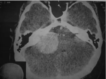
A contrast enhanced CT scan of the ears was or-dered to better assess the internal auditory meatus and the pontocerebellar angle. A globular tumor measuring 33.3mm in its largest dimension was found invading the right internal auditory meatus, where it grew larger and compressed the ipsilateral pontocerebellar transition zone (Figure 1).
The patient was referred to the Neurology and Neurosurgery service at Faculdade de Medicina da PUC SP to undergo BERA test and otoacoustic emissions tests. Dimenhydrinate was prescribed to deal with the patient’s symptoms.
The patient never came back to the ENT ward at PUC-SP, as he is currently in custody and could not un-dergo the tests mentioned above. He is waiting to receive preoperative care at the Neurology and Neurosurgery service at Faculdade de Medicina da PUC - SP.
DISCUSSION
Vestibular schwannomas amount to 80-90% of all
pontocerebellar angle tumors1,4,5 and may be diagnosed
in patients of all ages, although they are more commonly
found in people over the age of fifty1,2,8,9 and increased
prevalence among women at a ratio of 3:21,2,9,10 when
compared to males.
Even though progressive unilateral hypoacusis is the earliest and most frequent symptom, appearing in 75-95% of all patients, our patient was included in the 5% who have no hearing loss in spite of having vestibular
schwannoma3,13,14. His speech recognition rate was 100%,
a finding that goes against other papers in the literature, as voice discrimination is usually altered and affected by roll-over1-3,9.
The patient complained of right unilateral tinnitus, which prompted us to look for a possible retrocochlear expansive process that would explain the unilateral
hum-ming associated with other vestibular symptoms1,2,6,8,9.
631
Brazilian Journalof otorhinolaryngology 74 (4) July/august 2008
http://www.rborl.org.br / e-mail: [email protected]
According to the literature, tinnitus is the second most frequent complaint of vestibular schwannoma patients,
as it is present in 60-86% of the cases1,9.
The patient’s main complaint was a growing feeling of instability and unbalance that resulted from the presence of a tumor in the pontocerebellar angle, a finding in per-fect agreement with the literature, as approximately 70% of the tumors in this area measuring more than 30mm will
produce such symptoms15.
Ear contrast enhanced CT with emphasis on the internal auditory meatus and pontocerebellar angle alon-gside skull MRI scans are the diagnostic tests of choice for vestibular schwannomas, as their sensitivity rates are
95% and 100% respectively1-3,6,9.
Involvement of the V and VII cranial nerves led to right face hypoesthesia, reduced ipsilateral corneal reflex, reduced tearing, hypoesthesia of the posterior-superior portion of the ear in the involved side, and a slight devia-tion of the labial sulcus, all also mendevia-tioned in the literature. Facial paralysis is rarely reported, as it is found only in
case of very large tumors1,2,9.
Otoneurological tests indicated right ear arefle-xia, the most common finding in patients submitted to
electronystagmography1,2,9.
Brainstem evoked response audiometry could not be performed, as the patient is in custody. However, we would expect to find an interval greater than 2.3ms be-tween waves I and III; increased interval bebe-tween waves I and V above 4.4ms; interaural difference on wave V greater than 0.4ms; and absent wave I.
CONCLUSION
Vestibular schwannomas may be present and intro-duce only vestibular disorders such as instability during gait, either associated or not with unilateral tinnitus, in patients without complaints of hypoacusis. We should pay attention to such fact when diagnosing patients for vestibular schwannoma.
REFERENCES
1. Caldas Neto S. Tumores do VIII nervo. In: Sociedade Brasileira de Otorrinolaringologia, editor. Tratado de Otorrinolaringologia. 1ª ed. São Paulo: Roca; 2002. p. 564-80.
2. Jackler RK. Information on acoustic neuroma. [online] Apresenta informações, imagens, referências e links sobre neuroma do acústico. San Francisco: University of California; 1998. Disponível em: <http:// itsa.ucsf.edu/~rkj/IndexAN.html> (28 jun 2003)
3. Roland PS, Glasscock ME. Acoustic Neuroma. In: Paparella MM, Shu-mrick DA, Gluckman JL, editors. Otolaryngology. 3th.ed. Philadelphia: WB Saunders Company; 1991. p. 1775-87.
4. Bedavanija AA, Brieger J, Lehr HA. Association of proliferative activity and size in acoustic neuroma: implications for timing of surgery. J Neurosurg 2003;98(4):807-11.
5. Ogawa K, Kanzaki J, Ogawa S. Acoustic neuroma with normal hearing. Acta Otolaryngol Suppl 1991;487:144-9.
6. National Institutes of Health. Consensus Development Conference Statement. 1991;9(4):1-24.
7. Tos M, Thomsen J. Epidemiology of acoustic neuroma: has the inci-dence increased during the last years? In: Tos M, Thomsen J, editors. Acoustic neuromas and skull base surgery. New York: Kluger; 1992. p.3-6.
8. Pitts LH, Jackler RK. Treatment of acoustic neuromas. N Engl J Med 1998;339(20):1471-3.
9. Hungria H, editor. Neuroma do acústico. 7ª ed. Rio de Janeiro: Gua-nabara Koogan; 1995. Otorrinolaringologia; p.454-7.
10. Pertuiset B. Les neurinomes de l’acoustic développés dans l’angle pontocérébelleux. Neurochirurgie, 1970;16(1):1-147.
11. Gruskin P, Carberry JN, Chandrasekhar S. Pathology of acoustic tu-mors. In: House WF, Luetje CM, editors. Acoustic tumors - diagnosis and management. San Diego: Singular 1997; p.27-83.
12. Valente M, Peterein J, Goebel J. Four cases of acoustic neuromas with normal hearing. Am Acad Audiol 1995;6(3):203-10.
13. Shaan M, Vassalli L, Landolfi M. Atypical presentation of acoustic neuroma. Otolaryngo Head Neck Surg 1993;109(5):865-70. 14. Selesnick SH, Jackler RK. Atypical hearing loss in acoustic neuroma
patients. Laryngoscope 1993;103(4pt1):437-41.