Article
Printed in Brazil - ©2017 Sociedade Brasileira de Química0103 - 5053 $6.00+0.00*e-mail: [email protected]
Synthesis, Characterization and Dielectric Properties of New
5-(4-Hydroxyphenyl)-10,15,20-tri-4-[2-(3-pentadecylphenoxy)ethoxy]phenyl porphyrin and Their Ni, Co
and Cu Complexes
João P. F. Mota,*,a Antônio E. da Costa Júnior,a Viviane G. P. Ribeiro,a Samuel G. Sampaio,a
Nayane M. A. Lima,a Fernando L. F. da Silva,a Claudenilson S. Clemente,a
Giuseppe Mele,b Diego Lomonacoa and Selma E. Mazzettoa
aLaboratório de Produtos e Tecnologia em Processos (LPT), Departamento de Química Orgânica e Inorgânica,
Universidade Federal do Ceará, Campus do Pici, 60440-900 Fortaleza-CE, Brazil
bDepartment of Engineering for Innovation, University of Salento, Lecce, Italy
New asymmetric cardanol-based porphyrins, free-base and coordinated with Ni, Co and Cu, were synthesized and completely characterized as A3B type. Such porphyrins were obtained aiming improved solubility in polar solvents due insertion of an −OH phenolic group. Their thermal and dielectric properties were also evaluated. Changes in the synthetic route reduced the reaction time and improved the yields of the aldehyde precursor obtainment. Electronic absorption spectra of the new porphyrins in CH2Cl2, EtOH and acetone, indicated a decrease in the ε (molar absorptivity)
values with increasing solvent polarity, except for the nickel complex which, in acetone, showed a slight increase of 2% in the ε value. The dielectric measurements showed that the conductivity (σ) and the loss tangent (tan δ) increased with frequency, but the permittivity (ε’) decreased. The results showed that the coordination of the porphyrin promoted a significant change in thermal and dielectric properties, specially for to the Ni-complex compound, which presented the best dielectric properties with interesting values of permittivity and loss tangent at 100 MHz (19.46 and 0.011 a.u., respectively).
Keywords: asymmetric porphyrins, cardanol, dielectric properties
Introduction
Porphyrins are relatively planar aromatic system consisting of four pyrrole type rings joined by four methynic carbons. They have attracted scientists from many areas due to their biological importance, their fascinating
physical, chemical, and spectroscopic properties.1 These
compounds have a high electron density due to its
18 π-electrons delocalized on the macrocyclic ring, which
are responsible for their spectroscopic characteristics.2
Porphyrin and its derivatives play multiple roles in nature, such as energy transfer and light absorption in a wide range of the electromagnetic spectrum. Such features are of great interest to the industry of optoelectronic components, such as solar cells, due to its ability to act as photosensitizers
when irradiated under visible light.3 Thus, this is certainly
one of the most promising areas of application for these compounds. The structural modeling and synthesis of new
porphyrins has grown exponentially in order to control
their electronic and thermal properties.4 The addition
of substituents at the meso positions and metal ions in
the center of porphyrins can improve the electrical and magnetic properties, protecting against auto-oxidation and reducing the formation of dimers. However, these structural modifications can promote, in some cases, a significant
decrease in their planarity.5
A simple way to obtain porphyrins is the acid catalyzed condensation reaction of pyrrole with specific aldehyde, followed by oxidation of the porphyrinogen.
This procedure, originally developed by Rothemund,6
has been refined by Adler-Longo,7-9 due to the addition
of metal salts to the reaction. Despite the modest yields, the relative simplicity of this method has become suited for preparation of large amounts of tetraarylporphyrins. Higher yields and milder reaction conditions using
Lewis acid (TFA or BF3) as catalyst were obtained by
Lindsey et al.10 More recently, substituted asymmetrical
aldehyde condensations, using a mixture of two different
aldehydes as starting materials.11-13
Asymmetric porphyrins are molecules that exhibit some advantages over the symmetric porphyrins (A4) like: reduced aggregation, groups that allows the use of a wide range of solvents, and presence of dipole moment,
which may facilitate the electron injection into the TiO2
conduction band.14 In addition, asymmetric porphyrins have
been synthesized seeking a better efficacy in photodynamic therapy (PDT), since the cellular affinity depends on amphiphilic character, that is, dependent on the structural
arrangement of groups hydrophilic (phenolic −OH or
carboxylic −COOH) and hydrophobic (alkyl or aryl chains)
meso-substituents in macrocycle.15,16
Some studies mention the synthesis of porphyrins
using cardanol as starting material,17-19 which provides
interesting lipophilic properties, besides being a substance derived from a renewable raw material, the cashew nut
shell liquid (CNSL).20 Cardanol is a phenolic lipid and
the main constituent of the CNSL, a dark oil obtained as a by product from industrial processing of the cashew
nut (Anacardium occidentale L.).21 It is known that the
presence of an alkyl chain or an aryl substituents in porphyrins are important, not just for increase solubility in organic solvents and influence the aggregation of these materials, but also to modify its chemical characteristics and
photophysical properties.11 Here, we report the synthesis
and characterization of novel asymmetric cardanol-based
A3B porphyrins 4a-d (4a: metal-free, 4b: Ni, 4c: Co,
4d: Cu) as well as the study of their thermal and dielectric
properties. Scheme 1 shows the steps and reagents used in the preparation of asymmetric porphyrins.
From the thermogravimetric analysis (TGA) it was possible to determine that the thermal stabilities of the
porphyrins 4a-d, in inert atmosphere, was in the following
order: 4a > 4d > 4c > 4b. In TGA, the porphyrin 4a showed
initial temperature of degradation at 352 °C (Tonset) and
other parameters as the final and the half decomposition
temperature (Tendset and T*, respectively), the residual
mass and the numbers of degradation stages were also
evaluated.22 The dielectric properties, like the loss tangent
(tan δ), permittivity (ε’) and conductivity (σ) are the
most important parameters to determine the suitability
for electronics applications.23,24 According to Lukichev,25
materials presenting high dielectric constant associated with low loss tangent have important applications in
microelectronics. The measurements showed that 4b have
good dielectric permittivity (ca. 20 a.u.) and 4d had the
lowest permittivity (ca. 6 a.u.). These results suggest that the metal promoted a significant change in thermal and electrical properties of the asymmetric porphyrin.
Results and Discussion
Synthesis
The synthesis of porphyrins starting from cardanol
are mentioned by many papers since 2004,26-29 and they
employed a procedure which consists in producing an aldehyde derivative as precursor. Normally, this
precursor is compound 3 (4-[2-(3-pentadecylphenoxy)
ethoxy] benzaldehyde), obtained by a reaction between
compound 2 (1-(2-bromoethoxy)-3-pentadecylbenzene)
and 4-hydroxybenzaldehyde in acetone using anhydrous
potassium carbonate (K2CO3) as base, stirred for 24 h and
with 40% yield.
In this study, it was found that the replacement of the
base, K2CO3, by KOH, promoted a decrease on the yield
(about 25%), that can be associated with the deprotonation
of acetone present in the reaction as solvent.30 A new
reaction procedure using DMF as solvent and KOH as base
at 100 °C, provided a better yield (62%) of compound 3 in
only 6 hours of reaction. Based on the result of this new procedure, it is possible to conclude that the DMF is not affected by a strong base.
Characterization
The chemical structures of the asymmetric porphyrin 4a
and its metal complexes 4b-d (A3B type) were characterized
using 1H and 13C nuclear magnetic resonance (NMR),
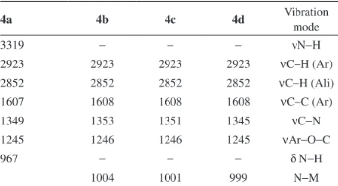
matrix-assisted laser desorption ionization-time of flight mass spectrometry (MALDI-TOF-MS), elemental analysis, UV-visible absorption, Fourier transform infrared (FTIR) and fluorescence emission spectroscopy. The FTIR data are listed in Table 1, which were consistent with the proposed structures of the corresponding compounds. For compound
4a, the absorption bands at 3319 and 967 cm-1 are assigned
to the N−H stretching and in plane bending vibrations of
the pyrrole group, respectively.2 These absorption bands
disappeared in the spectra of metal complexes (4b-d),
since the hydrogen atom of the N−H bond is replaced by
a metal ion to form N−M bond, and a new metal-sensitive
absorption band appeared at ca. 1000 cm-1, which further
confirmed the formation of metal complexes.31 Moreover,
the presence of aliphatic groups in porphyrins 4a-d, are
also evident by the presence of absorbance peaks at 2923
and 2852 cm-1, which are characteristic of asymmetric and
symmetric νC−H modes, respectively.17 In addition, the
compounds exhibit absorption bands of the phenyl groups
at ca. 1610 cm-1, assigned to the C=C stretching vibration.32
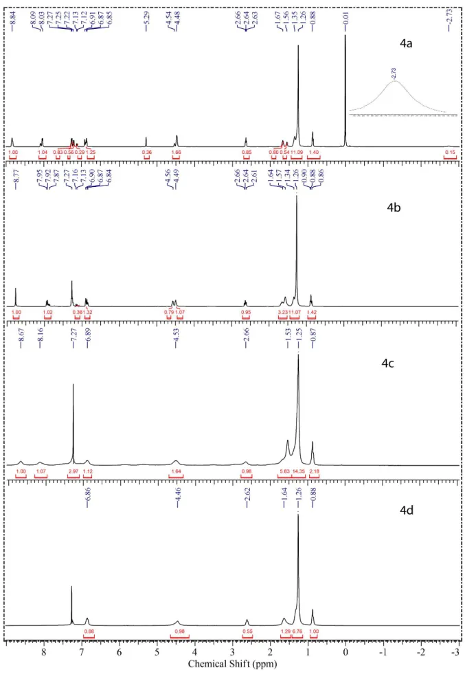
The 1H NMR and 13C NMR spectra were in accordance
with the assigned structures and presented all the expected
signals for asymmetrical meso-substituted porphyrins
derived from cardanol. The signals obtained in CDCl3
(1H NMR, Figure 1) for 4a-d exhibited the spectral signatures
of the porphyrin core and signals typical of the peripheral cardanol units, such as the singlet at 1.26 ppm corresponding
to the 72 protons of the aliphatic groups (−CH2−).
For free base porphyrin 4a, the characteristic chemical
shift of pyrrole N−H located into the porphyrin ring is
located at −2.73 ppm. If we assume equivalence 1 for the
peak at 8.84 ppm which was assigned to 8H (β-pyrrolic), the
integration of the other peaks can be related to the following
hydrogens: peak at 4.48-4.54 ppm to 12H (O−(CH2)2−O),
at 2.64 ppm to 6H (Ph−CH2), at 0.88 ppm to 9H (CH3) and
peak at 5.29 ppm to 1H (Ph−OH). These peaks indicated
that the porphyrin 4a is an asymmetric compound of A3B
type. After formation of complexes (4b-d), the signal
peak at −2.73 ppm disappeared, since the hydrogen atom
in the N−H bond is replaced by a metal ion.33 In some
paramagnetic metalloporphyrins the signals may be broad due to the hyperfine electron-nuclear interactions and by
relaxation processes.11 Figure 1 shows that compounds 4c
(cobalt porphyrin) and 4d (copper porphyrin) presented this
phenomenon, thus indicating the paramagnetic character
of these metals. 13C NMR data to 4a showed signals that
confirm the presence of alkyl (13.85-35.83 ppm), ethers (66.22 ppm) and aromatic groups (111.37-158.45 ppm).
Similar signals were observed for metalloporphyrins 4b-d
(Figure S1). The results indicated that compounds 4a-d
were obtained with good purity, which was confirmed by mass spectrometry (MALDI-TOF, Figure S2) and
elemental analysis. The m/z for the molecular ion presented
the following values: 1669.938 (4a), 1726.860 (4b),
1726.907 (4c) and 1731.889 (4d).
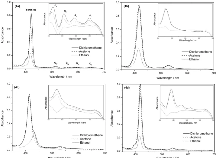
Electronic absorption and luminescence spectra
Electronic absorption spectra of porphyrins 4a-d in
CH2Cl2, EtOH and acetone (Figure 2), show one intense
band in the near-ultraviolet region of the spectrum (Soret or B band) and absorption bands at higher wavelengths in the visible region (Q bands). The free base porphyrin
4a have four Q bands denoted as IV, III, II, and I,32 while
the metallated porphyrins presented one (4b and 4c) or
two Q bands (4d) due to the increase of symmetry of
the complexes.34 Characteristic Q and B (Soret) bands of
metalloporphyrins are determined by transitions of the
π-electrons between two higher occupied and two lower
unoccupied orbitals.35 Nevertheless, the changes in the
molecular structure and the effect of the solvent may alter
the spectra of the porphyrins.36 The molar absorptivity
coefficient values (ε) of Soret band of the porphyrins 4a-d
are shown in Table 2, and the results indicated a decrease
in the values of ε with increasing solvent polarity, except
for the nickel porphyrin (4b), in which there was a slight
increase of 2% at the ε obtained in acetone. The low
values of the molar absorptivity coefficient (Table 2) of the compounds in ethanol clearly indicate the formation of
Table 1. FTIR data to asymmetric porphyrins and its vibrations mode in cm-1 (KBr)
4a 4b 4c 4d Vibration
mode
3319 − − − νN−H
2923 2923 2923 2923 νC−H (Ar)
2852 2852 2852 2852 νC−H (Ali)
1607 1608 1608 1608 νC−C (Ar)
1349 1353 1351 1345 νC−N
1245 1246 1246 1245 νAr−O−C
967 − − − δ N−H
aggregated species, due to, probably, the poor solubility in this solvent, evidenced by decrease in the intensity of the absorption bands (hypochromic effect).
Specifically for compound 4c, we observe the
formation of J-aggregates that showed a red-shifted
absorption band at 432 nm.37 This phenomenon indicated
that the molecules of compound 4c have side-by-side
interactions in an ethanolic solution. Usually, porphyrins derived from cardanol have good solubility in solvents with low polarity such as chloroform (µ = 1.04), dichloromethane (µ = 1.60) and THF (µ = 1.75), but they are insoluble in polar solvents like acetone (µ = 2.88).
Therefore, these asymmetric porphyrins 4a-d allowed
us to work with a greater variety of solvents, including acetone. This significant change on the solubility of these porphyrins probably occurs due to the presence of the hydroxyl group at the macrocyclic structure, which promotes a better interaction and affinity to polar solvents.
Fluorescence emission spectroscopy of porphyrin 4a
and metalloporphyrins 4b, 4c and 4d were investigated
in dichloromethane (10-5 M) at room temperature. The
emission spectrum of the porphyrin 4a showed two bands
in the red region, one intense at 658 nm (ca. 5 × 10⁷) and a
lower one at 715 nm (ca. 1 × 10⁷) after excitation at 421 nm
Table 2. Molar absorptivity coefficient (ε) of asymmetric porphyrins 4a-d in different organic solvents at 25 °C
Sample Dichloromethane Acetone Ethanol
λmax / nm ε / (105 L mol-1 cm-1) λmax / nm ε / (105 L mol-1 cm-1) λmax / nm ε / (105 L mol-1 cm-1)
4a 421 5.542 418 4.170 419 1.523
4b 417 1.181 416 1.215 415 0.391
4c 414 1.060 414 1.012 432 0.325
4d 418 3.986 415 3.965 415 0.909
(Figure 3A). This spectral behavior was similar to other
meso-porphyrins and indicates that these compounds can be
used in optoelectronic systems working in the red spectral region, for example as catalyst, solar cells, OLEDs (organic
light-emitting diode) and photodynamic therapy.38,39 The
fluorescence emission was attributed to the transition from the excited singlet state S1/S2 to the ground state S0 (S2→ S0,
S1 → S0). The spectra of the complexes 4b-d showed a
very weak fluorescence band, and when 4b is excited at
421 nm, this band appeared at 651 nm with intensity about
2 × 10³ a.u. (Figure 3B). The decrease in the emission band
intensity to porphyrins 4b-d probably occurred due to the
heavy atom effect, where the presence of atoms with higher atomic numbers increases the probability of non-radiative
transitions.40,41 The excitation spectrum of 4a (Figure 4)
showed bands responsible for emission with major wavelength at 658 nm. Comparing the excitation spectrum with the absorption spectrum is possible to observe that the emission happened from the absorption of the Soret and Q bands.
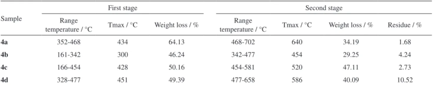
Thermal stability
In order to evaluate the thermal stability of the
compounds 4a-d, TGA studies were carried out in inert
atmosphere (N2) using a temperature range between
25 and 900 °C. In general, the results showed that the order of observed thermal stabilities of the porphyrins
4a-d were: 4a > 4d > 4c > 4b. Thus, we verified that
porphyrin 4a has the best thermal properties, probably
due to the absence of metal coordinated to the center of the macrocycle, that can act as promoter of degradation process. Table 3 summarizes the decomposition range and maximum temperature values, as well as the weight loss of the main stages of degradation and the residual
weight. The decomposition of the compounds 4a, 4c and
4d occurred in two different stages, while the porphyrin
4b presented three stages.
The first stage of degradation was attributed to the loss of the peripheral chains of cardanol attached to the macrocyclic ring of the porphyrins and the second was associated to the degradation of the macrocycle ring
4a-d.2 Metalloporphyrins showed initial temperature of
degradation lower than the free asymmetric porphyrin 4a.
Similar results was reported by Antina et al.,42 which
related the decrease in thermal stability of porphyrins
to changes in the aromaticity and π-polarization of the
macrocyclic system, due to the presence of the metal ion.
Clemente et al.17 showed that the thermal degradation
of the symmetric porphyrin, derived from cardanol,
started at 250 °C (Tonset). This information indicated
Figure 3. Fluorescence emission spectra of the free base porphyrin 4a (A) and the complex 4b, 4c and 4d (B) with a concentration of 10-5 M in CH2Cl2.
Figure 4. Excitation spectrum of the porphyrin 4a with emission band in 658 nm.
Table 3. Thermogravimetric parameters of the two first stages of degradation in N2 atmosphere
Sample
First stage Second stage
Range
temperature / °C Tmax / °C Weight loss / %
Range
temperature / °C Tmax / °C Weight loss / % Residue / %
4a 352-468 434 64.13 468-702 640 34.19 1.68
4b 161-342 300 46.24 342-477 454 29.25 4.24
4c 166-454 428 50.16 454-581 520 47.11 2.73
that the asymmetric porphyrin 4a is more thermally stable than its symmetric porphyrin analogue, due to
the thermal decomposition of 4a started only at 352 °C.
Therefore, these porphyrins are suitable for applications in technological areas that require luminescent compounds (red region) and elevated thermal stability, like OLEDs
and solar cells.43 During the second stage of thermal
degradation, all samples showed gradual mass loss between 29.25 and 47.11% in the temperature range of 342 to 702 °C.
Dielectric properties
Dielectric permittivity (ε’) is a measure that shows the
ability of materials to store energy and have their dipoles oriented when subjected to an alternating electric field. This capacity is associated with the electric susceptibility of these compounds. The extent of the polarization is directly related to the nature of the material, shape and their structures. Therefore, if the electrical properties (dielectric permittivity) are known, it is possible to predict the influence of an electric field in the polarization of the material studied.
The dielectric permittivity and dielectric loss tangent
(tan δ) of porphyrins 4a-d were obtained at room
temperature using the following relationship:44,45
Z = 1 / iwεC0 = 1 / iw(ε’ + iε’’)C0 (1)
where ε’ and ε’’ are the real (permittivity) and imaginary
parts (dielectric loss) of the dielectric function, respectively.
C0 (C0 = ε0A / d) is the empty cell capacitance, where, A
is the electrode area, d is the thickness sample, ε0 is the
permittivity of free space (ε0 = 8.854 × 10⁻¹² F m-1) and w
(w = 2πf) is the angular frequency. The components of the
complex dielectric function was derived using the relations,
ε’ = C / C0 and ε’’ = −iσ / wC (2)
where C is the capacitance and σ is the conductivity. The
imaginary part of the dielectric function can be calculated as
ε’’ = ε’tan δ. This expression determines the dielectric loss.
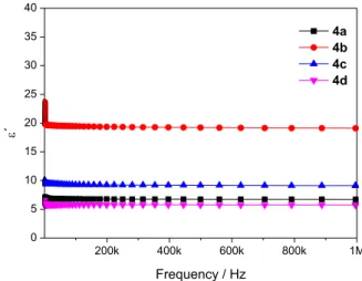
It was observed a small decrease in the dielectric
permittivity for all asymmetric porphyrins (4a-d),
when the applied frequency was increased (Figure 5). At frequencies greater than 1 MHz it was observed that the values practically did not change. The values of dielectric permittivity at low frequencies can be assigned to different types of polarization, such as dipole or
electronic.42 According to the data, it was observed that
this slight decrease in dielectric permittivity resulted
from the decrease in polarization of the molecules across electric field. When comparing the dielectric permittivity values of the analyzed molecules, we can see that the
porphyrin 4b had the highest permittivity (ca. 20 a.u.),
while 4d porphyrin had the lowest (ca. 6 a.u.). These
results indicated that the compound 4b is more susceptible
to polarization, since higher permittivity values are resulted from the dipole generation. The decrease in the dielectric permittivity value with increasing frequency can be attributed to dipole relaxation phenomenon in which the dipoles at low frequencies follow the frequency of
the applied field.46 However, with the increase frequency,
the oscillations in the dipoles cannot follow the applied field and tend to disappear due to the limited time for its
polarization.47
Dielectric loss tangent is characterized by the loss of energy due to the movement and orientation of electric dipoles of the material, where compounds that have low dielectric loss are considered suitable for use in
microelectronics.48 The dielectric loss tangent (tan δ,
Figure 6) was determined as a function of frequency and
the results obtained for compounds 4a-d indicated that
this parameter increases proportionally with the frequency. In general, the compounds showed low loss tangent and exhibited dispersion for high values in ca. 100 kHz.
Porphyrin 4b showed the best dielectric characteristics,
since it combines a high value of dielectric permittivity (19.46 at 100 kHz) and a low dielectric loss tangent (0.011 at 100 kHz).
The results in Table 4 shows that when a metal ion was added in the center of asymmetric porphyrin, the dielectric
properties were significantly altered.23 The increase in the
dielectric permittivity (ε’) of the 4b porphyrin (about three
times), compared with the free porphyrin 4a (permittivity
6.96), is a proof of this influence. Another important
parameter of the dielectric properties is the conductivity (σ),
and it is associated with migration of charge carriers.49 The
conductivity in alternating current (σac) of the asymmetric
porphyrins at room temperature was determined as
described by Deger et al.50 The porphyrin coordinated
with nickel (4b) had the highest conductivity values at
all frequencies: 1.3 × 10⁻⁸ Sm-1 (1 kHz), 2.8 × 10⁻⁸ Sm-1
(10 kHz), 1.9 × 10⁻⁷ Sm-1 (100 kHz) and 4.6 × 10⁻⁶ Sm-1
(1 MHz). For other porphyrins, the maximum conductivities
were observed in 1 MHz to 4a (8.6 × 10⁻⁷ Sm-1),
4c (1.6 × 10⁻⁶ Sm-1) and 4d (6.7 × 10⁻⁷ Sm-1), respectively.
These conductivities indicated that the compounds studied have low polarization when subjected to an external electric field.
Conclusions
The novel metal-free porphyrin 4a and their metallated
analogs 4b-d, were successfully synthesized and
characterized like asymmetric porphyrins A3B type. The results of TGA/DTA (differential thermal analysis) analysis showed that the metalloporphyrins have lower
initial temperature of degradation (4b, 4c and 4d)
compared to free asymmetric porphyrin (4a). The dielectric
measurements indicates that the asymmetric porphyrins 4b
have good dielectric properties, due to its better dielectric
permittivity and low loss tangent. This study indicates that the coordination of the porphyrin nucleus with a metal promoted a significant change in thermal and electrical properties of these compounds.
Experimental
Reagents
All the starting reagents used in this work were purchased from Sigma-Aldrich Chemical, Vetec, Dynamics, Synth and Acros Organic, and used as received. Solvents were previously distilled before their use.
Analyses
UV-Vis: analyses were performed on a Varian Cary 5000 spectrophotometer, where the spectrum was obtained
using quartz cells of 1.0 cm optical path (CHCl3) in the
following concentration: 4a 1.5 × 10⁻⁶ M, 4b 8.0 × 10⁻⁶ M,
4c 8.0 × 10⁻⁶ M and 4d 2.5 × 10⁻⁶ M. FTIR: spectra
were obtained using a PerkinElmer Spectrum One spectrophotometer, and the samples were prepared as
pellets (KBr). 1H NMR and 13C NMR: was obtained in
a nuclear magnetic resonance Bruker Avance DPX 300
spectrometer, operating at 300 (1H) and 75 MHz (13C)
(CDCl3). TGA/DTA: the thermal stability of the samples
was evaluated by thermogravimetric analysis It was used an equipment Mettler Toledo TGA/SDTA 851e. The decomposition analyses were performed under nitrogen
atmosphere in a constant flow of 50 mL min-1, with
heating rate of 10.0 °C min-1, sample mass of 5.0 mg and
a temperature program from 25 to 900 °C. MS: samples were analyzed on a Bruker Microflex LT (MaldiTof) using alphacyano, and dichloromethane as a matrix and 5% TFA as mobile phase. Elemental analysis (CNH): the percentual composition of the samples was analyzed on a PerkinElmer 2400 Series II. Dielectric measurements: the samples were shaped in a cylinder, with metal bases (Al). They were pressed by two electrodes connected in order to perform capacitance measurements, dielectric loss, impedance real and imaginary. Measurements were made at room
Figure 6. Frequency dependence of the dielectric loss tangent at room temperature.
Table 4. Values of the dielectric properties to asymmetric porphyrins at room temperature
Compound 1 kHz 10 kHz 100 kHz 1 MHz
ε’ tan δ ε’ tan δ ε’ tan δ ε’ tan δ
4a 6.96 0.007 6.87 0.003 6.80 0.007 6.73 0.015
4b 20.36 0.073 19.66 0.016 19.46 0.011 19.10 0.026
4c 9.65 0.014 9.43 0.011 9.30 0.012 9.12 0.020
temperature in atmospheric air through a precision Agilent 4294A impedance analyzer connected to a microcomputer in the 1 Hz-10 MHz frequency range.
Synthesis of the asymmetric porphyrins
Cardanol precursors of the porphyrins
Hydrogenated cardanol (1) was obtained in our
laboratory through chromatographic separation of the
constituents of CNSL followed by hydrogenation.51 The
compound 1-(2-bromoethoxy)-3-pentadecylbenzene (2)
derived from 1, was prepared and characterized by FTIR,
and 1H NMR according to the procedure previously
described.52
Aldehyde (3)
Compound 4-[2-(3-pentadecylphenoxy)ethoxy]
benzaldehyde (3) was obtained by a mix of 2 (3.0 g,
7.30 mmol) and 4-hydroxybenzaldehyde (1.3 g, 10.90 mmol) using KOH (1.3 g, 23.30 mmol) as base in 50 mL of DMF. The system was maintained in stirring at 100 °C for 6 h, and the progress of reaction was carried out by thin layer chromatography (TLC). The reaction mixture was subjected to a pretreatment by means of a liquid-liquid partition with distilled water (200 mL) and ethyl acetate (200 mL). The organic phase was dried with
anhydrous Na2SO4, concentrated and purified in column
chromatography with silica gel having as eluent hexane/
ethyl acetate (95:5). The compound 3 was obtained in 62%
yield (2.0 g), molecular weight 452.67 g mol-1 (C
30H44O3).
Metal-free porphyrin (4a)
The free base
5-(4-hydroxyphenyl)-10,15,20-tri-4-[2-(3-pentadecylphenoxy)ethoxy]phenyl porphyrin (4a) was
obtained by a mix of the pyrrole (0.88 mmol), 3 (0.88 mmol),
4-hydroxybenzaldehyde (0.22 mmol), NaCl (16.25 mmol),
BF3 (0.22 mmol) and DDQ (0.50 mmol) in chloroform/
ethanol (50 mL, ethanol 0.8%) at room temperature for 1 hour. The crude was packaged in a silica column chromatography and eluted with solvents of increasing polarity: dichloromethane, dichloromethane/ethanol
(9:1). The asymmetric free base porphyrins (4a) were
obtained in the second fraction eluted and the compound
was recrystallized with ethanol/hexane. 4a: yield 26%.
1H NMR (300 MHz, CDCl
3) d−2.73 (s, 2H, N−H), 0.88
(t, 9H, J 6.5 Hz, CH3), 1.26 (s, 72H, CH2−(CH2)12−CH3),
1.67 (m, 6H, Ph−CH2−CH2), 2.64 (t, 6H, J 8.0 Hz,
Ph−CH2), 4.52 (d, 12H, J 4.5 Hz, O−(CH2)2−O), 5.29 (s,
1H, Ph−OH), 6.85-8.09 (28H, Ph−H), 8.84 (s, 8H, H-β
pyrrolic); 13C NMR (75 MHz, CDCl
3) d 13.85 (CH3),
22.42 (CH2−CH3), 29.43-31.66 (CH2−(CH2)13−CH3),
35.83 (Ph−CH2), 66.22 (O−(CH2)2−O), 111.37-158.45
(C-aromatic); FTIR (KBr) ν / cm-1 3436, 3319, 2923,
2852, 1607, 1583, 1509, 1448, 1245, 1158, 1071, 967;
UV-Vis (CH2Cl2) λmax / nm 421, 516, 557, 592, 649; MS
(MALDI-TOF) m/z, for C113H144N4O7 [M+], observed:
1669.938; required: 1670.375; elemental analysis (CNH)
for C113H144N4O7, observed: C 81.29, N 3.65, H 8.76%;
required: C 81.25, N 3.35, H 8.69%.
Metalloporphyrins (4b-d)
4a (0.06 mmol) and acetate metal salts (0.60 mmol,
Ni, 4b; Co, 4c; and Cu, 4d) were added in a mix of the
chloroform/DMF (20 mL of each) for 3 hours at 90 °C. The product was purified by chromatography in a silica gel column using dichloromethane as eluent, and the product collected in the first fraction eluted corresponding
to compounds 4b-d. The metalloporphyrins were
recrystallized with a mix of ethanol/hexane.
4b
Yield 89%. 1H NMR (300 MHz, CDCl
3) d 0.88 (t, 9H,
J 6.3 Hz, CH3), 1.26 (s, 72H, CH2−(CH2)12−CH3), 1.64 (m,
6H, Ph−CH2−CH2), 2.64 (t, 6H, J 7.5 Hz, Ph−CH2), 4.55 (d,
12H, J 4.8 Hz, O−(CH2)2−O), 6.84-7.95 (28H, Ph−H), 8.77
(s, 8H, H-β pyrrolic); 13C NMR (75 MHz, CDCl
3) d 14.08
(CH3), 22.66 (CH2−CH3), 29.36-31.90 (CH2−(CH2)13−CH3),
36.06 (Ph−CH2), 66.88 (O−(CH2)2−O), 111.67-158.73
(C-aromatic); FTIR (KBr) ν / cm-1 3432, 2923, 2852,
1608, 1584, 1507, 1448, 1246, 1158, 1074, 1004; UV-Vis
(CH2Cl2) λmax / nm 416, 529; MS (MALDI-TOF) m/z,
for C113H142N4O7Ni [M+], observed: 1726.860; required:
1727.054; elemental analysis (CNH) for C113H142N4O7Ni,
observed: C 79.56, N 2.21, H 9.38%; required: C 78.59, N 3.24, H 8.29%.
4c
Yield 79%. 1H NMR (300 MHz, CDCl
3) d 0.87 (t, 9H,
J 5.4 Hz, CH3), 1.25 (s, 72H, CH2−(CH2)12−CH3), 1.53
(broad signal, 6H, Ph−CH2−CH2), 2.66 (broad signal, 6H,
Ph−CH2), 4.53 (broad signal, 12H, O−(CH2)2−O),
6.89-8.16 (broad signals, 28H, Ph−H), 8.67 (broad signal, 8H,
H-β pyrrolic); 13C NMR (75 MHz, CDCl
3) d 13.69 (CH3),
22.27 (CH2−CH3), 28.96-31.51 (CH2−(CH2)13−CH3),
35.78 (Ph−CH2), 67.41 (O−(CH2)2−O), 111.48-158.44
(C-aromatic); FTIR (KBr) ν / cm-1 3436, 2923, 2852,
1608, 1583, 1507, 1449, 1246, 1158, 1073, 1001; UV-Vis
(CHCl3) λmax / nm 414, 530; MS (MALDI-TOF) m/z, for
C113H142N4O7Co [M+], observed: 1726.907; [M+] required:
1727.298; elemental analysis (CNH) for C113H142N4O7Co,
4d
Yield 83%. 1H NMR (300 MHz, CDCl
3) d 0.88 (t, 9H,
J 6.0 Hz, CH3), 1.26 (s, 72H, CH2−(CH2)12−CH3), 1.64
(broad signal, 6H, Ph−CH2−CH2), 2.62 (broad signal,
6H, Ph−CH2), 4.46 (broad signal, 12H, O−(CH2)2−O),
6.86 (broad signal, 6H, H-aromatic); 13C NMR (75 MHz,
CDCl3) d 14.10 (CH3), 22.66 (CH2−CH3),
29.36-31.89 (CH2−(CH2)12−CH2), 36.07 (Ph−CH2), 66.45
(O−(CH2)2−O), 111.61-158.70 (C-aromatic); FTIR (KBr)
ν / cm-1 3434, 2923, 2852, 1608, 1583, 1504, 1447, 1245,
1158, 1072, 999; UV-Vis (CHCl3) λmax / nm 418, 542,
578; MS (MALDI-TOF) m/z, for C113H142N4O7Cu [M+],
observed: 1731.889; required: 1731.896; elemental analysis
(CNH) for C113H142N4O7Cu, observed: C 78.28, N 3.47, H
8.41%; required: C 78.37, N 3.23, H 8.26%.
Supplementary Information
Supplementary information is available free of charge at http://jbcs.org.br as PDF file.
Acknowledgments
The authors would like to thank the support from CAPES and CNPq (PVE 401359/2014-0 and 402566/2013-0), Almonds Company in Brazil by providing cashew nuts, CENAUREMN (Centro Nordestino de Aplicação e Uso da Ressonância Magnética Nuclear, Brazil) for the NMR analyses
References
1. Birin, K. P.; Gorbunova, Y. G.; Tsivadze, A. Y.; Bessmertnykh-Lemeune, A. G.; Guilard, R.; Eur. J. Org. Chem. 2015, 25, 5610.
2. Sun, E.; Shi, Y.; Zhang, P.; Zhou, M.; Zhang, Y.; Tang, X.; Shi, T.; J. Mol. Struct. 2008, 889, 28.
3. Li, K.; Lin, L.; Peng, T.; Guo, Y.; Li, R.; Zhang, J.; Chem. Commun. 2015, 51, 12443.
4. Henriques, C. A.; Gonçalves, N. P. F.; Abreu, A. R.; Calvete, M. J. F.; Pereira, M. M.; J. Porphyrins Phthalocyanines 2012,
16, 290.
5. Basova, T. V.; Çamur, M.; Esenpinar, A. A.; Tuncel, S.; Hassan, A.; Alexeyev, A.; Banimuslem, H.; Durmus, M.; Gurek, A. G.; Ahsen, V.; Synth. Met. 2012, 162, 735.
6. Rothemund, P.; J. Am. Chem. Soc. 1935, 57, 2010.
7. Adler, A. D.; Longo, F. R.; Shergalis, W.; J. Org. Chem. 1964,
86, 3145.
8. Adler, A. D.; Longo, F. R.; Finarelli, J. D.; Godmacher, J.; Assour, J.; Korsakoff, L.; J. Org. Chem. 1967, 32, 476. 9. Adler, A. D.; Sklar, L.; Longo, F. R.; Finarelli, J. D.; Finarelli,
M. G.; J. Heterocycl. Chem. 1968, 5, 669.
10. Lindsey, J. S.; Schreiman, I. C.; Hsu, H. C.; Kearney, P. C.; Marguerettaz, A. N.; J. Org. Chem. 1987, 52, 827.
11. Guo, Y.-C.; Xiao, W.; Mele, G.; Martina, F.; Margapoti, E.; Mazzetto, S. E.; Vasapollo, G.; J. Porphyrins Phthalocyanines
2006, 10, 1071.
12. Zhuang, C.; Tang, X.; Wang, D.; Xia, A.; Lian, W.; Shi, Y.; Shi, T.; J. Serb. Chem. Soc. 2009, 74, 1097.
13. Schiavon, M. A.; Iwamoto, L. S.; Ferreira, A. G.; Iamamoto, Y.; Zanoni, M. V. B.; Assis, M. D.; J. Braz. Chem. Soc. 2000, 11, 458.
14. Urbani, M.; Gratzel, M.; Nazeeruddin, M. K.; Torres, T.; Chem. Rev. 2014, 114, 12330.
15. Fagadar-Cosma, E.; Cseh, L.; Badea, V.; Fagadar-Cosma, G.; Vlascici, D.; Comb. Chem. High Throughput Screening 2007,
10, 466.
16. Peng, C.-L.; Lai, P.-S.; Shieh, M.-J.; Biomed. Eng.-App. Bas.
C.2008, 20, 9.
17. Clemente, C. S.; Ribeiro, V. G. P.; Sousa, J. E. A.; Maia, F. J. N.; Barreto, A. C. H.; Andrade, N. F.; Denardin, J. C.; Mele, G.; Carbone, L.; Mazzetto, S. E.; Fechine, P. B. A.; J. Nano Res.
2013, 15, 1739.
18. Sandrino, B.; Clemente, S. C.; Oliveira, T. M. B. F.; Ribeiro, F. W. P.; Pavinatto, F. J.; Mazzetto, S. E.; Neto, P. L.; Correia, N. A.; Pessoa, C. A.; Wohnrath, K.; Colloids Surf., A 2013, 425, 68. 19. Bloise, E.; Carbone, L.; Colafemmina, G.; D’Accolti, L.;
Mazzetto, S. E.; Vasapollo, G.; Mele, G.; Molecules 2012, 17, 12252.
20. Attanasi, O. A.; Mele, G.; Filippone, P.; Mazzetto, S. E.; Vasapollo, G.; Arkivoc 2009, viii, 69.
21. Júnior, A. E. C.; Barreto, A. C. H.; Rosa, B. S.; Maia, F. J. N.; Lomonaco, D.; Mazzetto, S. E.; J. Compos. Mater. 2015, 49, 2203.
22. Guan, C.; Li, L.; Chen, D.; Gao, Z.; Sun, W.; Thermochim. Acta 2004, 413, 31.
23. Saleh, A. M.; Hraibat, S. M.; Kitaneh, R. M.-L.; Abu-Samreh, M. M.; Musameh, S. M.; J. Semicond. 2012, 33, 082002-1. 24. Canlıca, M.; Altındal, A.; Nyokong, T.; J. Porphyrins
Phthalocyanines 2012, 16, 826.
25. Lukichev, A. A.; Chem. Phys. 2014, 428, 29.
26. Mele, G.; Vasapollo, G.; Mini-Rev. Org. Chem. 2008, 5, 243. 27. Mele, G.; Del Sole, R.; Vasapollo, G.; Garcia-Lopez, E.;
Palmisano, L.; Mazzetto, S. E.; Attanasi, O. A.; Filippone, P.;
Green Chem. 2004, 6, 604.
28. Vasapollo, G.; Mele, G.; Del Sole, R.; Pio, I.; Li, J.; Mazzetto, S. E.; Molecules 2011, 16, 5769.
29. Vasapollo, G.; Mele, G.; Del Sole, R.; Molecules 2011, 16, 6871.
30. Clayden, J.; Greeves, N.; Warren, S.; Wothers, P.; Organic
31. Xu, Z.; Mei, Q.; Hua Q.; Tian, R.; Weng, J.; Shi, Y.; Huang, W.;
J. Mol. Struct. 2015, 1094, 1.
32. Yu, M.; Chen, G.-J.; Liu, G.-F.; J. Phys. Chem. Sol. 2007, 68, 541.
33. Brem, B.; Gal, E.; Gaina, L.; Cristea, C.; Gabudean, A. M.; Astilean, S.; Silaghi-Dumitresco, L.; Dyes Pigments 2015, 123, 386.
34. Yu, M.; Zhang, Y. J.; Shi, J. H.; Liu, G. F.; Zhang, H. J.; Solid State Sci. 2009, 11, 2016.
35. Wei, L.; TongShun, S.; Sci. China, Ser. B: Chem. 2007, 50, 488. 36. Sun, E.-J.; Sun, Z.-Y.; Yuan, M.; Wang, D.; Shi, T.-S.; Dyes
Pigments 2009, 81, 124.
37. Fagadar-cosma, E.; Vlascici, D.; Fagadar-cosma, G.; Palade, A.; Lascu, A.; Creanga, I.; Birdeanu, M.; Cristescu, R.; Cernica, I.; Molecules 2014, 19, 21239.
38. Valicsek, Z.; Horváth, O.; Microchem. J. 2013, 107, 47. 39. Zheng, W.; Shan, N.; Yu, L.; Wang, X.; Dyes Pigments 2008,
77, 153.
40. Borisevich, E. A.; Solov’ev, K. N.; Phys.-Usp. 2005, 48, 231. 41. Nastasi, F.; Campagna, S.; Ngo, T. H.; Dehaen, W.; Maes, W.;
Kruk, M.; Photochem. Photobiol. Sci. 2011, 10, 143. 42. Antina, E. V.; Balantseva, E. V.; Berezin, M. B.; Russ. J. Gen.
Chem. 2011, 81, 1222.
43. Chevrier, M.; Kesters, J.; Blayo, C.; Richeter, S.; Van Der Lee, A.; Coulembier, O.; Surin, M.; Mehdi, A.; Lazzaroni, R.; Evans,
R. C.; Maes, W.; Dubois, P.; Clément, S.; Macromol. Chem.
Phys. 2016, 217, 445.
44. Yazici, A.; Ünüs, N.; Altindal, A.; Salih, B.; Bekaroglu, O.;
Dalton Trans. 2012, 41, 3773.
45. Yang, J.; Yang, X.; Pu, Z.; Chen, L.; Liu, X.; Mater. Lett. 2013,
93, 199.
46. Yu, L.; Zhang, Y.; Tong, W.; Shang, J.; Lv, F.; Ke, S.; Huang, H.; Proc. SPIE 2012, 8409, 1.
47. Boonlakhorn, J.; Kidkhunthod, P.; Thongbai, P.; J. Eur. Ceram.
Soc. 2015, 35, 3521.
48. Li, Y.; Yuan, J.; Xue, J.; Cai, F.; Chen, F.; Fu, Q.; Compos. Sci.
Technol. 2015, 118, 198.
49. Zou, Y.; Yang, J.; Zhan, Y.; Yang, X.; Zhong, J.; Zhao, R.; Liu, X.; J. Appl. Polym. Sci. 2012, 125, 3829.
50. Deger, D.; Ulutaş, K.; Yakut, Ş.; Kara, H.; Mater. Sci. Semicond.
Process. 2015, 38, 1.
51. Lomonaco, D.; Santiago, G. M. P.; Ferreira, Y. S.; Arriaga, A. M. C.; Mazzetto, S. E.; Mele, G.; Vasapollo, G.; Green Chem. 2009, 11, 31.
52. Attanasi, O. A.; Del Sole, R.; Filippone, P.; Mazzetto, S. E.; Mele, G.; Vasapollo, G.; J. Porphyrins Phthalocyanines 2004,
8, 1276.
Submitted: June 6, 2016