Artigo Original
Laura Davison Mangilli1 Fernanda Chiarion Sassi2 Renato Antônio Sernik3 Clarice Tanaka2 Claudia Regina Furquim de Andrade2
Descritores
Fonoaudiologia Músculos mastigatórios Mastigação Eletromiografia Ultrassom Keywords
Speech, language and hearing sciences Masticatory muscles Mastication Electromyography Ultrasonics
Correspondence address:
Claudia Regina Furquim de Andrade R. Cipotânea, 51, Cidade Universitária, Butantã, São Paulo (SP), Brasil, CEP: 05360-160.
E-mail: clauan@usp.br
Received: 6/3/2011
Accepted: 11/24/2011
Study conducted at the Speech-Language Pathology and Audiology Department, Central Institute, and at the Radiology Department, Radiology Institute, Clinical Hospital, School of Medicine, Universidade de São Paulo – USP – São Paulo (SP), Brazil.
(1) Graduate Program (Doctorate degree) in Rehabilitation Sciences, Department of Physical Therapy, Speech-Language Pathology and Audiology, and Occupational Therapy, School of Medicine, Universidade de São Paulo – USP – São Paulo (SP), Brazil.
(2) Department of Physical Therapy, Speech-Language Pathology and Audiology, and Occupational Therapy, School of Medicine, Universidade de São Paulo – USP – São Paulo (SP), Brazil.
(3) Department of Radiology, School of Medicine, Universidade de São Paulo – USP – São Paulo (SP), Brazil.
Conflict of interests: None
characterization of masticatory function in individuals
with normal occlusion
Caracterização eletromiográfica e ultrassonográfica da
função mastigatória em indivíduos com oclusão normal
ABSTRACT
Purpose: To characterize the motor control of the masseter and temporal muscles and the morphology of the masseter muscles during mastication in individuals with normal occlusion and to verify the consistency betwe-en surface electromyography (sEMG) and ultrasound (USD). Methods: Participants were 22 adults, of both genders, with no alterations of the oral myofuntional system. The procedures performed included sEMG of the masseter (MM) and temporal (TM) muscles and USD of the MM, each during three tasks: resting condition and maximum voluntary dental clench with and without cotton rolls. Results: The following statistical tests were used: Kolmogorov-Smirnov, paired t-test and Spearman correlation (significance level of 5%). The sEMG data indicated a significant difference between the MM and TM during the maximum voluntary clench with and without cotton rolls, and the TM was more active than the TM in both clenching tasks. No significant difference was observed between the sides of the face when assessed with sEMG or USD. A significant positive correlation between the exams was observed for the left maximum voluntary dental clench with and without cotton rolls, and a trend toward significance was found for the right maximum dental clench without cotton rolls. Conclusion: The comparison of sEMG to USD for the investigation of muscle function reveals important information about the physiology of skeletal muscles. The results of the present study suggest a correlation between sEMG and USD, i.e., between increased electrical activity and the corresponding increase in muscle thickness.
RESUMO
INTRODUCTION
Various methods to evaluate the masticatory function using clinical instruments and complementary exams have been described in the literature. Among the complementary exams are surface electromyography (sEMG), computed tomogra-phy, magnetic resonance imaging, masticatory efficiency test (with food, chewing gum or beads) and ultrasound (USD). All of these methods present advantages and disadvantages that may occasionally necessitate the performance of more than one method for proper diagnosis(1-4). The sEMG and
USD have increasingly been foci of research on changes in mastication(4-6).
The sEMG provides data to physiologists and clinicians about the anatomy and physiology of the skeletal muscle during voluntary contractions. Although there are questions about the selectivity, reliability and interpretation of the electromyogra-phic signal, efforts have been made to standardize this method(7).
Some authors have described sEMG as a valuable method for the characterization and diagnosis of patients with occlusal and orofacial motricity changes(1,4-6). The sEMG provides
in-formation on the muscle, or, in the case of mastication, on the muscle groups, regarding the contraction time and amplitude of their electrical activity(8,9). Some studies relating sEMG to
mastication suggest that this test is an effective tool to quantify muscle activity during mastication and thus to identify their functional changes(4,10,11).
Numerous studies have determined the reproducibility of sEMG findings in healthy subjects to determine the normal reference parameters for evaluating changes in the orofa-cial myofunctional system(10). In regard to the masticatory
muscles, the results have been contradictory. Some authors suggest greater electrical activity of the temporal muscle at rest and increased electrical activity of the masseter muscle on the side of masticatory preference(10). Others indicate
that the masseter muscle contributes significantly to the generation of force during isometric contraction in dental clenching, whereas the temporal muscle is a postural muscle that controls the jaw movements(11,12). There are also studies
that indicate that the temporal muscle is more active than the masseter not only during dental clenching, but also during mastication(13).
Due to the presence of bone deformities and occlusal problems, a qualitative difference in the functioning of the masticatory muscles is expected In individuals with skeletal malocclusion compared with individuals without malocclu-sion(14). The presence of posterior crossbites indicates a higher
likelihood of masticatory changes(14). Studies involving patients
with different types of dentofacial deformities indicate a low performance of the masticatory muscles, reduced maximal force of contraction, reduced maximal potential recorded during the sEMG and reduced electrical potentials recorded during mastication, compared to control groups(14,15).
Some authors have determined the sections and volumes of the masticatory muscles by computed tomography and mag-netic resonance imaging(1,2). The optical sections from these
imaging techniques have shown a correlation with the maximal
muscle strength (maximal isometric contraction) and with other parameters related to functionality and age(3,6).
Ultrasound (USD) has been used to assess the thickness of the mandibular elevator muscles, especially of the masseter muscle, and these data have been correlated with variations of facial morphology in normal subjects(5,16) to define measures of
normality for future diagnostic comparisons(5). USD is a proven
imaging method that provides information on muscle structural changes(6). Recent studies have used USD to measure muscle
sections and to correlate these data with pathologies such as temporomandibular disorder (TMD), pain on palpation, facial morphology, bite force and occlusal factors(5,17).
USD has considerable advantages over other imaging moda-lities, such as computed tomography and magnetic resonance, which make it a more suitable method for large-scale studies(18).
Compared with computed tomography, USD has no known cumulative biological effects. It is a simple and inexpensive method to measure muscle thickness, as long as the radiologist follows a specific protocol(19). However, the significance of USD
as a reproducible technique for the evaluation of the masseter muscle remains uncertain, and the reliability indices found in the literature are quite variable(5,6,16).
The present study aimed to characterize the motor control of the masseter and temporal muscles and the morphology of the masseter muscles during mastication in individuals with nor-mal occlusion. The study also aimed to verify the congruence between the sEMG and USD exams.
METHODS
Participants
The study included 22 volunteers aged between 20 and 29 years (mean age of 23 years and 7 months), including 10 males and 12 females, with no changes in the orofacial myofunctional system or the scapular region, with no signs of temporomandi-bular changes, with complete permanent dentition (the absence/ extraction of the third molars was allowed), a Class I facial pattern, an Angle Class I molar relationship(20), an absence of
severe malocclusion and no prior use of orthodontic appliances or speech therapy.
The absence of changes in the orofacial myofunctional system was determined by the application of the standard clinical protocol of orofacial myofunctional evaluation with scores (OMES)(21). The OMES protocol aims to evaluate the
components of the stomatognathic system in terms of appea-rance/posture, mobility and performance during deglutition and mastication and comprises 32 tasks for a total possible score of 100 points. Data collection was performed through visual inspection during the evaluation and by analysis of the photos and video footage recorded on a digital camera.
Material
The sEMG was performed with a 4-channel Miotool 400 electromyography device calibrated at 500 microvolts (µV) with a bandpass filter (20-500 Hz) and 100x gain, with low noise level (<5 µV RMS), which is recommended by the International Society of Electrophysiological Kinesiology (ISEK).
The 2.0 Miograph software (Miotec® Biomedical Equipment) was used to capture and process the sEMG exam. This software performs on-line acquisition, storage and process-ing of signals and runs under the Windows XP operatprocess-ing system. The electrical activity signals of the muscle movements were captured with disposable, bipolar surface Ag/AgCl electrodes, model SDS500, double, fixed with Transpore tape (3M).
The USD was performed with the Philips L12-5/MSK Gen device.
Procedures
This study was approved by the Committee for Analysis of Research Projects of the Clinical Hospital and of the School of Medicine of the Universidade de São Paulo (CAPPesq HCFMUSP 068 6/09). The participants were subjected to the procedures of the study only after signing the Informed Consent Form. The methodology and procedures used to assess the participants in this study included the following.
Surface electromyography
All sEMG exams were performed by the institution’s Speech Therapy Service and by the same experienced speech therapist, under the same environmental conditions. Prior to
data collection, the equipment was calibrated according to ISEK(22) standards. Electrodes were placed at the midpoint of the
abdominal muscle, in the longitudinal direction of the muscle bundle, at the mesodistal position, as suggested by Soderberg and Cook(23), where the greatest signal amplitude is achieved for
this type of electrode. To ensure the correct positioning of the electrodes, the masseter and temporal muscles were identified by palpation at rest, and the maximal intercuspal position was found at maximal contraction. Next, the muscle function was tested for possible positioning errors, and the electrodes were repositioned when necessary.
The simultaneous electrical activities of the temporal and masseter muscles in both hemifaces were evaluated during the following tasks(14,15): resting; at maximal voluntary dental
clenching with cotton rolls between the teeth (Al); and at ma-ximal voluntary dental clenching with mama-ximal intercuspal position (MIC).
For electromyographic data collection, all participants were comfortably seated in a chair, with back support, feet planted on the floor, hands resting on the lower limbs, head properly posi-tioned (Frankfurt Plane, parallel to the ground), eyes open and looking at a predetermined fixed point. All subjects were guided through the test. The facial skin was prepared using gauze soaked in 70% alcohol to remove oils and dead cells at the test site, and local trichotomy was performed, to ensure good impedance during the exam. The resulting signals were analyzed by root mean square (RMS) and expressed in microvolts (V). The ground cable was connected to the electrode and set on the right wrist. First, the electrical activity of the masseter and temporal muscles were assessed at rest for 30 seconds. Three collections were performed to obtain the mean electrical activity.
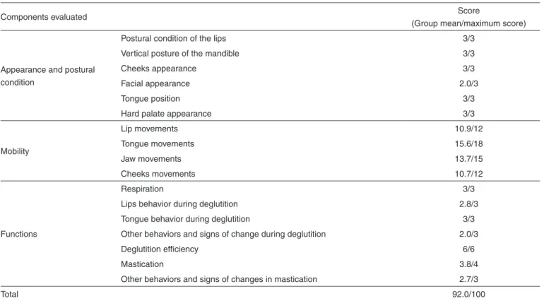
Table 1. Characterization of participants according to the OMES protocol
Components evaluated Score
(Group mean/maximum score)
Appearance and postural condition
Postural condition of the lips 3/3
Vertical posture of the mandible 3/3
Cheeks appearance 3/3
Facial appearance 2.0/3
Tongue position 3/3
Hard palate appearance 3/3
Mobility
Lip movements 10.9/12
Tongue movements 15.6/18
Jaw movements 13.7/15
Cheeks movements 10.7/12
Functions
Respiration 3/3
Lips behavior during deglutition 2.8/3
Tongue behavior during deglutition 3/3
Other behaviors and signs of change during deglutition 2.0/3
Deglutition efficiency 6/6
Mastication 3.8/4
Other behaviors and signs of changes in mastication 2.7/3
Subsequently, the participants were asked to remain at rest for 15 seconds, without recording. After this command, a 10 mm cotton roll was placed bilaterally between the first and second molars, and the participants were asked to apply the maximum bite force possible on the cotton roll for five seconds, three consecutive times, with a five second interval between trials. The same procedure was performed to obtain the electrical activity of the masseter and temporal muscles at maximal intercuspal position (maximal voluntary dental clenching without cotton).
Ultrasound
All ultrasounds were performed by the institution’s Radiology Service and by the same experienced radiologist. The evaluation of the thickness of the masseter muscle was performed using the methodology proposed by Satiroglu et al.(17). During image acquisition, the transducer was positioned
perpendicular to the skin surface, avoiding excessive pressure. Measurements were obtained from the bulkier portion of the masseter muscle, near the occlusal plane, approximately at the center of the mediolateral region of the branch distance.
The imaging and measurements were performed bilaterally, with the subjects in a supine position under three different conditions, as described above for the collection of sEMG: at rest (normal position); at maximal intercuspal position with 10 mm cotton rolls between the dental arches in the region of first and second molars (Al); and at the maximal intercuspal position without the cotton rolls (MIC).
The measurements were performed in real-time during imaging and were recorded in centimeters (cm). The imaging and measurements were performed three times with an interval of five seconds between each measurement (Figures 1 and 2).
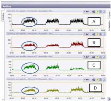
Analysis of surface electromyography
Temporal domain analysis was performed for the sEMG results. In this case, the information obtained describes the moment that the event occurred and the amplitude (i.e., an indicator of the magnitude of muscle activity) of the event. Under the resting condition, the obtained values represent the mean (RMS) of the electromyographic activity observed over 30 seconds. The duration of the muscle activity during voluntary
dental clenching tasks (Al and MIC) was obtained by selec-tion of the representative period of muscle activaselec-tion (i.e., on, peak and off). This period was selected with the cursor of the electromyography software and converted into µV (Figure 3).
Reliability
Based on the relevant literature, which suggests subjectivity in the sEMG measurement, a reliability analysis was performed to determine the index of agreement between the examiners and thus to ensure greater reliability of the measurements. Therefore, 20 electromyographic samples were randomly selected from a total of 198. These samples were analyzed independently by two researchers with experience in the field. The correlation coefficient was found to be high for all com-parisons (95%CI=0.9677-0.9956), indicating high consistency between examiners.
Figure 1. Ultrasound image during rest
Figure 2. Ultrasound image of the MIC
Note: A = electrical activity of the left temporal muscle; B = electrical activity of the right temporal muscle; C = electrical activity of the left masseter muscle; D = electrical activity of the right masseter muscle; MIC = voluntary dental clench-ing without cotton
Data analysis
The Kolmogorov-Smirnov test was initially used to test the normality of the distribution. The parametric tests were selected based on the adopted significance level (p<0.05). Paired t-tests were used for multiple comparisons between and within testing conditions, whereas Spearman’s correlation was used to assess the correlation between tests. The level of significance adopted for both tests was p<0.05.
RESULTS
A descriptive analysis including mean, standard deviation and confidence intervals was obtained from the ultrasound and surface electromyography data (Table 2).
The statistical analysis of the differences between the activation of the masseter and temporal muscles during sur-face electromyography revealed a difference in the maximal voluntary dental clenching without (MIC) and with (Al) cotton roll (Table 3). It was observed that the temporal muscle was
more active in both clenching conditions, as shown by the means in Table 2.
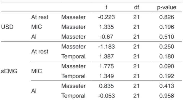
Statistical analysis was also performed to test for asymme-tries in muscle function between hemifaces as measured by ultrasound and surface electromyography. The data suggest no differences between the hemifaces for any testing condition in any exam performed (Table 4).
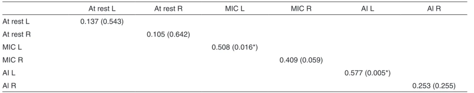
For the correlation between exams (Table 5), only the data from the masseter muscle were used because the ultrasound exam assessed only this muscle. A significant positive correla-tion between the exams was observed for the left maximal vo-luntary dental clenching without (p=0.016) and with (p=0.005) cotton rolls. There was also a trend toward significance for the right maximal voluntary dental clenching without cotton (p=0.059). The data showed high standard deviation values, and a possible increase in the sample size could result in a greater number of significant correlations (Table 1).
Table 2. Descriptive analysis of the data
Muscle Side Mean SD CI
At rest
USD Masseter Right 0.93 0.19 0.74 1.12
Left 0.93 0.19 0.73 1.12
sEMG
Masseter Right 4.09 0.90 3.19 4.99
Left 4.87 2.85 2.02 7.72
Temporal Right 5.75 4.32 1.43 10.07
Left 4.95 1.99 2.95 6.94
MIC
USD Masseter Right 1.19 0.20 0.99 1.39
Left 1.22 0.17 1.05 1.39
sEMG
Masseter Right 47.99 22.06 25.93 70.05
Left 54.83 28.99 25.84 83.83
Temporal Right 89.93 39.38 50.55 129.31
Left 102.17 56.13 46.03 158.30
Al
USD Masseter Right 1.26 0.17 1.09 1.43
Left 1.24 0.18 1.07 1.42
EMGs
Masseter Right 52.29 26.96 25.33 79.24
Left 52.06 21.00 31.05 73.06
Temporal Right 83.82 44.67 39.15 128.49
Left 89.40 63.69 25.72 153.09
Note: USS = ultrasound (measured in cm); sEMG = surface electromyography (measured in µV); MIC = voluntary dental clenching without cotton; Al = voluntary dental clenching with cotton; CI = confidence interval; SD = standard deviation
Table 3. Comparison between the masseter and temporal muscles in the surface electromyography
Temporal vs.
Masseter T df p-value
At rest Left 0.179 21 0.86
Right 1.949 21 0.065
MIC Left 4.294 21 <0.001* Right 6.817 21 <0.001*
Al Left 3.579 21 0.002*
Right 4.138 21 <0.001*
* Significant values (p<0.05) - Paired t-test
Note: MIC = voluntary dental clenching without cotton; AI = voluntary dental clenching with cotton
Table 4. Comparative analysis between hemifaces
t df p-value
USD
At rest Masseter -0.223 21 0.826 MIC Masseter 1.335 21 0.196 Al Masseter -0.67 21 0.510
sEMG
At rest Masseter -1.183 21 0.250 Temporal 1.387 21 0.180
MIC Masseter 1.775 21 0.090 Temporal 1.349 21 0.192
Al Masseter 0.835 21 0.413 Temporal -0.053 21 0.958
Paired t-test (p<0.05)
Thus, the results obtained in normal, young adults of both genders, for sEMG of the mastication muscles were (in µV): - At rest: right masseter - 3.19-4.99; left masseter - 2.02-7.72;
right temporal - 1.43-10.07; left temporal - 2.95-6.94; - Maximal voluntary dental clenching without cotton: right
masseter - 25.93-70.05; left masseter - 25.84-83.83; right temporal - 50.55-129.31; left temporal - 46.03-158.30; - Maximal voluntary dental clenching with cotton: right
masseter - 25.33-79.24; left masseter - 31.05-73.06; right temporal - 39.15-128.49; left temporal - 25.72-153.09. For the USD of the masseter muscle, the results ranged as
follows (in cm):
- At rest: right masseter – 0.74-1.12; left masseter – 0.73-1.12; - Maximal voluntary dental clenching without cotton: right
masseter – 0.99-1.39; left masseter – 1.05-1.39;
- Maximal voluntary dental clenching with cotton: right masseter – 1.09-1.43; left masseter – 1.07-1.42.
DISCUSSION
The present study involves sEMG and USD data collected in a group of healthy subjects who were selected based on rigid orofacial motricity characterization criteria. In a previous pilot study of the relationship between the electrical activation (sEMG) and the thickness (USD) of the masticatory muscles(24),
no correlation was observed between the sEMG and the USD, suggesting that the exams are complementary and not mutually exclusive. In the present study, increasing the sample size has allowed the observation of some positive correlations between the exams in the voluntary dental clenching activities, i.e., increased electrical activity with a corresponding increase in muscle thickness.
The physiology of the masticatory muscles suggests that the temporal, medial pterygoid and masseter muscles are activated during mandibular elevation(25). A previous study(26)
using sEMG found that among the masticatory muscles, only the masseter has a regular activity pattern, producing maximum contraction when the mouth is fully open during the masticatory cycle and then ceasing its activity when the mouth is almost closed. Another study(27) suggests that during sEMG, an
elec-tromyographic coordination exists between the masseter and the anterior temporal muscles bilaterally. Thus, there is a similar relationship between the mastication on the right and left side in terms of mastication forces and co-operation between muscles,
with higher muscle activity, especially that of the masseter, on the working side than on the balancing side.
The results of this study indicate that the temporal muscle was more active than the masseter in the voluntary dental clenching tasks, with and without cotton. According to Throckmorton and Dean(28), the determining factors for
mus-cle strength are the size and length of the musmus-cle fibers. For those authors, small/smaller muscles tend to be more active than large/bigger muscles because larger muscles are able to generate higher tension.
These results do not support the findings in the literature that during voluntary dental clenching with little force, the myoelectric activity of the temporal muscle does not exceed the activation of the masseter muscle. As the force increases, there is a corresponding increase in the activation of the masseter muscle. The temporal muscle activity tends to be higher when there is no posterior dental contact, and this muscle must control the mandibular excursions, thus maintaining dental occlusion. The participants in the present study were selected based on standard criteria for phonaudiological normality, without testing for dental occlusal contact. It is suggested that the authors of future studies should consider, in the inclusion criteria, specific odontological evaluation of occlusal contacts with load cells to measure the distribution of bite force across the teeth.
The USD results suggest increased thickness of the mas-seter muscle during voluntary dental clenching activities. In agreement with the literature, there was an increase of appro-ximately 30% in the thickness of the clenched masseter muscle compared to the masseter at rest. Prior work indicates that the masseter muscle at rest, measured in its medial portion, should be approximately 10 mm and should increase by 10 to 50% in thickness during maximal voluntary dental clenching(29).
One of the important contributions of this study was the repeated measure of the thickness of the masseter muscle that ensured a lower variation in the error and higher reliability of the measure of the actual variation in the muscle thickness. To control the measurement error variable, the position of the transducer was standardized(1-3,29). The variables related to
body posture, interocclusal relationship and rest time between voluntary dental clenching tasks were also controlled.
CONCLUSION
The results of the present study suggest a correlation
Table 5. Correlation values between the USD and sEMG exams
At rest L At rest R MIC L MIC R AI L Al R
At rest L 0.137 (0.543)
At rest R 0.105 (0.642)
MIC L 0.508 (0.016*)
MIC R 0.409 (0.059)
AI L 0.577 (0.005*)
Al R 0.253 (0.255)
* Significant values (p<0.05) – Spearman Correlation Coefficient
between sEMG and USD, i.e., that there is an increase in elec-trical activity and a corresponding increase in muscle thickness. The variation of the electrical activity and the thickness of the masseter muscle can be considered normal reference values for comparison with findings in future studies.
The combination of sEMG and USD in the investigation of muscle functionality provides important information on skeletal muscle physiology. It should be considered that during muscle activity there is variation in muscle physiology, with possible signs of fatigue. There are still no objective measures to assess the time required for muscle exhaustion or the numbers of repetitions that trigger fatigue.
REFERENCES
1. Hannam AG, Wood WW. Relationships between the size and spatial morphology of human masseter and medial pterygoid muscles, the craniofacial skeleton, and jaw biomechanics. Am J Phys Anthropol.1989;80(4):429-45 .
2. van Spronsen PH, Weijs WA, Prahl-Andersen B, van Ginkel FC. Relationship between jaw-muscle cross-section and craniofacial morphology in normal adults, studied with magnetic resonance imaging. Eur J Orthod. 1991;13(5):351-61.
3. Newton JP, Abel RW, Robertson EM, Yemm R. Changes in human masseter and medial pterygoid muscle with age: A study by computed tomography. Gerodontics.1987;3(4):151-4.
4. Felício CM, Couto GA, Ferreira CL, Mestriner Junior W. Reliability of masticatory efficiency with beads and correlation with the muscle activity. Pró-Fono. 2008;20(4):225-30.
5. Trawitzki LV, Dantas RO, Mello-Filho FV, Marques W Jr. Effect of treatment of dentofacial deformities on the electromyographic activity of masticatory muscles. Int J Oral Maxillofac Surg. 2006;35(2):170-3. 6. Benington PC, Gardener JE, Hunt NP. Masseter muscle volume measured
using ultrasonographic and its relationship with facial morphology. Eur J Ortho.1999;21(6):659-70.
7. Farina D, Merletti R, Enoka RM. The extraction of neural strategies from the surface EMG. J Appl Physiol. 2004;96(4):1486-95.
8. Sassi FC, Andrade CR. Eletromiografia de superfície e o tratamento da gagueira: uma perspectiva neuromotora. Rev Soc Bras Fonoaudiol. 2004;9(1):55-60.
9. Burkhead LM, Sapienza CM, Rosenbek JC. Strength-training exercises in dysphagia rehabilitation. Dysphagia. 2007;22(3):251-65
10. Felício CM, Sidequersky FV, Tartaglia GM, Sforza C. Electromyographic standardized indices in healthy Brazilian young adults and data reproducibility. J Oral Rehabil. 2009;36(8):577-83.
11. McCarroll RS, Naeije M, Hansson TL. Balance in masticatory muscle activity during natural chewing and submaximal clenching. J Oral Rehabil. 1989;16(5):441-6.
12. Kerstein RB. Combining technologies: a computerized occlusal analysis system synchronized with a computerized electromyography system. Cranio. 2004;22(2):96-109.
13. Moreno I, Sanchez T, Ardizone I, Aneiros F, Celemin A. Electromyographic comparisons between clenching, swallowing and chewing in muscle with varying occlusal parameters. Med Oral Patol Oral Cir Bucal. 2008;13(3):E207-13.
14. Nakata Y, Ueda HM, Kato M, Tabe H, Shikata-Wakisaka N, Matsumoto E, et al. Change in stomatognathic function induced by orthognathic surgery in patients with mandibular prognathism. J Oral Maxillofac Surg. 2007;65(3):444-51.
15. Sforza C, Peretta R, Grandi G, Ferronato G, Ferrario VF. Soft tissue facial planes and masticatory muscle function in skeletal Class III patients before and after orthognatic surgery treatment. J Oral Maxillofac Surg. 2008;66(4):691-8.
16. Kubo K, Kawata T, Ogawa T, Watanabe M, Sasaki K. Outer shape changes of human masseter with contraction by ultrasound morphometry. Arch Oral Biol. 2006;51(2):146-53.
17. Satiroglu F, Arun T, Isik F. Comparative data on facial morphology and muscle thickness using ultrasonography. Eur J Orthod. 2005;27(6):562-7. 18. Serra MD, Duarte Gavião MB, dos Santos Uchôa MN. The use of
ultrasound in the investigation of the muscle of mastication. Ultrasound Med Biol. 2008;34(12):1875-84.
19. Emshoff R, Bertram S, Brandlmaier I, Scheiderbauer G, Rudisch A, Bodner G. Ultrassonographic assessment of local cross-sectional dimension of masseter muscle sites: a reproducible technique? J Oral Rehabil. 2002;29(11):1059-62.
20. Moyers RE. Ortodontia.4a ed. Rio de Janeiro: Guanabara Koogan; 1991. 483p.
21. Felício CM, Ferreira CL. Protocol of orofacial myofunctional evaluation with scores. Int J Pediatr Otorhinolaryngol.2008;72(3):367-75. 22. Merletti R. Standards for reporting EMG data. J Electromyography
Kinesiol. 1999;9(1):III-IV.
23. Soderberg GL, Cook MT. Electromyography in biomechanics. Phys Ther. 1984;64(12):1813-20.
24. Mangilli LD, Sassi FC, Sernik RA, Tanaka C, Andrade CR. Avaliação eletromiográfica e ultrassonográfica do músculo masseter em indivíduos normais: estudo piloto. Pró-Fono. 2009;21(3):261-4.
25. Seikel JA, King DW, Darmright DG. Anatomy & physiology for speech, language and hearing. 4th ed. New York: Delmar Cengage Learning; 2010.
26. Palmer JB, Rudin NJ, Lara G, Crompton AW. Coordination of mastication and swallowing. Dysphagia. 1992;7(4):187-200.
27. Felício CM. Desenvolvimento normal das funções estomatognáticas. In: Fernandes FD, Mendes BC, Navas AL. Tratado de Fonoaudiologia. São Paulo: Ed. Roca; 2010.
28. Throckmorton GS, Dean JS. The relationship between jaw-muscle mechanical advantage and activity levels during isometric bites in humans. Arch Oral Biol. 1994;39(5):429-37.