ABSTRACT
Objective: To analyze tests used in routine clinical practice for the diagnosis of myocardial ischemia to predict the development of cardiac events in type 2 diabetic patients. Methods:The occurrence of cardiac events (new myocardial infarct, myocardial re-vascularization pro-cedures, congestive heart failure, acute pulmonary edema, sudden death, and death after myocardial infarction or pulmonary edema) were prospectively assessed in a cohort of 135 type 2 diabetic patients after up to seven years of follow-up. At baseline, coronary artery disease was assessed by the WHO cardiovascular questionnaire, resting electrocardiogram, and stress myocardial scintigraphy. Results: Forty-eight cardiac events were observed in 41 patients (10.5 events/100 patients-year). In a Cox’s proportional-hazard model only the presence of symptoms of coronary artery disease on the WHO cardiovascular questionnaire alone (RR= 2.13, 95% CI 1.11–4.07, P= 0.022) or in com-bination with abnormalities on resting ECG (RR= 2.03, 95% CI 1.05–3.92, P= 0.034) or on myocardial scintigraphy (RR= 1.89, 95% CI 1.001–3.57, P= 0.050) predicted cardiac events, adjusted for fasting plasma glucose, mean blood pressure, body mass index, peripheral vascular disease and diabetic nephropathy. Conclusion: The WHO cardiovascular ques-tionnaire, a simple tool for the diagnosis of coronary artery disease, is a significant predictor of cardiac events in type 2 diabetic patients. (Arq Bras Endocrinol Metab 2006;50/1:46-52)
Keywords:Coronary artery disease; Cardiac events; Risk factors; Type 2 diabetes
RESUMO
Valor dos Procedimentos Diagnósticos para Isquemia Miocárdica Usados na Prática Clínica de Rotina para Predição de Eventos Cardíacos em Pacientes com Diabetes Mellitus Tipo 2: Um Estudo Prospectivo. Objetivo: Analisar os testes usados na prática clínica de rotina para diagnóstico de isquemia miocárdica na predição do desenvolvimento de eventos cardíacos em pacientes com diabetes mellitus tipo 2 (DM2).
Métodos:A ocorrência de eventos cardíacos (novo infarto do miocárdio [IM], procedimentos de re-vascularização miocárdica, insuficiência cardíaca congestiva, edema agudo de pulmão, morte súbita e morte após IM ou edema pulmonar) foi avaliada prospectivamente em uma coorte de 135 pacientes com DM2 após até 7 anos de acompanha-mento. Na condição basal, a doença arterial coronariana (DAC) foi avaliada pelo questionário cardiovascular da OMS, eletrocardiograma de repouso e cintilografia do miocárdio sob stress. Resultados:48 even-tos cardíacos foram observados em 41 pacientes (10,5 eveneven-tos/100 pacientes-ano). No modelo de risco proporcional de Cox apenas a presença de sintomas de DAC no questionário cardiovascular da OMS isoladamente (RR= 2,13, 95% CI 1,11–4,07, P= 0,022) ou em combinação com anormalidades no ECG de repouso (RR= 2,03, 95% CI 1,05–3,92, P= 0,034) ou cintilografia do miocárdio (RR= 1,89, 95% CI 1,001–3,57, P= 0,050) predisseram eventos cardíacos, ajustados para a glicemia de
artigo original
to Predict Cardiac Events in Patients With
Type 2 Diabetes Mellitus: A Prospective Study
Mirela J. de Azevedo
André F.R. Neto
Maria Luiza A. Caramori
Maristela O. Beck
Juliano S.R. Moreira
Roberto Ludwig
Jorge L. Gross
Endocrine Division (MJA, AFRN, MLAC, MOB, JSRM, JLG) and Nuclear Medicine Division (RL), Hospital de Clínicas de Porto Alegre, Federal University of Rio Grande do Sul, Porto Alegre, RS, Brazil.
jejum, pressão arterial média, índice de massa cor-poral, doença vascular periférica e nefropatia dia-bética. Conclusão: O questionário cardiovascular da OMS, um procedimento simples para o diagnós-tico da DAC, é um preditor significativo de eventos cardíacos em pacientes com DM2. (Arq Bras Endocrinol Metab 2006;50/1:46-52)
Descritores: Doença arterial coronariana; Eventos cardíacos; Fatores de risco; Diabetes tipo 2
C
ORONARY ARTERY DISEASE (CAD) is the main cause of death in type 2 diabetic patients (1). The risk of macrovascular disease in patients with diabetes is greater for any given risk factor than in subjects without diabetes (2). Moreover, the risk of myocardial infarct is similar in patients with type 2 diabetes without overt evidence of CAD and non-diabetic patients with a previous myocardial infarct (3).As in non-diabetic subjects, the diagnosis of CAD in patients with diabetes is usually based on the presence of symptoms of ischemic myocardial disease and abnormalities suggestive of ischemia or infarction on resting electrocardiogram (ECG) or non-invasive stress tests. However, diabetic patients often present peculiarities that may interfere with the performance of these diagnostic tests. About 50% of type 2 diabetic patients are unable to perform an exercise test (4), and consequently nuclear perfusion imaging or echocar-diography after pharmacologically induced stress is an essential diagnostic tool for these patients. In addition, silent myocardial ischemia is more frequent in diabetic patients (5), and it has been associated with the pre-sence of diabetic nephropathy and autonomic neuro-pathy (6). Other tests used to evaluate CAD are inva-sive (coronary angiography) or have limited availability (coronary artery calcium as detected by electron beam computed tomography). Therefore, one should have a higher degree of suspicion for CAD in patients with diabetes and the use of non-invasive tests might be appropriate for screening purposes in these patients.
Prospective studies that analyzed the role of diagnostic tools to predict cardiac events in diabetic patients (7-10) did not include commonly used information as symptoms compatible with myocardial ischemia and resting ECG. At this stage, the best non-invasive test for the evaluation of CAD in diabetic patients remains unclear. Therefore, the aim of this study was to analyze the value of non-invasive tests used for the diagnosis of CAD in the routine clinical practice to predict the development of cardiac events in patients with type 2 diabetes.
PATIENTS AND METHODS
The original cohort was formed during the year of 1996, when one of the authors (MOB) performed a systematic evaluation for the presence of myocardial ischemia in consecutive patients with type 2 diabetes attending a once a week outpatient clinic of the Hos-pital de Clínicas de Porto Alegre, a university hosHos-pital. Patients with unstable angina, myocardial infarction in the previous six months, cognitive impairment due to neurological disease, history of alcohol abuse, chronic obstructive pulmonary disease, and chronic use of aminophylline or dipyridamole were not eligible. In-formed consent was obtained from each patient and the protocol was approved by the Ethics Committee of the Hospital. The patients were followed at every six to 12 months, up to seven years (mean: 3.4 ± 1.4 years). The following were considered as cardiac events: new myocardial infarct (Minnesota codes 1-1 to 1-3 or 7-1 on resting ECG (11), fixed non-perfused area on myocardial scintigraphy, or episode of chest pain with compatible ECG and enzyme alterations), myocardial re-vascularization procedures, congestive heart failure (class IV of the New York Heart Association plus compatible chest X-ray), acute pulmonary edema (typical clinical manifestations and compatible chest X-ray), sudden death (death occurring within six hours from the onset of symptoms [witnessed cardiac arrest] or within 24 hours from the time the patient was last seen alive in his/her usual state [12]), and death immediately after myocardial infarction or pulmonary edema.
Missing patients were contacted and invited to attend the clinic and information about the occurrence of cardiovascular events was checked. In positive cases medical records were reviewed. Eighteen patients were lost to follow-up and therefore 135 patients where included in this study. Patients lost to follow-up did not differ from the study group regarding proportion of males, smoking habit, CAD and hypertension, body mass index (BMI), diabetes duration, and lipid profile. Systolic blood pressure was higher among them (171.0 ± 24.1 vs. 153.2 ± 25.8 mmHg; P= 0.006).
Patients evaluation
levels ≥ 140/90 mmHg, or use of anti-hypertensive drugs. Retinopathy was assessed by fundoscopy after mydriasis. The presence of cerebrovascular disease was established if a history of stroke and/or compatible findings (sequelae) were present. Peripheral vascular disease was defined in the presence of intermittent claudication, as assessed by the WHO questionnaire for cardiovascular disease (11), and/or absence of posterior tibial pulse upon clinical examination. The diagnosis of CAD was based on the presence of at least one of the followings: angina or possible infarct according to the WHO questionnaire for cardiovas-cular disease (10; Appendix 1); resting ECG abnor-malities (Minnesota Codes: Q and QS patterns [1-1 to 1-3]; S-T junction [J] and segment depression [4-1 to 4-4]; T-wave items [5-1 to 5-3], and complete left bundle branch block [7-1]) (11), and perfusion abnormalities on myocardial scintigraphy at rest (fixed) or after infusion of dipyridamole (variable). The WHO cardiovascular questionnaire, resting ECG and myocardial scintigraphy were applied to all patients at baseline and during follow-up.
At baseline, the presence of any of the following were considered as cardiovascular risk factors: LDL cholesterol > 100 mg/dl, total cholesterol > 135 mg/dl, HDL cholesterol < 40 mg/dl for men and < 50 for women, triglycerides > 150 mg/dl, smoking habit, blood pressure levels ≥ 130/80 mmHg, and diabetic nephropathy.
Methods
Urinary albumin excretion rate (UAER) was measured in 24-h timed sterile urine collections by immuno-turbidimetry (Microalb, Ames-Bayer, Tarrytown, NY). The presence of diabetic nephropathy, UAER > 20 µg/min or 24-h proteinuria > 500 mg, was confirmed by at least two measurements in an interval of three to six months (13). Serum levels of glucose, creatinine, triglycerides, and cholesterol were measured by routine methods. Glycated hemoglobin data were not analyzed because three different methods were used during the study period. LDL cholesterol was estimated by the Friedewald equation.
Myocardial scintigraphy was obtained after dipyridamole stress and at rest. 30 mCi of technetium-99m sestamibi were injected intravenously, 3 minutes after the intravenous infusion of dipyridamole (0.56 mg/kg body weight). At baseline, planar images (anterior, left anterior oblique, and left lateral views) were acquired 1 hour after administration of the tracer using a single detector gamma-camera (OHIO-Nuclear VP 450) with a low-energy, all-purpose
APPENDIX. WHO CARDIOVASCULAR QUESTIONNAIRE
Section A: Chest Pain on Effort
1. Have you had any pain or discomfort in your chest? Yes
No
If "No" proceed to Section C.
If "Yes" ask next question. (If during the remainder of section A an answer is recorded in a box marked *, proceed to section B) 2. Do you get it when you walk uphill or hurry?
Yes No
Never hurries or walks uphill
3. Do you get it when you walk at an ordinary pace on the level? Yes
No
4. What do you do if you get it while you are walking? Stop or slow down
Carry on
(Record "Stop or slow down" if subject carries on after taking nitroglycerine)
5. If you stand still, what happens to it? Relieved
Not relieved 6. How soon?
10 minutes or less More than 10 minutes 7. Will you show me where it was?
Sternum (upper or middle) Sternum (lower)
Left anterior chest Left arm Other
(Record all areas mentioned) 8. Do you feel it anywhere else?
Yes No
(If "Yes", record additional information as above)
Section B: Possible Infarction
9. Have you ever had a severe pain across the front of your chest lasting for half an hour or more?
Yes No
Diagnostic Criteria for Angina Pectoris and Possible Infarction
"Angina" is defined as being present in subjects who answer as follows: Question 1: "Yes"
Question 2 or 3: "Yes"
Question 4: "Stop or slow down" Question 5: "Relieved" Question 6: "10 minutes or less"
Question 7: (a) Sternum (upper or middle, or lower), or (b) left anterior chest and left arm.
(If interviewing instructions are correctly observed throughout, it is sufficient to check the answer to Question 7)
"Pain of possible infarction" is defined as being present in subjects who answer as follows:
Question 9: "Yes"
collimator. Myocardial scintigraphy at rest was performed with a similar dose of the tracer 1 day after dipyridamole scintigraphy. At the end of the study, myocardial scintigraphy was performed by single-photon emission computed tomography (SPECT), non-gated, using a single detector gamma camera (GE Starcam 2000) with a low-energy, high-resolution collimator. Images were analyzed according to segmentation in the short, long vertical and horizontal axis of the left ventricle. Segmental myocardial perfusion patterns were classified as: 1) normal pattern; 2) reversible or partially reversible segmental defects; or 3) fixed pattern. Planar and SPECT myocardial scintigraphies presented a concordance rate of 83%, as previously reported (6) in a similar sample of diabetic patients. Cardioactive medications were suspended 7 days before the scintigraphy.
Statistical analysis
Comparisons between patients with and without CAD at baseline were performed by Student’s t test, Mann-Whitney U test, or chi-square test, as appropriate. Incidence density was calculated by multiplying the total number of outcomes by 100 days and dividing this result by the total number of days at risk. Kaplan-Meier curves and log rank test were used to compare the probability of cardiac events according to the presence of CAD at baseline and according to the test used at baseline (WHO cardiovascular questionnaire, resting ECG or myocardial scintigraphy, alone or in combination) to diagnose CAD. The multivariate Cox’s proportional hazard model was used to estimate and compare the risk of cardiac events (dependent variable) in patients according baseline diagnostic tests for CAD, alone or in combination, adjusted for age and other cardiac risk factors. The independent variables in these models were selected based on their significance in univariate analyses or according to their biological relevance. P values < 0.05 were considered statistically significant.
RESULTS
During the follow-up period, 41 patients presented 48 cardiac events, with an incidence of 10.5-events/100 patients-year. The cardiac events were: 25 myocardial infarcts, four re-vascularization procedures, eight episodes of heart failure, five sudden deaths, three deaths after myocardial infarct, and three deaths after pulmonary edema. Another five patients died during the study due to stroke (one patient), prostate cancer
(one patient), sepses (two patients), and car accident (one patient).
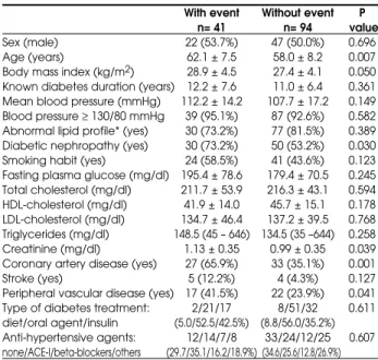
The baseline characteristics of patients with (n= 41) and without (n= 94) cardiac events at the end of the study are described in table 1. Patients with a car-diac event were older, and had higher BMI and serum creatinine levels than patients without cardiac events. They also had CAD, diabetic nephropathy, and peri-pheral vascular disease more often. The group with cardiac events did not differ from the group without cardiac events regarding the proportion of patients with LDL cholesterol > 100 mg/dl (75.7 vs. 81.3%; P= 0.486), total cholesterol > 135 mg/dl (92.7 vs. 98.9%; P= 0.164), HDL cholesterol < 40 mg/dl for men and < 50 for women (67.5 vs. 52.7%; P= 0.116) and triglycerides > 150 mg/dl (47.5 vs. 43.6%; P= 0.679).
CAD was present in 60 (44.4%) of all patients at baseline. The diagnosis was based on the WHO cardiovascular questionnaire alone in eight (5.9%), on resting ECG abnormalities in 13 (9.6%), and on myocardial perfusion scintigraphy in 10 (7.4%) patients. All three diagnostic tests were positive for CAD in nine (6.7%); the WHO questionnaire and the ECG in four (3.0%); the WHO questionnaire and the
Table 1.Baseline characteristics of type 2 diabetic patients with and without cardiovascular events at the end of the study.
With event Without event P n= 41 n= 94 value
Sex (male) 22 (53.7%) 47 (50.0%) 0.696
Age (years) 62.1 ± 7.5 58.0 ± 8.2 0.007 Body mass index (kg/m2) 28.9 ± 4.5 27.4 ± 4.1 0.050
Known diabetes duration (years) 12.2 ± 7.6 11.0 ± 6.4 0.361 Mean blood pressure (mmHg) 112.2 ± 14.2 107.7 ± 17.2 0.149 Blood pressure ≥130/80 mmHg 39 (95.1%) 87 (92.6%) 0.582 Abnormal lipid profile* (yes) 30 (73.2%) 77 (81.5%) 0.389 Diabetic nephropathy (yes) 30 (73.2%) 50 (53.2%) 0.030 Smoking habit (yes) 24 (58.5%) 41 (43.6%) 0.123 Fasting plasma glucose (mg/dl) 195.4 ± 78.6 179.4 ± 70.5 0.245 Total cholesterol (mg/dl) 211.7 ± 53.9 216.3 ± 43.1 0.594 HDL-cholesterol (mg/dl) 41.9 ± 14.0 45.7 ± 15.1 0.178 LDL-cholesterol (mg/dl) 134.7 ± 46.4 137.2 ± 39.5 0.768 Triglycerides (mg/dl) 148.5 (45 – 646) 134.5 (35 –644) 0.258 Creatinine (mg/dl) 1.13 ± 0.35 0.99 ± 0.35 0.039 Coronary artery disease (yes) 27 (65.9%) 33 (35.1%) 0.001
Stroke (yes) 5 (12.2%) 4 (4.3%) 0.127
Peripheral vascular disease (yes) 17 (41.5%) 22 (23.9%) 0.041 Type of diabetes treatment: 2/21/17 8/51/32 0.611 diet/oral agent/insulin (5.0/52.5/42.5%) (8.8/56.0/35.2%) Anti-hypertensive agents: 12/14/7/8 33/24/12/25 0.607 none/ACE-I/beta-blockers/others (29.7/35.1/16.2/18.9%) (34.6/25.6/12.8/26.9%)
scintigraphy in nine (6.7%); and the ECG and the scintigraphy in seven (5.2%) patients. Two or more of the evaluated cardiovascular risk factors were identified in 123 patients (91.1%).
Kaplan-Meier curves demonstrated higher rate of cardiac events in patients with CAD (27 [45.7%]) than in patients without CAD (14 [18.4%]; P= 0.035) at baseline. Presence of symptoms compatible with
CAD on the WHO questionnaire at baseline was signi-ficantly associated with the development of cardiac events (P= 0.016). The curves analyzing the predictive value of resting ECG (P= 0.038), WHO questionnaire and/or resting ECG (P= 0.014), and WHO ques-tionnaire and/or myocardial scintigraphy (P= 0.046) were also significant (figure 1). Abnormalities in the myocardial scintigraphy alone (P= 0.095), or in asso-ciation with resting ECG abnormalities (P= 0.064), were not predictive of cardiac events.
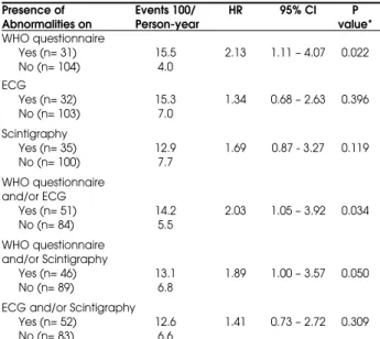
The results of the Cox regression models adjusted for age, fasting plasma glucose, mean arterial blood pressure, BMI, peripheral vascular disease and diabetic nephropathy at baseline are shown in table 2. The presence of CAD on the WHO questionnaire 1) alone (P= 0.022), 2) with or without resting ECG abnormalities (P= 0.034), and 3) with or without scintigraphy abnormalities (P= 0.05), was significantly associated with the development of cardiac events. Furthermore, when the protective role of the absence of symptoms on the WHO questionnaire and ECG abnormalities was analyzed, a relative risk (RR) of 0.45 (0.23 – 0.86; P= 0.016) for the development of cardiac events was observed. The association of a negative WHO cardiovascular questionnaire with a normal resting ECG had a negative predictive value for a new cardiac event of 82.1%.
Figure 1. Kaplan-Meier survival curves comparing the subset of diabetic patients with positive WHO cardiovascular questionnaire to patients with negative WHO cardiovascular questionnaire (Panel A), with positive WHO cardiovascular questionnaire and/or positive ECG to patients with negative WHO cardiovascular questionnaire and ECG (Panel B), and with positive WHO cardiovascular questionnaire and/or positive myocardial scintigraphy to patients with negative WHO cardiovascular questionnaire and myocardial scintigraphy (Panel C).
Table 2. Incidence of cardiac events during the follow-up period according to the diagnostic test used to define the presence of coronary artery disease at baseline.
Presence of Events 100/ HR 95% CI P Abnormalities on Person-year value*
WHO questionnaire
Yes (n= 31) 15.5 2.13 1.11 – 4.07 0.022
No (n= 104) 4.0
ECG
Yes (n= 32) 15.3 1.34 0.68 – 2.63 0.396
No (n= 103) 7.0
Scintigraphy
Yes (n= 35) 12.9 1.69 0.87 - 3.27 0.119
No (n= 100) 7.7
WHO questionnaire and/or ECG
Yes (n= 51) 14.2 2.03 1.05 – 3.92 0.034
No (n= 84) 5.5
WHO questionnaire and/or Scintigraphy
Yes (n= 46) 13.1 1.89 1.00 – 3.57 0.050
No (n= 89) 6.8
ECG and/or Scintigraphy
Yes (n= 52) 12.6 1.41 0.73 – 2.72 0.309
No (n= 83) 6.6
DISCUSSION
The presence of symptoms compatible with angina or possible infarct on the WHO cardiovascular questio-nnaire was the most important independent predictor of cardiac events in type 2 diabetic patients, either alone or in combination with ECG or myocardial scintigraphy abnormalities.
The WHO questionnaire for cardiovascular dis-ease was idealized by Rose (14) for the detection of ischemic heart pain in epidemiological studies. It is composed by nine questions that can be answered in one minute. This questionnaire has sensitivity of 83% and specificity of 91% for the diagnoses of angina, and sensitivity of 87% and specificity of 91% for myocardial infarction in non-diabetic subjects (14). However, the performance of this questionnaire may fluctuate across different ethnic groups (15), and it has not been vali-dated in diabetic patients. Moreover, the combination of symptoms of myocardial ischemia with ECG or scin-tigraphy abnormalities did not increase the risk for the development of a new cardiac event compared with the presence of symptoms of myocardial ischemia alone. Furthermore, when the WHO questionnaire is nega-tive and the ECG is normal, we observed a neganega-tive pre-dictive value for cardiac events of 82.1%. In this situa-tion, further additional tests are probably not necessary, reducing the economical costs of the cardiac evaluation. This study has possible limitations. Patients with angina equivalents, such as dyspnea, faintness, and fatigue, were not identified by the WHO cardio-vascular questionnaire. However the inclusion of these equivalents would have probably increased the dia-gnostic sensitivity of the WHO cardiovascular ques-tionnaire, although with a decrease in the specificity. Other limitation was the high cardiovascular risk profile of the participants included, since 97% had two or more cardiovascular risk factors, and almost 44% presented myocardial ischemia at baseline. This explains the higher proportion of cardiac events observed in the study period as compared to popu-lation based studies (16).
In this sample of diabetic patients, myocardial scintigraphy did not constitute a significant predictive factor for the occurrence of cardiac events. Myocardial scintigraphy has also been associated with a low pre-dictive value for significant CAD on angiography (17). However, other authors have described that scintigra-phy abnormalities could predict cardiac events in hig-hly selected diabetic patients (7,9,10,18). In the lar-gest of these studies, only patients who had symptoms suggestive of myocardial ischemia (9) or an abnormal
exercise treadmill test (10), and therefore an increased positive pre-test probability, underwent exercise dial scintigraphy. The low predictive value of myocar-dial scintigraphy for cardiovascular events in unselec-ted type 2 diabetic patients could be relaunselec-ted to the pre-sence of false positive results (19). These false positive results have been attributed to the presence of increa-sed ventricular mass (19) and microvascular disease (20). The WHO cardiovascular questionnaire, a simple tool used for the diagnosis of CAD, easily obtainable and without any cost, is a good predictor of cardiac events in type 2 diabetic patients. This finding is relevant to the clinical practice, since type 2 diabetic patients with a history of symptoms compatible with CAD, independently of abnormalities on resting ECG or in scintigraphy, should undergo a more complete cardiac evaluation. Although all patients with type 2 diabetes should be considered as potentially having cardiovascular disease (3), this aspect is not always appreciated, as demonstrated by the EUROASPIRE II survey (21). According to that study (21), a significant number of diabetic patients with known cardiovascular disease were still smoking, were not being treated with aspirin, and had not reached target blood pressure and cholesterol levels. Thus, the present study could increase the awareness of cardiovascular involvement in diabetic patients, motivating clinicians to adopt a more intensive multi-risk intervention strategy aiming to reduce mortality among these patients (22).
ACKNOWLEDGEMENTS
This study was partially supported by Projeto de Núcleos de Excelência do Ministério de Ciência e Tecnologia, Conselho Nacional de Desenvolvimento Científico e Tecnológico (CNPq) and Fundo de Incentivo à Pesquisa (FIPE) from Hospital de Clínicas de Porto Alegre.
REFERENCES
1. Panzram G. Mortality and survival in type 2 (non-insulin-dependent) diabetes mellitus. Diabetologia 1987;30:123-31. 2. Stamler J, Vacarro O, Neaton J, Wentworth D. Diabetes, other risk factors, and 12-year cardiovascular mortality for men screened for the Multiple Risk Factor Intervention Trial. Diabetes Care 1993;16:434-49. 3. Haffner SM, Lehto S, Ronnemaa T, Pyorälä K, Laakso M.
4. Janand-Delenne B, Savin B, Habib G, Bory M, Vague P, Lassman-Vague V. Silent myocardial ischemia in patients with diabetes: who to screen. Diabetes Care 1999;22:1396-404.
5. Nesto RW, Phillips RT, Kett KG, Hill T, Perper E, Young E, et al. Angina and exertional myocardial ischemia in diabetic and nondiabetic patients: assessment by exercise thallium scintigraphy. Ann Intern Med 1988; 108:170-5.
6. Beck MO, Silveiro SP, Friedman R, Clausell N, Gross JL. Asymptomatic coronary artery disease is associated with cardiac autonomic neuropathy and diabetic nephropathy in type 2 diabetic patients. Diabetes Care 1999;22:1745-7.
7. Vanzetto G, Halimi S, Hammoud T, Fagret D, Benhamou PY, Cordonnier D, et al. Prediction of cardiovascular events in clinically selected high-risk NIDDM patients. Prognostic value of exercise stress test and thallium-201 single-photon emission computed tomography. Diabetes Care 1999;22:19-26.
8. Marwick TH, Case C, Sawada S, Vasey C, Short L, Lauer M. Use of stress echocardiography to predict mortality in patients with diabetes and known or suspected coronary artery disease. Diabetes Care 2002;25:1042-8. 9. Giri S, Shaw LJ, Murthy DR, Travin MI, Miller DD,
Hachamovitch R, et al. Impact of diabetes on the risk stratification using stress single-photon emission compu-ted tomography myocardial perfusion imaging in patients with symptoms suggestive of coronary artery disease. Circulation 2002;105:32-40.
10. Faglia E, Favales F, Calia P, Paleari F, Segaline G, Gamba PL, et al. Cardiac events in 735 type 2 diabetic patients who underwent screening for unknown asymptomatic coronary heart disease: 5-year follow-up report from the Milan Study on Atherosclerosis and Diabetes (MiSAD). Diabetes Care 2002;25:2032-6. 11. Rose GA, Blackburn H, Gillum RF, Prineas RJ.
Cardio-vascular survey methods. WHO Monograph Series #56, 2nded. WHO, England, 1982, pp.123-65.
12. Burke AP, Farb A, Malcom GT, Liang YH, Smialek J, Virmani R. Coronary risk factors and plaque morphology in men with coronary disease who died suddenly. N Engl J Med 1997;336:1276-82.
13. American Diabetes Association: Nephropathy in Diabetes. Position Statement. Diabetes Care 2004; 27(suppl 1):S79-83.
14. Rose GA. The diagnosis of ischaemic heart pain and intermittent claudication in field studies. Bull World Health Organ 1962;27:645-58.
15. Fischbacher CM, Bhopal R, Unwin N, White M, Alberti KG. The performance of the Rose angina questionnaire in South Asian and European origin populations: A comparative study in Newcastle, UK. Int J Epidemiol 2001;30:1009-16.
16. Evans JMM, Wang J, Morris AD. Comparison of cardiovascular risk between patients with type 2 diabetes and those who had had a myocardial infarction: Cross sectional and cohort studies. Brit Med J 2002;324:939-42.
17. Koistinen MJ, Huikuri HV, Pirttiaho H, Linnaluoto MK, Takkunen JT. Evaluation of exercise electrocardio-graphy and thallium tomographic imaging in detecting asymptomatic coronary artery disease in diabetic patients. Br Heart J 1990;63:7-11.
18. Cohen MC, Curran PJ, L’Italien GJ, Mittleman MA, Zarich SW. Long-term prognostic value of preoperative dipyridamole thallium imaging and clinical indexes in patients with diabetes mellitus undergoing peripheral vascular surgery. Am J Cardiol 1999;83:1038-42.
19. Nesto RW. Screening for coronary artery disease in diabetes. Diabetes Care 1999;22:1393-5.
20. Nitenberg A, Ledoux S, Valensi P, Sachs R, Attali JR, Antony I. Impairment of coronary microvascular dilation in response to cold pressor-induced sympathetic stimulation in type 2 diabetic patients with abnormal stress thallium imaging. Diabetes 2001;50:1180-5. 21. EUROASPIRE I and II Group. Clinical reality of coronary
prevention guidelines: A comparison of EUROASPIRE I and II in nine countries. European Action on Secondary Prevention by Intervention to Reduce Events. Lancet 2001;357:995-1001.
22. Gaede P, Vedel P, Larsen N, Jensen GVH, Parving HH, Pedersen O. Multifactorial intervention and cardio-vascular disease in patients with type 2 diabetes. N Engl J Med 2003;348:383-93.
Endereço para correspondência:
Jorge L. Gross
Serviço de Endocrinologia
Hospital de Clínicas de Porto Alegre
Rua Ramiro Barcelos 2350, prédio 12, 4o. andar 90035-003 Porto Alegre, RS
Fax: (51) 3316-8777