Rev. bras. fisioter., São Carlos, v. 11, n. 2, p. 149-154, Mar./Apr. 2007 ©Revista Brasileira de Fisioterapia
ANALYSIS OF THE RELIABILITY OF A METHOD FOR MEASURING
HAMSTRING ACTIVE PEAK TORQUE ANGLE
A
QUINOCF
1, F
REIREMTF
2, N
EVESNM
2, F
ERREIRAPCA
2& F
ONSECAST
31 Divinópolis Educational Foundation, Divinópolis, MG - Brazil
2 Physical Education, Physical Therapy and Occupational Therapy School, Federal University of Minas Gerais - UFMG, Belo Horizonte, MG - Brazil
3 Physical Therapy Department, UFMG, Belo Horizonte, MG - Brazil
Correspondence to: Cecília Ferreira de Aquino, Av. Sete de Setembro, 949, Bairro Centro, CEP 35500-011, Divinópolis, MG – Brazil, e-mail: [email protected]
Received: 31/07/2006 - Revised: 01/12/2006 - Accepted: 26/01/2007
ABSTRACT
Introduction: Muscle length can be inferred from the length-tension relationship of the muscle. This relationship is traditionally investigated by measuring the peak torque produced by the muscle and the angle at which it is generated. Objective: The present study investigated the test-retest reliability of a method for measuring hamstring active peak torque angle in healthy young adults. Method: Twenty-five healthy individuals (22.88 ± 1.67 years) were assessed twice with an interval of three weeks. An isokinetic dynamometer was used in passive mode to assess hamstring passive torque. Muscle activity was monitored to ensure electromyographic silence. The dynamometer was also used in concentric mode to determine hamstring total torque. The active torque was obtained as the difference between total torque and passive torque. The active peak torque angle was used for the analysis. Results: There was no significant difference between the two measurements (t= 1.009; p= 0.323). The intraclass correlation coefficient for the active peak torque angle values obtained was 0.948 (p= 0.0001; 95% CI: 0.883 – 0.977). Conclusion: This study has shown that the method described is reliable for the quantification of active peak torque angle, thus suggesting that this method can be used to evaluate shifts in the torque-angle curve produced by muscle length changes.
Key words: muscle, length, reliability, torque.
INTRODUCTION
Skeletal muscles alter their capacity to produce torque throughout the range of joint movement1. Production of torque along the movement arch is determined by the variation of muscle levers (the perpendicular distance of the action vector of the muscle to the joint axis) as the joint moves and by the muscle–length relationship.1,2. The muscle-length relationship demonstrates the physiologic property of muscles to change the capacity to produce tension as it length varies3. Experimental procedures with animals involving surgery to remove connective tissue and electrical stimulation of the muscle with electrodes informs about the length-tension curve trough the analysis of peak tension produced by muscles4. However, such procedure cannot be performed in experiments with humans. Therefore, in research contexts where muscle levers does not vary, maximum muscle torque and the angle in which such torque is produced have been assessed as a means to make inferences about the length-tension curve5,6,7.
Alterations in muscle length can change the optimum point in which the muscle generates maximum tension5,9,13. The objective determination of the joint angle in which maximum muscle torque is produced would allow inferences about possible muscle length changes5,6. However, information obtained with this procedure in humans corresponds to the sum of passive and active forces of the muscle9,14. Therefore, the peak torque angle may not reflect muscle length in which active components of muscle generate maximum tension through superposition of actin and myosin filaments. Considering that total force produced by a muscle corresponds to the sum of active and passive forces, both being influenced by muscle length14, the determination of active muscle torque should be done subtracting the passive force from the total force produced1,9. Therefore, the objective of the present study was to verify the test-retest reliability of a method to determine the hamstrings angle of peak torque in healthy individuals.
MATERIALS AND METHODS
Subjects
Twenty-five healthy college students took part in this study. The convenience sample was comprised of three man and 22 women with a mean age of 22.88 ± 1.67 years, mean weight of 55.38 ± 9.88 kg and mean height of 1.65 ± 0.06 meters (Table 1). The only inclusion criterion was the absence of any history of injuries in the lower limbs. Although no volunteer was practicing physical activities regularly, there were no restrictions regarding activity level.
Volunteers signed up an informed consent form and agreed to undergo assessments of this research. Both the research project and the consent form were approved by the Ethics Committee of the University where the study was conducted (Document n° 074/04).
Instruments
An isokinetic dynamometer (Biodex Medical System Inc., Shirley, NY) was used to assess hamstrings passive torque and isokinetic performance.
In order to monitor electromyographic activity of lateral vastus (LV) and biceps femoris (BF) during registration of hamstrings passive torque, a MP150WSW electromyographer (Biopac System Inc., Goleta, CA) connected to a micro-computer was used. The equipment has an input impedance of two mega ohms (MΩ) and a common-mode rejection ratio of 1000MΩ. Two pairs of active surface electrodes with a diameter of 11.4 mm e inter-electrode distance of 20 mm (TSD150A, Biopac System Inc., Goleta, CA) were used. A reference electrode (LEAD110A, Biopac System Inc., Goleta, CA) was placed over a bony surface (acromion).
Procedures
Initially, research procedures were explained to volunteers and they signed a consent form agreeing to take part in the study. Assessments were performed only in the non-dominant leg. The dominant leg was considered to be the leg chosen by the participant to kick a ball15. Two assessments were made three weeks apart. All participants completed assessments as planned.
After determination of the lower limb to be assessed, the skin was shaved and cleaned with alcohol. Pairs of active electrodes were placed over the area of greatest muscle bulk of LV and BF of the non-dominant leg, according to direction of muscle fibers. A reference electrode was placed over the acromion.
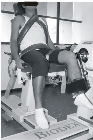
After electrode placement, participants were positioned in the dynamometer seat with 90 degrees of hip flexion. Knee joint axis was aligned with the axis of rotation of the dynamometer and the adaptation to support the knee was positioned right above the lateral maleoli (Figure 1). Movement amplitude was limited to 0 to 90 degrees of knee flexion for all volunteers in the initial and final assessments.
Figure 1. Positioning of volunteers during active peak torque evaluation
The dynamometer operated in the passive mode at a speed of 60°/s for the measure of hamstrings passive torque. Volunteers were requested not to resist movement of the dynamometer lever. Movement of the dynamometer lever begun at 90 degrees of knee flexion, proceeded until complete extension and then passively returned to flexion. During the passive return, the isokinetic dynamometer software registered the torque generated by leg weight, isokinetic lever weight and the force produced by energy conserved in the elastic elements of muscles around the joint. Such torque was defined as the passive torque of the flexion return movement.
During evaluation of passive torque, electromyographic data from LV and BF were registered simultaneously at a frequency of 1000 Hz to monitor of muscle activity and ensure that the test was performed passively. The two muscles were monitored because they are representative of activity of quadriceps and hamstrings. Only trials in which muscle activity did not exceed two standard deviations from the mean activity registered during rest were considered for analysis. A program was developed in Matlabâ to compare muscle activity during the test with the rest electomyographic signal at each 50 milliseconds interval. The program was executed right after each measure in order to guide the decision to accept or discard the test.
After measurement of passive torque, a hamstring isokinetic performance test (total isokinetic torque) was conducted with equal positioning and test amplitude. This evaluation was also performed at 60°/s, in the concentric mode, with seven repetitions of knee flexion and extension movements. Only data referring to concentric contraction of hamstrings during knee flexion were used for analysis. Volunteers performed three sub maximal practice trials before data collection. After the test, data provided by the software regarding coefficients of variation of knee flexors torque and angle of peak torque measures in each repetition were analyzed. Repetitions presenting the greater and smaller values for angle of peak torque were discarded, and thus only five values were left for analysis. When the test demonstrated torque values with a coefficient of variation above 10% and the five remaining angle values demonstrated variation greater than 10 degrees the measure was discarded. This procedure intended to guarantee greater consistency of the data. Participants that needed to repeat the measure had a rest time of 10 minutes before performing the new trial.
Data reduction
Torque and angle values obtained from passive torque and total isokinetic torque assessments at the speed of 60º/s and the frequency of 100 Hz were used to determine hamstrings angle of peak active torque. In the total isokinetic torque assessment, the dynamometer software provides torque data with or without correction of the effects of gravity on the lever and body segments masses. As the torques generated by the effect of gravity are present in the passive torque and total isokinetic torque measurements, it would be annulated
during data reduction by the subtraction of passive torque form total isokinetic torque. Therefore, data were collected without corrections for the effects of gravity. Data collected in the passive knee flexion measure and in the five most consistent repetitions of isokinetic hamstring assessment were transferred to a computer for analysis. A program developed in Matlab® was used to determine the angle of active peak torque of the hamstrings. Torque data were filtered with a fourth-order Butterworth filter and a 1.25Hz cut-off. Passive torque values for each joint angle were subtracted from the total torque curve obtained in the isokinetic assessment. The remaining torques represented the hamstring active torque. The Matlab® program identified the angle of peak active torque for each of the five repetitions of total torque assessment, and the mean of these values was used for analysis.
Statistical Analysis
Paired t tests were used to analyze differences between mean peak active torque values of hamstrings between the two assessments (test and retest). Test-retest reliability of the measures described in this study was determined through Intraclass Correlation Coefficients (ICC) and corresponding confidence intervals. (CI 95%). ICC values vary between 0.00 and 1.00, with values close to 1.00 indicating higher reliability. According to Portney and Watkins17, ICC values greater than 0.75 indicate adequate test-retest reliability. For clinical measures, reliability indices should exceed 0.9017. The level of significance of 0.05 was used for all analyses. Statistical tests were performed with the software Statistical Package for Social Sciences (SPSS® – Version 13.0).
RESULTS
The mean of peak active torques obtained in the test was 26.40 degrees (DP= 6.97) and 25.76 in retest (DP= 7.24). Table one presents the values of active peak torque of all volunteers for the first and second assessments. No significant differences were found between the two measurements performed three weeks apart (t= 1.009; p= 0.323). This result did not change when male volunteers were excluded (t= 1.170; p= 0.255). The Intraclass Correlation Coefficient (ICC), that was used to analyze test-retest reliablity of the hamstring active torque values obtained with the methods described in this study, demonstrated a correlation of 0.948 (p< 0.001; CI 95% 0.881 - 0.977). Once again results were not changed when male subjects were excluded from analysis. In this case the ICC was 0.952 (p< 0.001; IC 95% 0.884 - 0.980).
DISCUSSION
Volunteer Gender Age Body Mass Height Test-Angle Retest-Angle
01 Female 24 53.8 1.68 27.8 26.0
02 Female 21 47.4 1.63 34.8 27.8
03 Female 21 54.5 1.71 31.4 26.8
04 Female 24 45.0 1.65 27.2 23.2
05 Female 24 57.0 1.70 41.8 43.8
06 Female 20 46.4 1.60 17.6 15.4
07 Male 23 63.8 1.71 29.0 26.0
08 Male 20 57.2 1.74 20.4 21.8
09 Female 23 62.5 1.69 34.0 28.4
10 Female 24 53.6 1.64 38.5 40.3
11 Female 21 52.4 1.60 26.0 27.0
12 Female 23 76.0 1.63 36.0 37.2
13 Male 22 88.5 1.77 22.3 25.5
14 Female 22 45.0 1.58 31.8 28.8
15 Female 23 55.5 1.53 28.6 29.0
16 Female 25 52.2 1.62 20.4 14.8
17 Female 23 50.2 1.60 18.4 17.6
18 Female 22 55.3 1.66 21.0 20.4
19 Female 23 44.3 1.56 27.6 34.2
20 Female 27 61.2 1.72 21.6 22.2
21 Female 25 46.5 1.56 14.8 16.4
22 Female 23 51.2 1.60 22.4 22.2
23 Female 22 59.4 1.68 22.6 24.8
24 Female 25 52.5 1.62 20.0 21.2
25 Female 22 53.2 1.65 24.0 23.2
Table 1. Sample description (gender, age, body mass and height) and active peak torque angles for hamstrings in the firstevaluation (test) and second evaluation (retest).
provide objective information about changes of the hamstrings torque-angle curve caused by alterations in muscle length. Objective determination of muscle length is necessary in order to document the efficacy of therapeutic interventions aimed at modifying the structure of muscles5,18. Changes in the alignment of body segments and available range of movement of a joint are associated with alterations in muscle length8,19. Therefore, the good reliability indices found for
hamstring active peak torque values indicates that this method can be useful in studies evaluating the efficacy of treatment programs designed to change muscle length in order to correct posture or increase range of movement.
increased tolerance to stretch, and not to changes in muscle length18,21. With increased tolerance to stretch, the individual allows the application of greater forces on the joint during a flexibility assessment, and thus greater values are obtained for range of movement22. Additionally, results obtained in the flexibility assessment can vary according to the force applied during the examination. If the forces applied during examination are not controlled, factors as increased tolerance or a desire of the participant to demonstrate progress can interfere in the results of maximum range of movement.18,21. Therefore, alterations of this measure may not reflect a real change in the structure of muscle tissue.
As the analysis of the length-tension curve of a muscle does not have the inherent limitations of flexibility measures, it can indirectly inform about muscle length5,6,9. However, it is impossible to obtain direct measures of the length tension relationship in humans. Therefore, angle of peak torque measures obtained through analysis of the torque-angle curve were used in studies that investigated the effects of interventions such as muscle strengthening5,23 and stretching7,24 on muscle length. These studies used the measure of angle of peak torque to infer about muscle length, but reliability estimates were not reported, and the passive torque was not subtracted from total torque5,7. However, as passive and active torques are both influenced by muscle length14 and in these studies an intervention protocol was used with the objective of increasing muscle length7, it is possible that no modifications in total torque would be observed at the end of the study, although passive resistance and the length-tension relationship may have suffered changes. In case a decrease in passive torque and a shift of the length-tension curve (active component) in the direction of greater muscle length occur simultaneously, the angle of peak total active torque may not change because the two effects could cancel each other when total active torque is considered. Therefore, if the objective of a study is to evaluate the impact of an intervention on muscle length, the utilization of a method that does not take off passive torque to calculate the angle of peak torque can obscure study results.
One limitation of the present study refers to restrictions in the clinical applicability of the method to measure the angle of active peak torque, since it demands an isokinetic dynamometer and such equipment is not common in clinical settings. Although the methodology proposed in this study minimizes patient and examiner interferences in the determination of measures, the investigation of muscle length assessment procedures that are suitable not only to scientific research but also to the clinical routine is necessary. Another possible limitation is related to the characteristics of the sample, which had few male subjects. However, as demonstrated in statistical analysis, this factor did not have any interference in results.
Results of the present study indicate that hamstrings produce maximum active torque around 25 degrees of knee
flexion. In the determination of angle of peak torque, the torque-angle curve profile is influenced by mechanical factors, related to muscle lever arms, and physiological factors, related to the length-tension relationship13. For some muscles, such as elbow flexors, there is a predominance of mechanical factors (leverage) in the determination of the torque-angle curve profile. Conversely, the hamstrings torque-angle curve has a profile that reflects the contribution of the physiological component without predominance of mechanical factors25. In this case, it is not possible to determine if the high reliability indices obtained are specific for hamstrings or if the investigated methodology can be applied to assess other muscles. Therefore, the utilization of the angle of peak active torque assessment to analyze changes in muscle length of muscles in which the mechanical component predominates over physiological factors should be investigated in future studies.
CONCLUSION
Results demonstrate that the method described in this study is reliable in the determination of the angle of active peak torque of the hamstrings. This method can be used in studies with the objective of investigating changes in the torque-angle curve due to alterations in the length of this muscle group after different intervention protocols. However, it is not possible to conclude that the method proposed in this study is adequate to measure the angle of active peak torque of other muscles.
Supported by: CNPq (grant PIBIC/ CNPq) and CAPES (grant PROF/
CAPES).
REFERENCES
1. Mohamed O, Perry J, Hislop H. Relationship between wire EMG activity, muscle length, and torque of the hamstrings. Clin Biomech. 2002;17(8):569-79.
2. Rassier DE, Macintosh BR, Herzog W. Length dependence of active force production in skeletal muscle. J Appl Physiol. 1999;86(5):1445-57.
3. Gordon AM, Huxley AF, Julian FJ. The variation in isometric tension with sarcomere length in vertebrate muscle fibres. J Physiol. 1966;184(1):170-92.
4. Jones C, Allen T, Talbot J, Morgan DL, Proske U. Changes in the mechanical properties of human and amphibian muscle after eccentric exercise. Eur J Appl Physiol Occup Physiol. 1997;76(1):21-31.
5. Brockett CL, Morgan DL, Proske U. Human hamstring muscles adapt to eccentric exercise by changing optimum lengt. Med Sci Sports Exerc. 2001;33(5):783-90.
6. Lynn R, Talbot JA, Morgan DL. Differences in rat skeletal muscles after incline and decline running. J Appl Physiol. 1998; 85(1):98-104.
8. Gossman MR, Sarhmann SA, Rose SJ. Review of length-associated changes in muscle: Experimental evidence and clinical implication. Phys Ther. 1982;62(12):1799-2008.
9. Savelberg HHCM, Meijer K. Contribution of mono and biarticular muscles to extending knee joint moments in runners and cyclists. Journal of Applied Physiology. 2003;94:2241-8.
10. Tabary JC, Tabary C, Tardieu C, Tardieu G, Goldspink G. Physiological and structural changes in the cat’s soleus muscle due imobilization at different lengths by plaster casts. J Physiol. 1972;244:231-44.
11. Williams PE, Goldspink G. Changes in sarcomere length and physiological properties in immobilized muscle. J Anat. 1978;127(3):459-68.
12. Williams PE, Catanese T, Lucey EG, Goldspink G. The importance of stretch and contractile activity in the prevention of connective tissue accumulation in muscle. J Anat. 1988;158:109-14.
13. Koh TJ. Do adaptations in serial sarcomere number occur with strength training? Hum Mov Sci. 1995;14:61-77.
14. Gajdosik R. Passive extensibility of skeletal muscle: review of the literature with clinical implications. Clin Biomech. 2001; 16(2):87-101.
15. Witvrouw E, Danneels L, Asselman P, D’Have T, Cambier D. Muscle flexibility as a risk factor for developing muscle injuries in male professional soccer players. A prospective study. Am J Sports Med. 2003;31(1):41-6.
16. Lamontagne A, Malouin F, Richards CL. Contribution of passive stiffness to ankle plantarflexor moment during gait after stroke. Arch Phys Med Rehabil. 2000;81(3):351-8.
17. Portney LG, Watkins MP. Statistical measures of reliability. In: Portney LG, Watkins MP. Foundations of clinical research: application to practice. 2ª ed. New Jersey: Prentice Hall Health; 2000. p. 557-86.
18. Halbertsma JP, Goeken L. Stretching exercises: effect on passive extensibility and stiffness in short hamstrings of healthy subjects. Arch Phys Med Rehabil. 1994;75(9):976-81. 19. Sahrmann SA. Concepts and principles of movement. In:
Sahrmann SA, editor. Diagnosis and treatment of movement impairment syndromes. St. Louis: Mosby; 2002. p. 9-50. 20. Hartig DE, Henderson JM. Increasing hamstring flexibility
decreases lower extremity overuse injuries in military basic trainees. Am J Sports Med. 1999;27(2):173-6.
21. Magnusson SP, Simonsen EB, Aagaard P, Sorensen H, Kjaer M. A mechanism for altered flexibility in human skeletal muscle. J Physiol. 1996;497(Pt 1):291-8.
22. Halbertsma JP, Van Bolhuis AI, Goeken LN. Sport stretching: effect on passive muscle stiffness of short hamstrings. Arch Phys Med Rehabil. 1996;77(7):688-92.
23. Mchugh MP, Tetro DT. Changes in the relationship between joint angle and torque production associated with the repeated bout effect. J Sports Sci. 2003;21(11):927-32.
24. Cramer JT, Housh TJ, Weir JP, Johnson GO, Coburn JW, Beck TW. The acute effects of static stretching on peak torque, mean power output, electromyography, and mechanomyography. Eur J Appl Physiol. 2005;93(5-6):530-9.