Evaluation of the vertical alterations of the upper second
molars after the alignment and leveling phase
using the MBT technique
Alael Barreiro Fernandes de Paiva Lino1, Julio Wilson Vigorito2
Objective: This study aimed at comparing the distocervical angulations of upper second molars crowns of groups with normal occlusion and with Angle Class I and II malocclusions. Additionally, it aimed to analyze the changes oc-curring ater alignment and leveling performed with MBT technique.
Methods: The sample of normal occlusions comprised 32 lateral teleradiographs, while the sample of malocclusions comprised 38 initial and post-leveling lateral teleradiographs.
Results: The results did not show statistically signiicant diferences between morphological characteristics of the nor-mal occlusion group and the nor-malocclusion group. On the other hand, ater alignment and leveling, statistically signii-cant reduction was observed in distocervical inclinations with extrusion of the distal portion of upper second molars.
Keywords:Dental occlusion. Design of orthodontic appliances. Radiography. Corrective orthodontics.
How to cite this article: Lino ABFP, Vigorito JW. Evaluation of the vertical alterations of the upper second molars ater the alignment and leveling phase using the MBT technique. Dental Press J Orthod. 2013 Sept-Oct;18(5):115-20.
Submitted: December 11, 2009 - Revised and accepted: December 29, 2010
Contact address: Alael Barreiro Fernandes de Paiva Lino Av. Professor Lineu Prestes, 2.227, Cidade Universitária CEP: 05.508-000 – São Paulo – SP – Brazil
E-mail: [email protected]
1 Resident of Orthodontics, School of Dentistry - University of São Paulo (USP). 2 Full Professor of Orthodontics, School of Dentistry - University of São Paulo
(USP).
» The patient displayed in this article previously approved the use of her facial and intraoral photographs.
» The authors report no commercial, proprietary or inancial interest in the products or companies described in this article.
Objetivo: a proposta do presente trabalho foi comparar as angulações distocervicais das coroas dos segundos molares superiores de um grupo com oclusão normal e outro composto de más oclusões de Classes I e II de Angle, e analisar as modiicações ocorridas após o alinhamento e nivelamento com a técnica MBT.
Métodos: a amostra de oclusão normal foi composta por 32 telerradiograias laterais; e a amostra de má oclusão constituiu-se de 38 telerradiograias laterais iniciais e pós-nivelamento.
Resultados: os resultados não mostraram diferença estatisticamente signiicativa entre as características morfológicas dos grupos com oclusão normal e com má oclusão; porém, após o alinhamento e nivelamento, observamos redução estatistica-mente signiicativa nas inclinações distocervicais, com extrusão da porção distal dos segundos molares superiores.
INTRODUCTION
The University of São Paulo College of Dentistry postgraduate program ofers a course that provides clinical practice of research about MBT pre-adjust-ed appliances. It aims to qualify students to perform the Straight-Wire MBT technique on the treatment of malocclusions, with or without teeth extractions, based on the researches developed in the program and the material that has been documented. By means of observing the clinical results and the elements of diag-nosis obtained ater orthodontic alignment and leveling of treated groups, it was noticed, in some cases, prema-ture contacts and occlusal interferences on the region of second molars. This fact encouraged the study of the characteristics of this area and any potential altera-tions occurring ater dental alignment and leveling.
MBT appliances were assembled according to what the technique suggests,11 regarding height and angulation of the orthodontic accessories. These vari-ables are described in the orthodontic literature with the purpose of maintaining and preparing both func-tional characteristics and anchorage, in order to ob-tain normal occlusion free of occlusal interferences. Teeth angulations are obtained by means of changing the brackets position or inserting second-order bends on stainless steel arches12 on the upper second molars. These variations aim to control the anchorage, matain the initial tooth position and prevent occlusal in-terferences on mandibular movement.6,10,12,14,16
In order to prepare the anchorage, a previous study12 determine that the upper second molar move-ment be performed with cervicodistal angulation of 20°, with the orthodontic arch in 10° of angulation on this tooth’s region. Another possibility to perform angulation on the upper second molar is inserting the accessory with angulation of 15 degrees14 or bond-ing the tube on the same plane after basic levelbond-ing of first molars to upper canines.10 On the other hand, some authors recommend inclusion on the mechan-ics of second molars in cases of extraction of four first premolars, third molars or in cases of buccolingual crossing. In normal clinical situations, it is necessary to await the natural establishment of the occlusion.18 In 1972, after studying 120 models of individuals with characteristics of normal occlusion, Andrews1 presented an article suggesting six keys of normal occlusion that should be obtained after orthodontic
therapy. These occlusal characteristics served as a ba-sis for introducing the Straight-Wire appliance. This appliance has on its design, brackets slots with pre-angulation following the mesiodistal pre-angulation of the vestibular surface. In relation to the upper second molar, Andrews’ findings showed, on average, a me-sial angulation of the clinical crown in 0.39 degrees.4 The literature about preadjusted appliances proposes several values such as 5 degrees,11 3 degrees,7 4.09 de-grees,17 0 degrees,15 -0.30 degrees17 and -5 degrees.5 In addition to angulation, the height in which the orthodontic accessories are positioned on the teeth is a recurrent variable in orthodontic clinics.
Vigorito16 mentions in his book that the occlusal-cervical height of the bracket on the upper second molar must be 0.5 mm lower than that on the upper first molar, with 0° of angulation and insertion of cau-dal angle on the orthodontic arch on the upper second molar region. As for the MBT technique,11 it is rec-ommended to use the accessories installation table in which it is stated that the mean height for the upper second molar is 2.0 mm and 3.0 mm for the first mo-lars. As verified in the studies previously mentioned, there are different prescriptions concerning the an-gulation to be expressed on the upper second molar, varying from the MBT prescription, that indicates 5° of mesial angulation, to the researches indicating that the upper second molars present and must keep, dur-ing and after the orthodontic therapy, a cervicodis-tal angulation.5,6,9,10,12,14,16 The present study aimed to compare the characteristics of mesiodistal angulation and vertical positioning9 of the upper second molars between the normal occlusion group, which had the control function, and the malocclusion group. In ad-dition, it aimed at verifying the alterations occurring after alignment and leveling performed with MBT preadjusted appliances. Based on the aforementioned objectives, this study proposed:
1. To verify potential differences in vertical and angular positions of the upper second molars, observed in lateral teleradiographs, by means of comparing Class I and II malocclusion to nor-mal occlusion.
MATERIAL AND METHODS
Sample
The control group consisted of a normal occlu-sion sample comprising 32 lateral teleradiographs obtained from Brazilian, leukoderma individuals, 16 of which were males and 16 females, aged between 12 and 17 years and 1 month old, with permanent dentition classified as normal dental occlusion, and with no previous orthodontic treatment. The maloc-clusion sample comprised 76 lateral teleradiographs obtained from 38 Brazilian, leukoderma patients of both genders, with permanent teeth and Angle Class I and Class II malocclusions, under treatment at, Cor-rective Orthodontic MBT preadjusted Clinic, at the University of São Paulo (USP) College of Dentistry. The radiographs were divided into two groups: T1r which corresponds to initial teleradiographs and T2r which corresponds to the final phases of alignment and leveling.
Appliance assemblage
MBT appliances were assembled according to what the technique suggests,11 regarding height and angulation of the orthodontic accessories in the re-gion of second molars.
Image digitizing
Radiograph digitizing
The radiographs were scanned and the images were imported by the Radiocef Studio 4.0 software,
kindly granted by Radio Memory Ltda (www.ra-diomemory.com.br). On this software, cephalomet-ric landmarks were marked and the magnitudes, nec-essary for this research, were defined.
Landmarks determination
Landmarks were directly marked on the images of the structures of interest. The landmarks used were:
Cephalometric landmarks (Fig 1)
» Landmark D7: Most distal landmark of the up-per second molar image.
» Landmark M7: Most mesial landmark of the up-per second molar image.
» Landmark PTMs: Most upper point of the ptery-gomaxillary fossa.
» Landmark PTM: Most lower point of the ptery-gomaxillary fossa which corresponds to the pterygo-maxillary fissure.
Determination of the lines of orientation.
Radiographic lines and planes (Fig 2)
» Pterygomaxillary fossa long axis (vertical PTM): line that joins landmarks PTMs to PTM.
» Perpendicular to the long axis of the pterygo-maxillary fossa (horizontal PTM): Line traced from the landmark PTM perpendicular to the pterygo-maxillary fossa long axis.
» D7-horizontal PTM: Orthogonal to horizontal line PTM up to landmark D7.
Figure 1 - Cephalometric landmarks demarcated on the lateral teleradio-graph. 1) PTMs, 2) PTM, 3) M7, 4) D7.
Figure 2 - Lines demarcated on the lateral teleradiograph. 1) Vertical line PTM. 2) Horizontal line PTM. 3) M7-Horizontal line PTM. 4) D7-Horizontal line PTM. 5) md7 line.
1
2
4 3
1
2 4
5
measurement process were submitted to t test for
sys-tematic error verification, while the Dahlberg meth-od was used for random error verification.
RESULTS
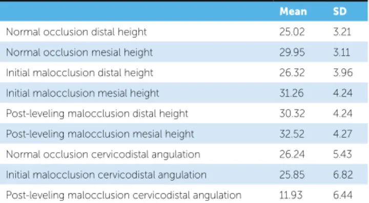
The descriptive statistics obtained from the telera-diographs are presented in Table 1.
Comparisons between the means of the lin-ear magnitudes observed in the teleradiographs are shown in Table 2.
Comparisons between the means of the angular magnitudes observed in the teleradiographs are pre-sented in Table 3.
DISCUSSION
» M7-horizontal PTM: Orthogonal to horizontal line PTM between landmark M7.
» Mesio-distal line of the upper second molar (line md7): Line that joins landmarks M7 to D7.
2.5 Determining the magnitudes.
Radiographic magnitudes (Fig 3)
» D7-horizontal PTM Height (distal height): Or-thogonal distance between landmark D7 with hori-zontal line PTM.
» M7-horizontal PTM Height (mesial height): Orthogonal distance between landmark M7 with horizontal line PTM.
» md7 - horizontal PTM Angle line (cervicodis-tal angle): Formed by intersection of horizon(cervicodis-tal line PTM with line md7.
Statistical methods
Descriptive statistics were obtained by means of verifying both mean and standard deviation of all magnitudes studied. Normal distribution of data was assessed through Shapiro-Wilks test. The means of the magnitudes obtained from the teleradiographs and after alignment and leveling were compared through one-way ANOVA test complemented by Tukey test at 5%. To verify the method error, the samples were reevaluated and the values obtained from the second
Figure 3 - Linear and angular magnitudes determined no the lateral telera-diograph. 1) mesial height. 2) distal height. 3) cervicodistal angle.
Mean SD
Normal occlusion distal height 25.02 3.21
Normal occlusion mesial height 29.95 3.11
Initial malocclusion distal height 26.32 3.96
Initial malocclusion mesial height 31.26 4.24
Post-leveling malocclusion distal height 30.32 4.24
Post-leveling malocclusion mesial height 32.52 4.27
Normal occlusion cervicodistal angulation 26.24 5.43
Initial malocclusion cervicodistal angulation 25.85 6.82
Post-leveling malocclusion cervicodistal angulation 11.93 6.44
Table 1 - Descriptive statistics of cephalometric magnitudes values.
Table 2 - Comparison between the means of linear magnitudes ob-served in the teleradiograph by one-way ANOVA test complemented by Tukey at 5%.
Table 3 - Comparison between the means of angular magnitudes ob-served in the teleradiograph by one-way ANOVA test complemented by Tukey at 5%.
A) Normal occlusion distal height. B) Normal occlusion mesial height. C) Ini-tial malocclusion distal height. D) Initial malocclusion mesial height. E)
Post-leveling malocclusion distal height. F) Post-leveling malocclusion mesial
height *: non-signiicant diference. **: signiicant diference.
G) Normal occlusion cervicodistal angle. H) Malocclusion initial time cervico-distal angle. I) Post-leveling malocclusion cervicodistal angle. *) non-signii-cant diference. **) signiinon-signii-cant diference.
A B C D E F
A - ** * ** ** **
B * - ** * * *
C * * - ** ** **
D * * * - * *
E * * * * - **
G H I
G - * **
H * - **
We also found authors indicating negative inclina-tion6,8,9,10,12,14 to preserve and prepare the anchorage during orthodontic mechanics as well as keep the functional position of the upper second molar aim-ing to avoid premature contacts and interference in mandibular eccentric movements.
Thus, we can conclude that angular alterations performed on the upper second molars for prepar-ing and maintainprepar-ing the anchorage or for levelprepar-ing and alignment, must be corrected in order to restore the characteristics of this region.
Additionally, the results obtained from this research demonstrate that the use of pre-adjusted accessories with MBT prescription signiicantly reduces mesio-distal angulation6,10 and changes the vertical position-ing of the teeth observed in both normal occlusion and malocclusion groups. Aiming to minimize any possi-ble errors while installing the accessories, this research carefully followed, by means of the suggested bonding table, the recommendations of the MTB technique.11
Clinically, the obtained results allow us to suggest that the accessory positioning should be performed on the second molar with disto-occlusal angle and with a height lower than that performed on the irst molar. This way, extrusion would be controlled and angula-tion would be preserved. Furthermore, we observed the need of installing and modifying the accessories in order to determine disto-occlusal angulation around 5°. With such a prescription, the characteristics of both Systematic error evaluation demonstrated
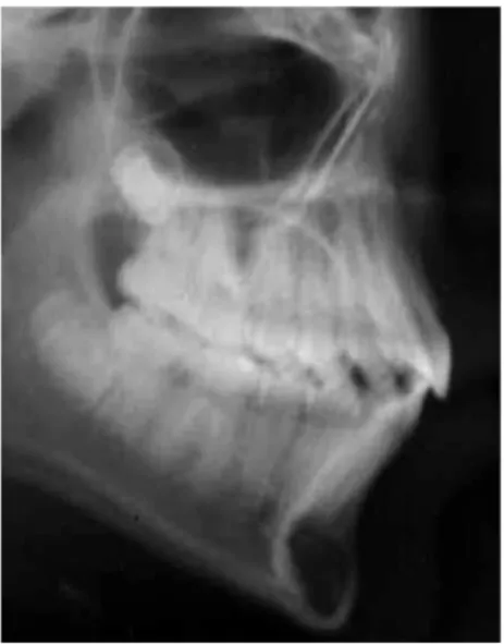
repro-ducibility (p < 0.01), while random error variability ranged from 0.62 for mesial height to 0.70 for distal height of the normal occlusion group. The results of the method error validated the results obtained from the analysis of cephalometric magnitudes, thus, sup-porting discussion about the clinical implications of installing orthodontic accessories on the upper sec-ond molars. Distocervical angulation of the upper second molars is a characteristic found in individu-als with normal occlusion (Fig 4).6,9 According to the results of this research, it was possible to observe that the group with malocclusion showed the same char-acteristics regarding the vertical positioning of upper second molars in relation to the control group (Fig 5). Ater alignment and leveling, the characteristics ob-served in the second molars were signiicantly altered (Fig 6). The cervicodistal angle of the upper second molar was signiicantly reduced from 25.85° to 11.93° due to extrusion of the distal surface and maintenance of the vertical position of the mesial surface. Signii-cant alteration was observed in the vertical positioning of the upper second molar distal surface. At the ini-tial phase, it was 26.32 mm, while ater leveling it was 30.32 mm On the other hand, the vertical position of the mesial face did not show signiicant diferences af-ter alignment. Relevant liaf-terature provides researches that indicate lower angulations on upper second mo-lars than what has been suggested in this study.4,7,11,15,17
Figure 4 - Lateral teleradiograph – normal occlu-sion.
Figure 5 - Pre-treatment lateral teleradiograph of the malocclusion group.
position and angulation of the upper second molar would be preserved. We believe that further researches must be carried out in order to indicate modiications in the prescription of accessories for upper second mo-lars and thus, keep the functional characteristics pres-ent in the region of these teeth, minimizing the risk of premature contacts and occlusal interferences as well as enabling vertical control to be managed.
CONCLUSIONS
1. No statistically significant differences were observed in the angular characteristics of the second molars when both the normal occlusion and the malocclusion groups were compared. The clinical crowns of the second molars are always angulated in distocervical direction. 2. The upper second molars presented diferences
in the angulation of the crowns when compared to the beginning and to the group of normal oc-clusion, with a decrease due to extrusion of the distal portion. Signiicant reduction of the me-siodistal angulation of upper second molars and extrusion of the distal portion were observed.
1. Andrews LF. Six keys to normal occlusion Am J Orthod. 1972;62(2):296-309. 2. Andrews LF. Straight-wire appliance, explained and compared. J Clin Orthod,
1976;10(3):174-95.
3. Andrews LF. Straight-wire appliance, origin, controversy, commentary. J Clin Orthod. 1976;10(2):99-114.
4. Andrews LF. Straight-wire: the concept and appliance. 1st ed. San Diego:
K-W; 1989.
5. Baptista JM. Ortodontia personalizada. 1ª ed. São Paulo: Ed. Santos; 2004. 6. Carvalho DS. Avaliação da posição morfo-funcional dos segundos
molares permanentes superiores em pacientes: tratados
ortodonticamente e dotados de oclusão normal [tese]. São Paulo (SP): Faculdade de Odontologia da USP; 1989.
7. Currim S, Wadkar PV, Objective assessment of occlusal and coronal characteristics of untreated normals: a measurement study. Am J Orthod Dentofacial Orthop. 2004;125(5):582-8.
8. Holdaway RA, Bracket angulation as applied to the edgewise appliance. Angle Orthod. 1952;22(4):227-326.
9. Lino ABFP, Vigorito JW. Avaliação cefalométrica da posição vertical das faces mesial e distal dos segundos molares superiores em indivíduos com oclusão normal. In: Dominguez GC, organizador. Nova visão em ortodontia e ortopedia funcional dos maxilares. 1a ed. São Paulo: Ed. Santos; 2006. v. 1, p. 51-6.
10. Lino AP. Ortodontia corretiva técnica MD3. 1ª ed. São Paulo: Artes Médicas; 2001.
11. McLaughlin RP, Bennett JC, Trevisi HJ, Mecânica sistematizada de tratamento ortodôntico. 1ª ed. São Paulo: Artes Médicas; 2004. 12. Merrifield LL, The sequential directional force edgewise technique. In:
Johnston LE Jr. New vistas in orthodontics. 1ª ed. Philadelphia: Lea & Febiger; 1985.
13. Parker WS. Consideration of pure Begg technique. Angle Orthod. 1969;39(1):1-10.
14. Root TL. Level anchorage edgewise. In: Johnston LE Jr. New vistas in orthodontics. 1ª ed. Philadelphia: Lea & Febiger; 1985.
15. Roth RH. Functional occlusion for the orthodontist - Part I. J Clin Orthod. 1981;15(1):32-51.
16. Vigorito JW. Ortodontia clínica, diagnóstico e terapêutica. 1ª ed. São Paulo: Santa Madonna; 2004.
17. Watanabe K, Koga M. A morphometric study with setup models for bracket design. Angle Orthod. 2001;71(6):499-511.
18. Zielinsky L. El plan de tratamiento ortodoncico y la oclusión como objetivo. 1a Parte. Ortodoncia,1978;33(88):137-47.