CLINICAL SCIENCE
The role of the resistive index in Hashimoto’s
thyroi-ditis: a Sonographic pilot study in children
Basar Sarikaya,I,IIIHuseyin Demirbilek,IIDeniz Akata,I Nurgun KandemirII
IHacettepe University Medical Faculty Department of Radiology, Ankara, Turkey.IIHacettepe University Medical Faculty, Department of Pediatrics,
Ankara, Turkey.IIIUniversity of Minnesota, Department of Radiology, Minneapolis/MN, USA.
OBJECTIVE:The role of Doppler ultrasonography in the diagnosis of diffuse thyroid diseases is not well established. In particular, Doppler ultrasonography findings in children with Hashimoto’s thyroiditis are very limited. We examined gray-scale and Doppler ultrasound findings in Hashimoto’s thyroiditis in children in an attempt to understand the feasibility of future prospective controlled studies.
MATERIALS AND METHODS:Twenty-one children with newly diagnosed Hashimoto’s thyroiditis were recruited in the study. The patients were euthyroid or had subclinical hypothyroidism at the time of the ultrasonography examination. According to the color Doppler scale developed by Schulz et al., thyroid glands were classified into four patterns based on visual scoring and the mean resistive index (RI), which was calculated via measurements from both lobes, and these results were compared with gray-scale findings.
RESULTS:The mean RI value, calculated as the mean of the RI values of both lobes obtained from each patient, was found to be 0.57¡0.05 (range 0.48-0.67) cm/sn. The distribution of thyroid classifications was as follows: Pattern 0, n = 7; Pattern I, n = 6; Pattern II, n = 4; and Pattern III (‘‘thyroid inferno’’), n = 4. The mean RI values in patients with normal or near-normal gray-scale findings (n = 10) and patients with more substantial gray-scale changes (n = 11) were not significantly different and were lower than the values in normal children previously presented in the literature.
CONCLUSION: The results indicated that the RI may be more sensitive than other ultrasound parameters for the diagnosis of Hashimoto’s thyroiditis.
KEYWORDS: Doppler Ultrasound; Hashimoto’s Thyroiditis.
Sarikaya B, Demirbilek H, Akata D, Kandemir N. The role of the resistive index in Hashimoto’s thyroiditis: a Sonographic pilot study in children. Clinics. 2012;67(11):1253-1257.
Received for publication onJune 10, 2012;First review completed onJuly 10, 2012;Accepted for publication onJuly 10, 2012
E-mail: [email protected]
Tel.: 612 626-7741
INTRODUCTION
Ultrasonography has been used in the diagnosis of diffuse thyroid diseases for many years (1-10). The diagnostic role of Doppler ultrasonography (US) in diseases of the thyroid was evaluated in studies on thyroid nodules during the 1980s and 1990s (11,12). However, the use of color Doppler US in thyroid disease is a relatively new and promising concept. Previously published studies mainly focused on the detection of adenomas and the differentiation of adenomas from carcinomas in cases of cold nodules with different points of view (13,14).
Limited information on the role of color Doppler US in diagnosing diffuse thyroid diseases, such as Hashimoto’s disease, exists in the literature. Ralls et al. initially described
a color Doppler US pattern in Graves’ disease that was not observed in normal individuals or in patients with other thyroid diseases and named it the ‘‘thyroid inferno’’. This pattern results from continuous multiple intrathyroidal flows during systole and diastole (15).
However, to the best of our knowledge, no previous study has described Doppler US findings in children with Hashimoto’s disease. In this preliminary study, we aimed to compare conventional ultrasonography (gray scale) with color Doppler US findings in newly diagnosed patients with Hashimoto’s disease.
MATERIALS AND METHODS
This study was conducted by a retrospective review of digitally stored US images of newly diagnosed Hashimoto’s thyroiditis patients. The subject population consisted of a small portion of a larger cohort of patients referred to the Ultrasound Unit from the Pediatric Endocrinology Unit of Hacettepe University Medical Center, Ankara, Turkey (16). Twenty-one patients were included over an 18-month period, including 19 females and two males, with ages
Copyrightß2012CLINICS– This is an Open Access article distributed under the terms of the Creative Commons Attribution Non-Commercial License (http:// creativecommons.org/licenses/by-nc/3.0/) which permits unrestricted non-commercial use, distribution, and reproduction in any medium, provided the original work is properly cited.
an adjunct to routine clinical sonographic evaluation of the patients.
Each subject underwent ultrasound examination using a Sonoline Elegra (Siemens, Erlangen, Germany) sonographic machine with a 7.5-MHz transducer. Ultrasound examina-tions were performed by the same researcher for all patients. Gray-scale ultrasonography parameters included echogenicity and size of the thyroid gland. The total thyroid volume in each patient was compared with upper level values determined for specific age groups by the World Health Organization (WHO), and patients with thyroids larger than the reference values were classified as having thyromegaly (17). The echogenicity of each individual thyroid gland was noted and used in the classification of gray-scale findings based on a classification system origin-ally created by Sostre and Reyes (Table 1) (4). However, a modification to the original classification was required to account for patients with completely normal gray-scale findings (Grade 0).
Color Doppler US examination was performed by setting the pulse repetition frequency (PRF) and color Doppler gain to appropriate levels (the maximum gain and minimum PRF at which no aliasing in the carotid artery or internal jugular vein was observed) in all patients. The vascularity of both lobes was determined based on a visual scale according to the classification previously created by Schulz et al. (Table 2) (18).
RI measurements were performed within each lobe of the thyroid at a location close to the center, where vascularity could still be observed. The values obtained for each lobe were averaged for each patient, and a mean RI value for the entire patient group was calculated. Mean RI values were also calculated for each Doppler pattern. The RI values of patients with normal gray-scale findings or minimal changes (Grade 0 or 1) were compared with those of patients with strongly positive gray-scale findings (n = 11).
dies). Two patients had negative values at the time of US examination but had previously tested positive for thyroid antibodies. Sixteen patients were euthyroid during US exami-nation, and five patients had subclinical hypothyroidism.
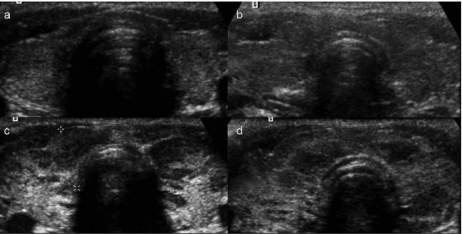
The thyroid gland volume was within normal limits (according to the WHO reference) in 13 patients, and eight patients had thyromegaly. According to the modified Sostre and Reyes gray-scale classification, thyroid glands were scored as Grade 0 in seven patients, Grade 1 in three patients, Grade 2 in seven patients, Grade 3 in two patients and Grade 4 in two patients (Table 3; also see Figure 1).
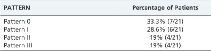
According to the color Doppler scale of Schulz et al. (17), seven patients had Pattern 0, six patients had Pattern I, five patients had Pattern II, and four patients had Pattern III (‘‘thyroid inferno’’) thyroid glands (Figure 2 and Table 4). The mean RI value, calculated as the mean of the mean RI values of both lobes in all patients, was 0.57¡0.05 (range
0.48-0.67) cm/sn.
Thyroid glands that were classified as having normal or near-normal gray-scale findings (n = 10) and those with substantial gray-scale changes (n = 11) demonstrated no statistically significant difference in the RI. Patients with minimal or no US gray-scale findings were found to have a mean RI value of 0.58¡0.056 cm/sn, and patients with
substantial gray-scale changes had a mean RI value of 0.56¡0.059 cm/sn.
In addition, the mean RIs calculated for each Doppler pattern were found to range from 0.56 to 0.58 (Table 5).
DISCUSSION
Hashimoto’s thyroiditis is the most common cause of goiter and hypothyroidism in children (19-21). Nearly all cases of thyroiditis seen in children are cases of Hashimoto’s disease (21). In the diagnosis of Hashimoto’s thyroiditis, two different diagnostic criteria exist, namely one developed by
Table 2 -Color Doppler classification in hypothyroidism, by Schulz et al.
Pattern 0 Blood flow limited to the peripheral thyroid arteries, while parenchymal flow is absent
Pattern I Presence of mildly increased parenchymal flow Pattern II Clearly increased color flow with a diffuse homogenous
distribution
Pattern III Markedly increased color flow with a homogenous distribution, including the so-called ‘‘thyroid inferno’’
Table 3 -Grading of the gray-scale findings and
distribution of patients within the grades. (Note: we have added Grade 0 to the original grading system created by Sostre and Reyes).
US GRADE Percentage of Patients
Grade 0 33.3% (7/21)
Grade 1 14.3% (3/21)
Grade 2 33.3% (7/21)
Grade 3 9.5% (2/21)
Fischer et al. and the other developed by the Japan Thyroid Association. These two criteria, while different, are both based on clinical and laboratory findings (22). Imaging of the thyroid gland (ultrasonography and scintigraphy) is not included in the diagnostic criteria because of its low specificity. The most objective finding in thyroid ultrasono-graphy is the quantitative measurement of thyroid volume. The WHO has defined the standard normal upper limits of ultrasonographically measured thyroid volumes according to age and gender because only 50% of goiter classifications can be accurately made by palpation. In an article published in 1999, the authors evaluated intraobserver and inter-observer differences and found no significant differences, therefore concluding that thyroid ultrasonography was reliable for evaluating thyroid dimensions and volume (23). US is valuable for determining the presence of nodular goiter in patients with Hashimoto’s disease and can enable the characterization and surveillance of these nodules. Furthermore, US also helps to position and guide fine-needle aspiration biopsy (24,25).
Hayashi et al. investigated ultrasound findings in 53 histologically confirmed patients with diffuse thyroid dis-eases and classified the thyroid echogenity into groups A and B, as iso-, hypo-, or hyperechoic compared with adjacent
muscles. Because the thyroid gland is expected to be more echogenic than adjacent muscles in normal individuals, the authors emphasized that a hypoechoic gland may be suggestive of hypothyroidism and that ultrasonography may be an important easy and noninvasive method for diagnosing Hashimoto’s disease (2).
In the absence of an ideal test for Hashimoto’s thyroiditis, Sostre and Reyes proposed that thyroid US could be an appropriate diagnostic test. They used a grading system to classify ultrasonographic patterns into four groups, with the sternomastoid muscle chosen as a reference (4). However, because Sostre and Reyes’ classification is based solely on the presence of ultrasonographic changes, there is no group in the classification that corresponds to clinical and laboratory findings of Hashimoto’s disease or to findings of completely normal ultrasonographic examinations. Therefore, in this study, it was deemed appropriate to add Grade 0 to their original grading system. In the present study, we found that a high grade was correlated with thyroid gland destruction and hypothyroidism. Five patients with subclinical hypothyroidism were found to have Grade 3 or Grade 4 disease.
Bogazzi et al. investigated the cause of the increase in thyroid blood flow in untreated Graves’ disease patients,
Figure 1 -Gray-scale grading. Grades 1-4 are shown in a-d, respectively. Grade 0, representing a completely normal thyroid gland, is not
shown in this figure.
Figure 2 -Color Doppler patterns.a.Pattern 0 (normal thyroid vascularity);b.color Doppler Pattern I (minimally increased thyroid
receptor antibodies or TSH may be the cause. This hypothesis was based on the fact that intrathyroidal vascularity and flow velocity increase in spontaneous hyperthyroidism but not in hyperthyroidism secondary to thyroid hormone intake or thyroid gland destruction. In addition, an increase in vascularity and flow velocity is also seen in Hashimoto’s disease patients with hypothyroidism (26). In the same study, intrathyroidal peak systolic flow velocity was thought to be a better index of thyroid disease because it demonstrated a more significant increase in patients with Graves’ disease than in those with Hashimoto’s disease. Iitaca et al. examined vascular endothelial growth factor (VEGF), which is an antigenic growth factor, and concluded that a significant relationship existed between intrathyroidal flows and VEGF levels (27). Once it was realized that hypervascularity was not unique to hyperthyroidism, Caruso et al. evaluated flow velocity in autoimmune thyroid diseases and concluded that the inferior thyroid arterial peak systolic flow velocity exceeded 150 cm/sec in patients with these diseases. However, the velocity remained within normal limits in patients with other thyroid diseases and did not exceed 65 cm/sec. These authors emphasized the importance of inferior thyroid artery peak systolic flow velocity in the differential diagnosis of diffuse thyroid disease and follow-up care of patients with Graves’ disease (28).
Schulz et al. investigated the role of color Doppler US in hypothyroidism and, in reference to previous studies, classified the vascularity (Table 2) (18). In their study, it was reported that the hypervascularity found in patients with Graves’ disease was also present in patients with hypothyroidism to some extent.
There is no consensus regarding normal values for Doppler parameters measured in the thyroid gland or inferior thyroid artery, namely the resistivity index (RI), pulsatility index (PI) and peak systolic flow velocities. In addition, no common guidelines exist on how to obtain these parameters (29-31). Mahmutyazicioglu and Turgut
sonographically normal thyroid glands.
Conversely, subjective color Doppler grading of the thyroid gland did not yield the expected results. Speci-fically, a small group of patients (38%, 8/21) was found to have markedly increased vascularity of the thyroid.
This study has several limitations. The major limitation is the lack of a control group. In future studies designed to test the efficacy of RI, the presence of a control group of age- and gender-matched subjects free of thyroid disease is crucial. Another limitation is the small size of the patient group, which limited our ability to perform any statistical analysis. Of the different spectral Doppler US parameters, only the RI was tested in this study. Other parameters, such as the peak systolic velocity, end diastolic velocity and pulsatility index, could be examined in future studies. The lack of assessment of the interobserver and intraobserver variability, due to the retrospective nature of the study, is another important limitation. This particular issue is extremely important for studies based on US imaging, a method known to result in significant interobserver and intraobserver variability.
Color Doppler imaging in Hashimoto’s disease appears to be a promising diagnostic imaging modality. In particular, the changes in RI values in patients with relatively normal gray-scale findings prompt us to suggest adding Color Doppler imaging to routine ultrasound examination of those patients. Further blinded, controlled studies with a sufficient number of patients are required to determine measures of test performance of RI in Hashimoto’s disease and would aid in determining the cut-off point for a normal RI value.
AUTHOR CONTRIBUTIONS
Sarikaya B participated in the radiological evaluations, data collection and analysis, literature review, and manuscript preparation. Demirbilek H participated in the patient selection, clinical assessment, data collection and analysis, and manuscript preparation. Akata D contributed to the radiological evaluations, manuscript preparation, and proofreading. Kandemir N contributed to the clinical assessment, manuscript prepara-tion, and proofreading.
REFERENCES
1. Espinasse F. Thyroid echography in chronic autoimmune thyroiditis. Journal de radiologie. 1983;64(10):537-44.
2. Hayashi N, Tasaki N, Konishi J, Yonekura Y, Senda M, Kasagi K, Yamamoto K, et al. Sonography of Hashimoto’s thyroiditis. JCU. 1986;14(2):123-6.
3. Lai SM, Chang TC, Chang CC, Kuo SH, Chen FW. Sonographic presentation in autoimmune thyroiditis. J Formos Med Assoc. 1990;89(12):1057-62.
4. Sostre S, Reyes MM. Sonographic diagnosis and grading of Hashimoto’s thyroiditis. Journal of endocrinological investigation. 1991;14(2):115-21 Table 5 -Mean RI values in patients with different
Doppler patterns.
DOPPLER US PATTERN Mean RI value
Pattern 0 0.58
Pattern I 0.56
Pattern II 0.58
Pattern III 0.57
5. Marcocci C, Vitti P, Catalano F, Concetti R, Pinchera A. Thyroid ultrasonography helps to identify patients with diffuse lymphocytic thyroiditis who are prone to develop hypothyroidism. 1991;72(1):209-13. 6. Chang TC, Hong CT, Chang SL, Hsieh HC, How SW. Correlation between sonography and pathology in thyroid diseases. J Formos Med Assoc. 1990;89(9):777-83.
7. Ivarsson SA, Ericsson UB, Fredriksson B, Persson PH. Ultrasonic imaging in the differential diagnosis of diffuse thyroid disorders in children. Am J Dis Child. 1989;143(11):1369-72.
8. Boi F, Loy M, Piga M, Sera A, Mariotti S. The usefulness of conventional and echo colour Doppler sonography in the differential diagnosis of toxic multinodular goitres. Eur J Endocrinol. 2000;143(3):339-46, http:// dx.doi.org/10.1530/eje.0.1430339.
9. Klima G, Berenek M, Rothlauer W. Definite distinction between thyroidal inflammatory conditions and other soft tissue inflammations of the neck using ultrasound. Acta Med Austriaca. 1996;23(1-2):80-2. 10. Yousem DM, Scheff AM. Thyroid and parathyroid gland pathology. Role
of imaging. Otolaryngol Clin North Am. 1995;28(3):621-49.
11. Eaton Se, Euinton HA, Newman CM, Weetman AP, Bennet WM. Clinical experience of amiodarone-induced thyrotoxicosis over a 3-year period: role of colour-flow Doppler sonography. Clin Endocrinol (Oxf). 2002;56(1):33-8, http://dx.doi.org/10.1046/j.0300-0664.2001.01457.x. 12. Shimamoto K, Endo T, Ishigaki T, Sakuma S, Makino N. Thyroid
nodules: evaluation with color Doppler sonography. J Ultrasound Med. 1993;12(11):673-8.
13. Stern WD, Laniado M, Vogl W, Weisser G, Tolksdorf A, Kaiser B, et al. The color-coded duplex sonography and contrast-enhanced magnetic resonance tomography of scintigraphically cold thyroid nodules. Rofo. 1994;160:3(1)-10, http://dx.doi.org/10.1055/s-2008-1032364.
14. Rago T, Vitti P, Chiovato L, Mazzeo S, De Liperi A, Miccoli P, et al. Role of conventional ultrasonography and color flow-doppler sonography in predicting malignancy in ‘‘cold’’ thyroid nodules. Eur J Endocrinol. 1998;138(1):41-6.
15. Ralls PW, Mayekawa DS, Lee KP, Coletti PM, Radin DR, Boswell WD, et al. Color flow Doppler sonography in Graves’ Disease: ‘Thyroid Inferno’. AJR Am J Roentgenol. 1988;150(4):781-4.
16. Demirbilek H, Kandemir N, Gonc EN, Ozon A, Alikasifoglu A, Yordam N. Hashimoto’s thyroiditis in children and adolescents: a retrospective study on clinical, epidemiological and laboratory properties of the disease. J Pediatr Endocrinol Metab. 2007;20(11):1199-205, http:// dx.doi.org/10.1515/JPEM.2007.20.11.1199.
17. Recommended normative values for thyroid volume in children aged 6-15 years. World Health Organization & International Council for Control of Iodine Deficiency Disorders. Bull World Health Organ. 1997;75(2):95-7. 18. Schulz SL, Uwe S, Ju¨rgen HH. Color Doppler sonography in
hypothyr-oidism. Eur J Ultrasound. 2003;16(3):183-9, http://dx.doi.org/10.1016/ S0929-8266(02)00072-1.
19. Dayan CM, Daniels GH. Chronic autoimmune thyroiditis. N Engl J Med. 1996;335(2):99-107, http://dx.doi.org/10.1056/NEJM199607113350206. 20. Maenpaa J, Raatikka M, Rasanen J, Taskinen E, Wager O. Natural course
of juvenile autoimmune thyroiditis. J Pediatr. 1985;107(6):898-904. 21. Bachrach LK, Foley TP. Thyroiditis in children. Pediatr Rev.
1989;11(6):184-91, http://dx.doi.org/10.1542/pir.11-6-184.
22. Fisher DA, Oddie TH, Johnson DE, Nelson JC. The diagnosis of Hashimoto’s thyroiditis. J Clin Endocrinol Metab. 1975;40(5):795-801, http://dx.doi.org/10.1210/jcem-40-5-795.
23. Ozgen A, Erol C, Kaya A, Ozmen MN, Akata D, Akhan O. Interobserver and intraobserver variations in sonographic measurement of thyroid volume in children. Eur J Endocrinol. 1999;140(4):328-31, http:// dx.doi.org/10.1530/eje.0.1400328.
24. Khurana KK, Richards VI, Chopra PS, Izquierdo R, Rubens D, Nesonero C. The role of ultrasonography-guided fine needle aspiration biopsy in the menagement of nonpalpable thyroid nodules. Thyroid. 1998;8(6):511-5, http://dx.doi.org/10.1089/thy.1998.8.511.
25. Solymosi T, Toth GL, Bodo M. Diagnostic accuracy of fine needle aspiration cytology of the thyroid: Impact of ultrasonography and ultrasonographically guided aspiration. Acta Cytologica. 2001;45(5):669-74, http://dx.doi.org/10.1159/000328285.
26. Bogazzi F, Bartalena L, Brogioni S, Burelli A, Manetti L, Tanda ML, et al. Thyroid vascularity and blood flow are not dependent on serum thyroid hormone levels: studies in vivo by color flow Doppler sonography. Eur J Endocrinol. 1999;140(5):452-6, http://dx.doi.org/10.1530/ eje.0.1400452.
27. Iitaca M, Miura S, Yamanaka K, Kawasaki S, Kitahama S, Kawakami Y, et al. Increased serum vascular endothelial growth factor levels and intrathyroidal vascular area in patients with Graves’ Disease and Hashimotos thyroiditis. J Clin Endocrinol Metabol. 1990;83(11):3908-12. 28. Caruso G, Attard M, Caronia A, Lagalla R. Color Doppler measurement
of blood flow in the inferior thyroid artery in patients with autoimmune thyroid diseases. Eur J Radiol. 2000;36(1):5-10, http://dx.doi.org/ 10.1016/S0720-048X(00)00147-9.
29. Vitti P, Rago T, Mazzeo S, Brogioni S, Lampis M, De Liperi A, et al. Thyroid blood flow evaluation by color flow Doppler sonography distinguished Graves’ Disease from Hashimoto’s Thyroiditis. J Endocrinol Invest. 1995;18(11):857-61.
30. Burns PN. Interpreting and analyzing the Doppler examination. In: Taylor KJW, Burns PN, Wells PNT, editors. Clinical application of Doppler examination. 2nd edition. New York: Raven Pres; 1995. 31. Budo RO, Rubin JM. Relationship between the resistive index and
vascular compliance and resistance. Radiology. 1999;211(2):411-7. 32. Mahmutyazicioglu K, Turgut M. Doppler evaluation of the thyroid in