Neurotization of free gracilis transfer with the brachialis
branch of the musculocutaneous nerve to restore finger
and thumb flexion in lower trunk brachial plexus injury:
an anatomical study and case report
Yi Yang,IXue-jun Zou,IIGuo Fu,IBen-Gang Qin,IJian-Tao Yang,IXiang-Ming Li,IIIYi Hou,IJian Qi,IPing Li,I Xiao-Lin Liu,ILi-Qiang GuI,*
IThe First Affiliated Hospital of Sun Yat-sen University, Department of Microsurgery and Orthopedic Trauma, Guangzhou, China. IINaval-Hospital,
Department of Orthopedic Trauma, Guangzhou, China.IIIThe First Affiliated Hospital of Henan University of Science and Technology, Department of
Orthopedic Surgery, Luoyang, China.
OBJECTIVE:To investigate the feasibility of using free gracilis muscle transfer along with the brachialis muscle branch of the musculocutaneous nerve to restore finger and thumb flexion in lower trunk brachial plexus injury according to an anatomical study and a case report.
METHODS:Thirty formalin-fixed upper extremities from 15 adult cadavers were used in this study. The distance from the point at which the brachialis muscle branch of the musculocutaneous nerve originates to the midpoint of the humeral condylar was measured, as well as the length, diameter, course and branch type of the brachialis muscle branch of the musculocutaneous nerve. An 18-year-old male who sustained an injury to the left brachial plexus underwent free gracilis transfer using the brachialis muscle branch of the musculocutaneous nerve as the donor nerve to restore finger and thumb flexion. Elbow flexion power and hand grip strength were recorded according to British Medical Research Council standards. Postoperative measures of the total active motion of the fingers were obtained monthly.
RESULTS:The mean length and diameter of the brachialis muscle branch of the musculocutaneous nerve were 52.66±6.45 and 1.39±0.09 mm, respectively, and three branching types were observed. For the patient, the first
gracilis contraction occurred during the 4th month. A noticeable improvement was observed in digit flexion one year later; the muscle power was M4, and the total active motion of the fingers was 209o
.
CONCLUSIONS:Repairing injury to the lower trunk of the brachial plexus by transferring the brachialis muscle branch of the musculocutaneous nerve to the anterior branch of the obturator nerve using a tension-free direct suture is technically feasible, and the clinical outcome was satisfactory in a single surgical patient.
KEYWORDS: Inferior Trunk of the Brachial Plexus Injury; Brachialis Muscle Branch of the Musculocutaneous Nerve; Gracilis; Muscle Transfer.
Yang Y, Zou X, Fu G, Qin BG, Yang JT, Li XM, et al. Neurotization of free gracilis transfer with the brachialis branch of the musculocutaneous nerve to restore finger and thumb flexion in lower trunk brachial plexus injury: an anatomical study and case report. Clinics. 2016;71(4):193-198 Received for publication onOctober 19, 2015;First review completed onDecember 1, 2015;Accepted for publication onJanuary 28, 2016 *Corresponding author. E-mail: [email protected]
’ INTRODUCTION
Traumatic brachial plexus injuries (BPI) are among the most severe peripheral nerve injuries in clinical practice and can cause permanent disability of the upper extremities (1). Several treat-ments are available for repairing upper trunk or total BPIs (2,3), whereas few methods exist for repairing injuries to the lower
trunk of the brachial plexus. Among these treatments, nerve transfer and tendon transfer are two common approaches (4-7). Free gracilis muscle transfer can be used to restore elbow and finger function after devastating BPIs (8-11). The contralateral C7 (CC7) nerve and the intercostal nerve are mainly used as donor nerves to restore hand function when transferring the gracilis muscle (12). However, the outcomes of using these donor nerves are controversial. Due to the limited availability of usable donor nerves and unsatisfactory results, restoration of sufficient hand function following injury to the inferior trunk of the brachial plexus is still challenging. Recently, one surgeon successfully reconstructed the function of digit flexion by transferring the brachialis muscle branch of the musculocutaneous nerve (BMBMCN) to the posterior part of the median nerve in patients
DOI:10.6061/clinics/2016(04)03
Copyright&2016CLINICS–This is an Open Access article distributed under the terms of the Creative Commons License (http://creativecommons.org/licenses/by/ 4.0/) which permits unrestricted use, distribution, and reproduction in any medium or format, provided the original work is properly cited.
with C8–T1 avulsions (13). These patients showed low donor site
morbidity after transection of the BMBMCN.
Due to the insufficient recovery of finger flexion when nerve transfer or tendon transfer is used after injury to the lower trunk of the brachial plexus, we present a new, effective treat-ment applied in one clinical case. This treattreat-ment is based on the anatomical finding that transferring the BMBMCN as a donor nerve to the anterior branch of the obturator nerve (ABON) of the gracilis can be used to re-enable thumb and finger flexion.
’ METHODS
Anatomical Study
Thirty formalin-fixed upper extremities from 15 adult cadavers (12 male and 3 female; 15 left and 15 right) donated to Southern
Medical University in China were used in this study. The average age of the cadavers was 65 years (range, 31–73 years), and the
average height was 170 cm (range, 154–175 cm). None of the
specimens showed evidence of gross pathology, previous surgical procedures, or traumatic injuries to the upper extremities. The specimens were placed in the supine position with the upper limb in the abducted position. All the specimens were dissected by the same trained surgeon.
A longitudinal incision extending to the infraclavicular region was made to expose the musculocutaneous nerve and the BMBMCN at the middle and distal levels of the medial upper arm. The length and diameter of the BMBMCN and the musculocutaneous nerve were measured using Vernier calipers (Figure 1). The following additional measurements were obtained: (1) the distance from the origin of the BMBMCN to the midpoint of the humeral condylar; (2) the length and diameter of the BMBMCN; and (3) the course and the branch types of the BMBMCN. All dissections and observations were performed under 10x magnification.
Clinical study
An 18-year-old male sustained a left BPI in a motorcycle accident and was transferred to our hospital 1 week after the injury. A physical examination showed near normal shoulder and elbow function, but the Horner sign was positive. Three months later, the patient recovered extension and partial flexion of his wrist. However, five months after the injury, no significant recovery of hand function was observed.
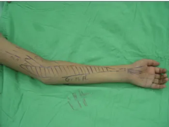
The range of wrist flexion and extension was near normal but with radial deviation. Flexion, extension, opposition, abduction, and adduction of the digits were absent, and the involved muscles had a strength of M0 according to the British Medical Research Council (BMRC) grading system (Figure 2). The patient had decreased sensation in his radial three digits, and sensation was completely absent in the ring and little fingers. Magnetic resonance imaging (MRI) and electromyogram (EMG) Figure 1 - Photograph showing the nerve branching from the
musculocutaneous nerve that innervates the brachialis muscle. Musculocutaneous nerve; Brachialis muscle branch of musculocuta-neous nerve; Median nerve; Lateral antebrachial cutamusculocuta-neous nerve.
testing indicated a C8 and T1 nerve root avulsion. Based on the results of the MRI evaluation, EMG testing and physical examination, the patient was diagnosed with a C8–T1 avulsion
of the left brachial plexus.
Surgical procedures
The surgical plan was discussed with the family and the patient before the operation (Figure 3). The operation was conducted under general anesthesia with the patient in the supine position. A 15-cm longitudinal incision was made along the medial and inferior surface of the medial upper extremity. The BMBMCN was located 14 cm proximal to the midpoint of the humeral condylar and was detached after local blockade with 1% lidocaine. The gracilis (appro-ximately 46 cm in length) was harvested along with the ABON and the vascular supply from a branch of the profunda femoris artery and vein. A skin paddle (approxi-mately 154 cm in length and width) was harvested to
facilitate postoperative flap monitoring as well. The entire gracilis was taken and positioned in the recipient site.
The proximal part of the gracilis muscle was attached to the medial brachial intermuscular septum of the arm and sutured to the flexor digitorum profundus and flexor pollicis longus distally with double interlacing sutures. The gracilis artery originating from the profunda femoris artery was anastomosed to the brachial artery. Two veins of the gracilis pedicle were anastomosed to the basilica and deep brachial vein of the brachial artery in an end-to-end manner.
The nerve of the gracilis was coapted to the BMBMCN using 9-0 nylon sutures with the assistance of a 10x surgical microscope (Figure 4).
The patient was required to wear a cast with 90o
flexion of the elbow and fingers for 6 weeks after the surgery. He was advised to perform rehabilitation exercises, undergo elec-trical stimulation therapy, and take neurotrophic drugs. Elbow flexion power and hand grip were recorded according to BMRC standards, and the total active motion of the fingers was measured each month postoperatively. Our institutional review board (the First Affiliated Hospital of Sun Yat-sen University, China) approved this study, and informed consent was obtained from the participant.
Statistical analysis
Data are presented as the means±SD. A paired t-test was
used for continuous variables and to analyze differences between the left and right sides of the cadavers. Statistical significance was considered at a value of po0.05. SPSS 13.0 (SPSS Inc., Chicago, IL, USA) was used to perform all statistical analyses.
’ RESULTS
Anatomical study
The microanatomical study of the 30 limbs of 15 adult cadavers showed that the brachialis muscle was innervated by the musculocutaneous nerve.
The mean lengths of the left and right sides of the BMBMCN were 52.60±6.62 and 52.72±6.50 mm,
respec-tively. The overall mean length of the BMBMCN was Figure 3 - The preoperative surgical plan. Musculocutaneous
nerve; Brachialis muscle; Gracilis muscle; flexion.
52.66±6.45 mm (range, 41.16–62.08 mm). The mean
dia-meters of the left and right sides of the BMBMCN were 1.38±0.09 and 1.39±0.10 mm, respectively. The overall
mean diameter of the BMBMCN was 1.39±0.09 mm (range,
1.20–1.56 mm). Based on the midpoint of the humeral
condylar, the level of the BMBMCN origin ranged from 110.0 mm to 160.0 mm with a mean of 139.4±15.1 mm. The
mean lengths were 139.3±15.3 and 139.5±15.4 mm for the
left and right side, respectively (Table 1). No significant differences were observed between the sides.
Three branching types were observed in this anatomical study. Type I included a single branch and was present in 25 limbs (83.33%), whereas type II included two branches and was present in 1 limb (3.33%). Type III included multiple branches and was present in 4 limbs (13.33%) (Table 2).
Clinical study
The patient’s cast was removed after the first month. Four months after the surgery, an initial sign of gracilis contrac-tion was observed. The muscle power was M3 at the 6th month. One year after the operation, a noticeable improve-ment (the muscle power was M4) was observed in digit flexion. The total active motion of the finger joints was excellent (209o
). Transection of the BMBMCN did not cause functional impairment to the elbow or the wrist. The sensation levels in the radial 3 digits and the elbow flexion power involving the biceps, flexor carpi radialis (FCR), extensor carpi radialis longus (ECRL), and extensor carpi radialis brevis (ECRB) were similar to the preoperative levels (Figure 5).
’ DISCUSSION
Motorcycle accidents are the leading cause of BPI, accounting for 54% of all BPI cases (14). Several different classification systems have been used for grading the severity of BPIs (15,16). BPIs are generally categorized as upper (C5-C7), lower (C8-T1) or total BPI (C5-T1). The incidence rates of C5-C7 and C5-T1 injuries are 42% and 48%, respectively, whereas the incidence of C8-T1 injuries is less frequent, with a rate of 3% (7).
Rationale for the transfer of the BMBMCN for finger flexion reconstruction
The musculocutaneous nerve innervates the two elbow flexors, the biceps and the brachialis. The biceps covers the anteromedial 2/3 of the brachialis, whereas the brachior-adialis covers the anterolateral 1/3 of the brachialis.
The brachialis is innervated by the musculocutaneous nerve as well as the radial or median nerve, or both of these nerves. Therefore, the brachialis potentially receives double or triple innervation (17). This multiple innervation may explain the preservation of normal elbow flexion function after BMBMCN transection.
Anatomical basis for the surgical design of transfer of the BMBMCN for finger flexion reconstruction
One concern for transferring the BMBMCN to the ABON of the gracilis is the available length of the donor and recipient nerves. In the present study, the anatomical examination showed that the major branching pattern of the BMBMCN was type I single branching (83.33%). The mean length and diameter of the BMBMCN were 52.66±6.45 mm and
1.39±0.09 mm, respectively. These results are similar to those of
other studies that have reported a mean length and diameter of the ABON of 8.7±2.1 cm and 2-2.6 mm (18,19), respectively. In
this anatomical study, the minimum length of the BMBMCN was 41.16 mm. In addition, the gracilis tendon is of sufficient length, and the recipient area is abundantly supplied with arteries and veins; therefore, the position of the harves-ted gracilis can be adjusharves-ted to ensure a tension-free nerve anastomosis, even if the BMBMCN is relatively short in a patient due to anatomical variation. Ideally, the diameter of the donor and recipient nerve should match precisely. The minimum and average diameters of the BMBMCN are 1.20 mm and 1.39 mm; the diameter of the ABON is 2-2.6 mm (18,19). Thus, the BMBMCN is at least 46-60% of the ABON diameter, whereas the cross-sectional area of the BMBMCN is at least 21-36% of the ABON. According to the literature, the cross-sectional area of the three intercostal nerves is 17%-27% of the musculocutaneous nerve (20,21). Tötösy et al. also found that normal muscle force could be achieved with a minimum of 30% of the original motor neuron pool (22). Therefore, the length and diameter of the BMBMCN is not a limiting factor for suturing the nerve.
Another concern was the axon count of the donor nerve. Bhandari et al. reported that the number of myelinated fibers in the spinal accessory nerve was 1,671 (23). Another study reported that 2090±462 myelinated fibers were present in
the BMBMCN (24). Because more myelinated fibers are present in the BMBMCN than in the spinal accessory nerve and the spinal accessory nerve was used as the main donor nerve to reinnervate the gracilis, the BMBMCN has the potential for powerful reinnervation of the gracilis.
The methods for repairing the inferior trunk after BPI mainly include nerve transfer and tendon transfer. Unfortu-nately, neither of these treatments can achieve an ideal recovery. Gu Y et al. transferred the BMBMCN to the posterior 1/4 to 1/3 of the median nerve to restore finger flexion (13). However, this can jeopardize other fascicles of the median nerve, and this technique should be limited to early injury cases. Using the BMBMCN as a donor for gracilis transfer to restore finger and thumb flexion could avoid such unnecessary injury to the median nerve and reduce the potential for axon misrouting. Yang J et al. transferred the Table 1-The measurements of the brachialis muscle branch of
the musculocutaneous nerve (mean±SD, mm).
Items Left (n=15) Right (n=15)
Total (n=30)
t-value p-value
Length 52.60±6.62 52.72±6.50 52.66±6.45 0.52 0.609 Diameter 1.38±0.09 1.39±0.10 1.39±0.09 0.397 0.698 Distance* 139.3±15.3 139.5±15.4 139.4±15.1 1.226 0.242 * Distance of the brachialis muscle branch of the musculocutaneous nerve origin to the midpoint of the humeral condylar.
Table 2-Branching type of the brachialis muscle branch of the musculocutaneous nerve.
Types Left (n=15) Right (n=15) Total (n=30)
Type I, single branch 14 11 25(83.33%)
Type II, double branches 0 1 1(3.33%)
Type III, multiple branches 1 3 4(13.33%)
pronator teres branch to innervate the anterior interosseous nerve in a patient with a C8-T1 avulsion, which fully restored the range of motion of the fingers (5). The patient’s finger flexor muscles regained grade 4 power; however, this method would not be feasible if only one branch was innervating the pronator teres. In another study, Bertelli et al. reported that transferring the supinator motor nerve to the posterior interosseous nerve effectively restored thumb and finger extension in patients with lower BPIs, but the outcome showed limited improvement. They also reported that transferring the brachialis muscle to the flexor digitorum profundus and flexor pollicis longus restored finger and thumb flexion (4). All of the patients described in that report achieved partial finger flexion, but the outcomes were not satisfactory. Goubier et al. transferred the ECRL to the flexor digitorum profundus and the brachioradialis tendon to the flexor pollicis longus tendon to restore thumb flexion in patients with lower BPI; all patients regained finger flexion (7). After the ECRL tendon transfer, wrist extension strength was decreased in some cases, but the ECRB muscle remained functional. In addition, initial recovery of shoulder and elbow movements may lead to a false expectancy for the recovery of hand function, which may result in late referral for treatment. Consequently, muscle atrophy will restrict the outcome of tendon transfer and nerve reconstruction. In this case, free muscle transfer might be the only choice for reconstructing hand function.
The BMBMCN has been used previously by different surgeons to repair C8–T1 brachial plexus avulsions and
re-establish hand function (4,13,25,26). In this study, we used the BMBMCN as a donor nerve to reinnervate the gracilis, and the patient gained noticeable improvements in digit flexion and muscle power, which was graded as M4 one year after the operation. Transection of the BMBMCN did not cause functional impairment of the elbow or the wrist, which makes this technique useful for repairing injuries to the inferior trunk
of the brachial plexus. Compared to nerve transfer or tendon transfer, a gracilis transfer can increase the number of functional muscles in the forearm without sacrificing the remaining hand and elbow functions in lower trunk BPI. In addition, from the time the patient was injured until he underwent the surgery, the muscles underwent a period of denervation and atrophy. Using the newly harvested gracilis could overcome such periods of muscle atrophy and provide better outcomes. This technique expanded the range and shortened the regeneration distance of the donor nerve in free muscle transfer. This procedure can be used as an initial treatment rather than a remedial treatment after primary nerve or tendon transfer fails. In addition, this method can be used in patients with late hospital referrals.
However, this technique should be restricted to inferior trunk BPIs because the musculocutaneous nerve originates at Figure 5 -Photographs showing that the transection of the brachialis muscle branch of the musculocutaneous nerve did not cause functional impairment of the elbow when the cast was removed after the first month (a, b). One year after the operation, a noticeable improvement was observed in digit flexion due to gracilis contraction (arrow). The muscle power was M4 (c, d).
C5-C7. After middle trunk injury, the donor BMBMCN is likely to be affected, and the outcome of free gracilis transfer may not be satisfactory.
This study also had some limitations. First, we performed this technique on only one patient; more cases are needed for future tests of its clinical application. Second, although the patient showed satisfactory recovery of function after the relatively short follow-up period, long-term follow-up is needed to estimate the final outcome. Third, we did not perform a histomorphometric study to measure the number of myelinated fibers in the BMBMCN. In addition, muscle strength less than M3 cannot be measured by quantitative muscle power assessment and therefore is not conducive to continuous observation of the recovery of the transplanted muscle. As a result, the study used the BMRC for outcome assessment. Other objective measurements (including using a dynamometer to measure muscle strength, quantitative sensory testing, etc.) described in previous reports (27,28) should be used to assess the safety and effectiveness of this technique in the future. Finally, because this technique is technically demanding, it should be applied only for strict indications.
Based on this anatomical study, transferring the BMBMCN directly to the ABON with a tension-free suture is technically feasible. This surgical procedure successfully achieved ade-quate recovery of muscle power in the reinnervated gracilis and restoration of digit flexion without compromising elbow and wrist flexion following injury to the inferior trunk of the brachial plexus in a single surgical patient.
’ ACKNOWLEDGMENTS
The authors acknowledge thefinancial support from the National Natural Science Foundation (NO.30973059) and the National Key Clinical Specialist Construction Programs of China (201402016). We thank Dr. Sarah E. Poliquin (Vanderbilt University) and Qing Zhang (Vanderbilt University) for the English language improvements.
’ AUTHOR CONTRIBUTIONS
Yang Y and Zou X were responsible for conception and design, acquisition of data and drafting the manuscript. Fu G and Qin BG revised the manuscript. Yang JT, Li XM and Hou Y contributed to analysis and interpretation of data. Qi J, Li P and Liu XL acquired data. Gu LQ was responsible for conception and design and acquisition of data. We declare that all the listed authors have participated actively in this study and meet the requirements of the authorship.
’ REFERENCES
1. Terzis JK, Kostas I, Soucacos PN. Restoration of shoulder function with nerve transfers in traumatic brachial plexus palsy patients. Microsurgery. 2006;26(4):316-24, http://dx.doi.org/10.1002/micr.20245.
2. Tu YK, Tsai YJ, Chang CH, Su FC, Hsiao CK, Tan JS. Surgical treatment for total root avulsion type brachial plexus injuries by neurotization: a prospective comparison study between total and hemicontralateral C7 nerve root transfer. Microsurgery. 2014; 34(2):91-101, http://dx.doi.org/10.1002/micr.22148. 3. Tsai YJ, Su FC, Hsiao CK, Tu YK. Comparison of objective muscle strength in
C5-C6 and C5-C7 brachial plexus injury patients after double nerve transfer. Microsurgery. 2015;35(2):107-14, http://dx.doi.org/10.1002/micr.22283. 4. Bertelli JA, Ghizoni MF. Transfer of supinator motor branches to the
posterior interosseous nerve in C7-T1 brachial plexus palsy. J Neurosurg. 2010;113(1):129-32, http://dx.doi.org/10.3171/2009.10.JNS09854. 5. Yang J, Jia X, Yu C, Gu Y. Pronator teres branch transfer to the anterior
interosseous nerve for treating C8T1 brachial plexus avulsion: an ana-tomic study and case report. Neurosurgery. 2014; 75(4):375-9; discussion 379, http://dx.doi.org/10.1227/NEU.0000000000000435.
6. Bertelli JA, Ghizoni MF. Brachialis muscle transfer to reconstruct finger flexion or wrist extension in brachial plexus palsy. J Hand Surg Am. 2006;31(2):190-6, http://dx.doi.org/10.1016/j.jhsa.2005.09.020. 7. Goubier JN, Teboul F. Management of hand palsies in isolated C7 to T1 or
C8, T1 root avulsions. Tech Hand Up Extrem Surg. 2008; 12(3):156-60, http://dx.doi.org/10.1097/BTH.0b013e318172d72b.
8. Adams JE, Kircher MF, Spinner RJ, Torchia ME, Bishop AT, Shin AY. Complications and outcomes of functional free gracilis transfer in brachial plexus palsy. Acta Orthop Belg. 2009;75(1):8-13.
9. Chuang DC, Hattori Y, Ma AH, Chen HC. The reconstructive strategy for improving elbow function in late obstetric brachial plexus palsy. Plast Reconstr Surg. 2002;109(1):116-26; discussion 127-9, http://dx.doi.org/ 10.1097/00006534-200201000-00020.
10. Dodakundi C, Doi K, Hattori Y, Sakamoto S, Fujihara Y, Takagi T, et al. Outcome of surgical reconstruction after traumatic total brachial plexus palsy. J Bone Joint Surg Am. 2013;95(16):1505-12, http://dx.doi.org/ 10.2106/JBJS.K.01279.
11. Terzis JK, Kostopoulos VK. Free Muscle Transfer in Posttraumatic Plexo-pathies Part II: The Elbow. Hand (N Y). 2010; 5(2):160-70.
12. Terzis JK, Kostopoulos VK. Free muscle transfer in posttraumatic plexo-pathies: part III. The hand. Plast Reconstr Surg. 2009;124(4):1225-36, http://dx.doi.org/10.1097/PRS.0b013e3181b5a322.
13. Gu Y, Wang H, Zhang L, Zhang G, Zhao X, Chen L. Transfer of brachialis branch of musculocutaneous nerve for finger flexion: anatomic study and case report. Microsurgery. 2004;24(5):358-62, http://dx.doi.org/10.1002/ micr.20053.
14. Flores LP. [Epidemiological study of the traumatic brachial plexus injuries in adults]. Arq Neuropsiquiatr. 2006;64(1):88-94, http://dx.doi.org/10.1590/ S0004-282X2006000100018.
15. Terzis JK, Vekris MD, Soucacos PN. Outcomes of brachial plexus reconstruction in 204 patients with devastating paralysis. Plast Reconstr Surg. 1999;104(5):1221-40, http://dx.doi.org/10.1097/00006534-199910000-00001.
16. Yang J, Qin B, Fu G, Li P, Zhu Q, Liu X, et al. Modified pathological classification of brachial plexus root injury and its MR imaging char-acteristics. J Reconstr Microsurg. 2014;30(3):171-8, http://dx.doi.org/ 10.1055/s-0033-1357498.
17. Won SY, Cho YH, Choi YJ, Favero V, Woo HS, Chang KY, et al. Intra-muscular innervation patterns of the brachialis muscle. Clin Anat. 2015; 28(1):123-7, http://dx.doi.org/10.1002/ca.22387.
18. Houdek MT, Wagner ER, Wyles CC, Moran SL. Anatomical feasibility of the anterior obturator nerve transfer to restore bowel and bladder function. Microsurgery. 2014; 34(6):459-63, http://dx.doi.org/10.1002/ micr.22256.
19. Goubier JN, Teboul F, Yeo S. Transfer of two motor branches of the anterior obturator nerve to the motor portion of the femoral nerve: an anatomical feasibility study. Microsurgery. 2012; 32(6):463-5, http://dx.doi.org/10.1002/ micr.22012.
20. Asfazadourian H, Tramond B, Dauge MC, Oberlin C. Morphometric study of the upper intercostal nerves: practical application for neuroti-zations in traumatic brachial plexus palsies. Chir Main. 1999;18(4):243-53. 21. Ogino T, Naito T. Intercostal nerve crossing to restore elbow flexion and sensibility of the hand for a root avulsion type of brachial plexus injury. Microsurgery. 1995;16(8):571-7, http://dx.doi.org/10.1002/micr.1920160812. 22. Totosy DZJ, Zung HV, Erdebil S, Gordon T. Innervation ratio is an important determinant of force in normal and reinnervated rat tibialis anterior muscles. J Neurophysiol. 1992;67(5):1385-403.
23. Bhandari PS, Deb P. Dorsal approach in transfer of the distal spinal accessory nerve into the suprascapular nerve: histomorphometric analysis and clinical results in 14 cases of upper brachial plexus injuries. J Hand Surg Am. 2011;36(7):1182-90, http://dx.doi.org/10.1016/j.jhsa.2011.02. 025.
24. Palazzi S, Palazzi JL, Caceres JP. Neurotization with the brachialis muscle motor nerve. Microsurgery. 2006;26(4):330-3, http://dx.doi.org/10.1002/ micr.20247.
25. Ray WZ, Yarbrough CK, Yee A, Mackinnon SE. Clinical outcomes fol-lowing brachialis to anterior interosseous nerve transfers. J Neurosurg. 2012;117(3):604-9, http://dx.doi.org/10.3171/2012.6.JNS111332. 26. Zheng XY, Hou CL, Gu YD, Shi QL, Guan SB. Repair of brachial plexus
lower trunk injury by transferring brachialis muscle branch of musculo-cutaneous nerve: anatomic feasibility and clinical trials. Chin Med J (Engl). 2008;121(2):99-104.
27. Li XM, Yang Y, Hou Y, Yang JT, Qin BG, Fu G, et al. Diagnostic accuracy of three sensory tests for diagnosis of sensory disturbances. J Reconstr Microsurg. 2015;31(1):67-73, http://dx.doi.org/10.1055/s-0034-1384819. 28. Li XM, Yang JT, Hou Y, Yang Y, Qin BG, Fu G, et al. Donor-side morbidity