Vol. 23, No.1, Winter-2011
Otoacoustic Emissions in Sudden Sensorineural Hearing Loss: Changes of
Measures with Treatment
*ShadmanNemati1, Seyyed-EbrahimNaghavi2, EhsanKazemnejad3, RoozbehBanan4
Abstract
Introduction:
To identify changes in OAEs parameters in treatment course of idiopathic sudden sensorineural hearing loss (iSSNHL).
Materials and Methods:
In aprospective studyfromAugust 2005 to January 2009, 26 patients with iSSNHL underwent conventional audiometry/tympanometry and two types of OAEs (TEOAEs and DPOAEs) before and after the completion of standard drug therapy.The changes in pre- and post- treatment parameters were compared with each other and with normal-contralateral ears.
Results:
In TEOAEs, the mean overall correlation (reproducibility) and the mean overall strength in involved ears were 10.96±23.36 and 0.99±3.45 dB, respectively, before the treatment, which reached 22.88±36.55 and 1.85±5.3, respectively, after the treatme nt (P>0.05). Significant
difference between “correlation score” (average of correlations at 3-4 involved frequencies)
before and after treatment was found: 6.52 ±18.19 vs. 21.67±37.8 (P<0.034). The difference between pre- and post-treatmentoverall correlation and correlation score in the “response group” were significant (P<0.031). In DPOAEs of the involved ears, the mean DP1 level and the DP1 signal-to-noiseratio changes were not significant with the treatment (P>0.05).
Conclusion:
Evoked OAEs, especially TEOAEs, are objective, rapid, and sensitive tools in the treatment course of iSSNHL.
Keywords:
Idiopathic sudden sensorineural hearing loss, Otoacoustic emissions, Response,Treatment
Received date: 15Jun 2010 Accepted date: 16 Nov 201
1
Department of otorhinolaryngology, Guilan University of Medical Sciences, Rasht, Iran 2Department of otorhinolaryngology, Guilan University of Medical Sciences, Rasht, Iran 3
Department of biostatistics, Guilan University of Medical Sciences, Rasht, Iran 4
Ear, Nose, Throat, Head and Neck surgery Research Center, Guilan University of Medical Sciences, Rasht, Iran *Corresponding author:
Ear, Nose, Throat, Head and Neck surgery Research Center, Amiralmomenin Hospital, Rasht, Guilan, Iran E-mail: [email protected], Tel: +989111370488
Introduction
David Kemp in 1978 described some types of sound waves of cochlear origin which can be detected with a microphone in the external auditory canal. Since then, many studies have been performed in the clinical applicability of these “emissions” named “otoacoustic emissions (OAEs)”.Out of the various types of OAEs, transient evoked OAEs (TEOAEs) and distortion product OAEs (DPOAEs) may be detected in nearly all subjects with normal cochlear and middle ear function. While TEOAEs will be absent in sensorineural hearing losses with less severities, DPOAEs are absent in sensorineural hearing loss exceeding 50 dB Hearing Level, but are measurable in inflammatory conditions causing HL secondary to cochlear nerve involvement (1-3). OAEs areobjective and non- invasive testing of the cochlear outer hair cell (OHC) function and have a direct relationship to hearing threshold sensitivity.With high reproducibility, high test-retest stability, and with temporal and spectral properties unique to each individual, OAEs are performed conveniently and rapidly and aremore sensitivein comparison with routine audiometric tests. They can be applied in difficult-to-test cases and inorganic hearing losses and are able to show “subclinical” events in the cochlea (1,4-9).
Many researchers have shown that evoked OAEs can successfully separate normally hearing and hearing impaired populations. Normative measurements have been studied, but more studies should be performed on the clinical applications of OAEs and on optimizing current protocols, especially in hearing- impaired populations. (3,10,11). Sudden sensorineural hearing loss (SSNHL) is the loss of hearing more than 30 dB in three contiguous frequencies thatoccurs in less than three days. It is fairly uncommon and has an overall incidence of 5-20 per 100,000 individuals per year. SSNHL is a controversial topic in
otolaryngology, with more than 100 different etiologies,yet its etiology remains unknown: "idiopathic" SSNHL (4,12,13). There are increasing evidences in the literature that in some cases SSNHL only has psychogenic causes (14-16). Since TEOAEs and DPOAEs seem to reflect the activity of the OHCs, it is reasonable to hypothesize that in most idiopathic SSNHLcases, OHC function deteriorates when the hearing threshold is raised, and it recovers as hearing improves.In this study we tried to identify the changes in measures of these „objective‟ tests during the recovery process of iSSNHL.
Materials and Methods
mode. The TEOAE waveform was analyzed in 500-1000-Hz-width frequency bands, and the signal-to- noise ratio and the reproducibility of signals(correlations)- in percent-and emission strength- in db-were
obtained in 5 different
contiguousfrequency bands (750 Hz to 4500 Hz). For DPOAEs, primary tones f1 and f2 were presented at 70dB and 60-dBSound Pressure Levels (SPL). The f2/f1 ratio was kept at approximately 1.2 (ranging from 1.21 to 1.23) and the frequency of f2 was changed in 1/4-octave steps from 500 Hz to 8000 Hz. The levels of the DPOAEs at 2fl- f2 were recorded. In nine different frequencies (ranging between 500 and 8000 Hz), DP-gram showed DP1 level (dB) and DP1 signal- to-noise ratio/dB.Forall the patients, necessary tests for the disease were performed, and they were then treated with oral steroids (prednisone1mg/kgoral daily for 10 days and then tapered) andacyclovir (800mg qid for 7 days). PTA and Speech Discrimination Score (SDS) were performed every 3-5 days during the treatment, and post-treatment PTA, SDS, TEOAE, and DPOAE were performed two weeks after termination. According to the treatment response, the patients were classified into three groups: the complete- or good-response group (≥30dB recovery in affected frequencies in PTA or ≥30% increase in SDS), the partial- or moderate-response group (≥10dB and ≤30 dB recovery in affected frequencies or ≥10% and <30% increase in SDS), and the poor- or no-response group (≤9 dB recovery in
PTA or ≤9% increase in SDS) (3,4). Then
we analyzed the data (various parameters of pre- and post-treatment DPOAE and TEOAE) from affected ears in the three study groups and in comparison with those of contralateral non-affected ears as controls. The data were analyzed by Chi-square test, Levene‟s test for equality of variances, T-test, one-way ANOVA, and Wilcoxon Signed Ranked test using
SPSS-16 software, and the level of significance was considered 0.05.
Results
contiguous, involved frequency bands. Therefore, we found significant difference between the “correlation scores” before and after the treatment: 6.52±18.19 vs. 21.67±37.8, (P<0.034). However, no significant difference was found in the
“emission strength scores” before and after
the treatment (P=0.44).We enrolled all the patients who responded to treatment (i.e. complete and partial response) in one group: response group (n=20 cases), and not responding patients in the other group (n=6). The difference between pre- and post-treatment parametersof the affected ears in the“response group”wassignificant for the correlation score (P<0.007) and the overall correlation (P<0.031), but there was no statistically significant difference inother parameters such as DP1 signal- to-noise ratio (P<0.075)or in theoverall strength, the emission strength scores, and the DP1 levels. Further, none of these parameters showed any statistically significant changes in the “no response group”.
Using the Receiver Operating Characteristics Curve (ROC curve), we found some cut-off points in the pre-treatment “overall correlation” and “correlation scores” indicating abnormality (Fig.1).
Fig 1:Receiver operating characteristics curve
(ROC curve) indicates “correlation score” below 11
as abnorma l (sensitivity=87%, specificity 68%, (P<0.001).
Therefore, we can regardthe pre-treatment “overall correlation” below 12, and
“correlation scores” below 11as abnormal
(sensitivity=82.5% and 87%, specificity=60% and 68%, respectively; (P<0.001). Also, we found that the difference between pre- and post-treatment
“correlation scores” and “overall
correlation”, in contrast to “DP1signal- to-noise”, may yield valuable measures for
defining “response to treatment in sudden
deafness” (Fig2).
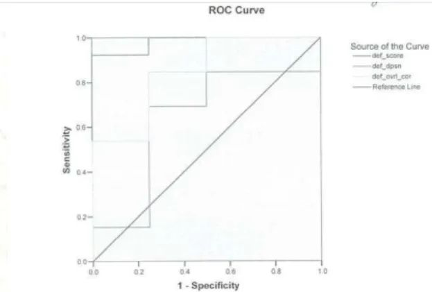
Fig 2: Rece iver operating characteristics curve (ROC curve) indicates that difference between
pre-treatment “overall correlation” and its value during
treatment course (def_ovr_score) as high as 1.5 (61.5% sensitivity and 75% specific ity) as an inde x
for “response” (P<0.042). For “correlation score” (def_score), the diffe rence as high as 3.1, will tell
us about “response” with 92% sensitivity and 100%
specificity (P<0.005).
In this regard, we can regardthe difference between pre-treatment “overall correlation” and its value during the course of treatmentas high as 1.5(61.5% sensitivity and 75% specificity) as an index for
“response” (P<0.042). Also, for
“correlation scores”, a difference of up to
3.1 will tell us about “response” with 92%
sensitivity and 100% specificity(P<0.005). Discussion
noninvasive differential diagnosis of hearing loss by adding the measurement of evoked OAEs growth functions over the range of frequencies to a standard audiometric evaluation(6,17). Also, there are many studies in the literature that demonstrate a prognostic role for OAEs in the iSSNHL (1,3,12,18,19), although there are some studies not agreeing with this (11,20),how are the changes in different parameters of OAEs, and, in principal, what parameters are more suitable, more stable, and more conforming to routine audiometries?In a previous study on ears with long-standing idiopathic sensorineural HL, evoked OAEs could not be recorded in ears with a hearing loss exceeding 35 dB at minimum hearing level of four audiometric frequencies: 500, 1,000, 2,000 and 4,000 Hz (4 MHL). In other words, although four MHLs were greater than 35 dB in most of the ears, evoked OAEs could be detected in about one- half of the ears with idiopathic sudden SNHL (18).Ishida et al published their study on eight SSNHL patients with good hearing improvement, and eight SSNHLpatients with poor hearing improvement in an attempt to elucidate the behavior of ear fullness, tinnitus and OAEs in the recovery course of the disease. SSNHLpatients with good hearing improvement tended to have OAE responses and the sensations of the ear fullness and tinnitus improved almost simultaneously with hearing level improvement. When hearing recovery was not full, OAEs did not reappear for these frequencies. Patients with poor hearing improvement tended to have absent OAEs and persistent ear fullness and tinnitus (1). Our study is in agreement with this study overall, although in this studythe changes of parameters had not beenelucidated, and only DPOAE had been performed.In 15 cases of idiopathic SD, Nakamura et al demonstrated that the amplitudes of TEOAEs and DPOAEs increased concurrently with the recovery of the
hearing threshold, and suggested that the function of outer hair cells had deteriorated when the hearing threshold was elevated and their activity recovered as hearing improved to nearly normal levels in cases with good outcome (13). Lalakiet alperformed pure-toneaudiometry (PTA) and TEOAE recordings in 30 SSNHL patientson the admission day, and at least three measures on the next eight days. The audiometric threshold improvement at each frequency was correlated with the TEOAE parameters on each measure (19). These two studies are inagreement with our results, and in fact, we had performedour study in a better way (e.g. with more cases and more OAE parameterscompared with Nakamura's study, and performing both TE
and DPOAEs compared with
study, Zhang et al investigated the basic characters of DPOAEs in 60 ears of 30 cases with SSNHLbefore and after treatment. In the recovery course, the amplitude and threshold of DPOAE were improved with the restoration of auditory threshold, but the restoring rate (RR) of auditory threshold was higher, and they concluded that the amplitude and threshold of DPOAE werebeyond that of the pure-tone behavioral thresholdin SSNHLrecovery course, which implies that DPOAE sensitively and directlyreflects the function of the cochlea (21). This study is in agreement with ours; although our study contains both DP- and TEOAEs.Perhaps one novelty of our study is calculating "correlation scores" in these cases, which are average of correlations of consecutive affected frequencies. This parameter will be more sensitive in reflecting response to treatment. Also using ROC curves, we offered some cut-off points for defining "abnormality" and "response to treatment" in sudden deafness;however, the sample size in our study is not enough for sensitivity and specificity estimation, and these cut-off points may be used only for future studies with larger sample sizes.
Conclusion
Evoked OAEs, especially TEOAEs, can be used as an objective, sensitive, and specific test in SSNHL, especially in difficult-to-test cases, for monitoring the results of the treatment. We suggest calculating “correlation scores” before and after the completion of treatment in all SSNHL cases.
Acknowledgment
Table 1: Some demographic, audiometric, and otoacoustic (TEOAE) characteristics of 25 patients with sudden deafness
*Audiogram pattern of sensorineural hearing loss **Cor. = Corre lation (reproducibility)
***Cor. Score = Co rre lation Score : this score is calculated by averaging corre lations of "involved frequencies"
References:
1.Ishida IM, Sugiura M, Teranishi M, Katayama N, Nakashima T.Otoacoustic emissions, ear fullness and tinnitus in the recovery course of sudden deafness. Auris Nasus Larynx 2008; 35(1): 41-6.
2. Lonsbury MB, Martin GK. Otoacoustic emissions. Curr Opin Otolaryngol Head Neck Surg 2003; 11(5): 361-6.
Case
No. (Years) Age Sex Involved ear Type*
Severity
Response to treatment
Overall cor**.
pre-treat (%)
Overall cor**. post
treat (%)
Cor. Score***
pre-treat (%)
Cor.
Score***
post-treat
(%)
1 15.00 female left high tone profound no response 8.0 -20.0 -12.6 -13.6
2 26.00 female left flat severe complete 33.0 97.0 6.6 96.0
3 19.00 male left flat severe no response -17.0 -12.0 -19.33 -34.0
4 56.00 female left flat moderate complete 38.0 48.0 45.0 65.0
5 29.00 male left low tone mild complete 28.0 86.0 29.0 81.8
6 61.00 male left low tone moderate complete -2.0 1.0 5.5 18.5
7 69.00 male right low tone moderate complete -12.0 -5.0 10.5 11.5
8 53.00 female left low tone moderate complete -6.0 91.0 0.5 89.5
9 30.00 male left flat profound no response -3.0 -9.0 1.0 -15.6
10 36.00 female right flat moderate no response 10.0 4.0 18.2 -4.2
11 52.00 female left low tone moderate partial 86.0 81.0 11.5 31.0
12 50.00 male left low tone moderate partial -9.0 7.0 -15.75 13.25
13 47.00 male right high tone mild complete 46.0 57.0 52.8 75.6
14 25.00 female right flat severe no response 22.0 5.0 9.8 8.4
15 19.00 male left flat moderate partial 25.0 16.0 -6.8 13.6
16 54.00 female left flat profound complete 18.0 18.0 13.4 29.6
17 44.00 male right flat profound partial 2.0 22.0 -3.6 3.6
18 49.00 male left flat severe complete -18.0 77.0 -7.6 81.0
19 38.00 male left flat moderate no response -3.0 -3.0 -8.4 1.6
20 50.00 male right high tone profound partial 21.0 -8.0 20.0 -18.75
21 24.00 male right flat profound partial 14.0 1.0 -0.8 -11.4
22 39.00 female right flat moderate complete -1.0 2.0 -5.8 1.8
23 52.00 female right low tone severe complete -11.0 5.0 -5.6 5.6
24 56.00 male left high tone mild no response 5.0 0.0 28.3 27.0
3. Schweinfurth JM, CacaceAT, Parnes SM. Clinical applications of otoacousticemissions in sudden hearing loss. Laryngoscope 1997; 107(11): 1457-63.
4. Arts HA. Sensorineuralhearing loss: Evaluation and management in adults. In: Cummings CW, Flint PW, Harker LA,Haughey BH, Richardson MA, Robbins KT, et al. (editors). Cumming‟s otolaryngology, head and neck surgery. 4thed. Philadelphia: Elesvier Mosby; 2005: 3550-5.
5. Borka C. Otoacoutic emissions. In: Luxon L. (editor). Textbook of audiological medicine, clinical aspects of hearing and balance.USA: Martin Dunitz; 2003: 259-66. 6. Mills DM. Determining the cause of hearing loss: Differential diagnosis using a comparison of audiometric and otoacousticemission responses. Ear Hearing 2006; 27(5): 508-25.
7. Kemp DT.Otoacoustic emissions, their origin in cochlear function, and use. Br Med Bull 2002 Vol. 63, 2002: 223-41.
8. Granjeiro RC, Kehrle HM, Bezerra RL. Transient and distortion product evoked oto-acoustic emissions in normal hearing patients with and without tinnitus. Otolaryngol Head
and Neck Surg 2008; 138(4): 502-6.
9. Lynne M, Judi A, Lapsley M. Detecting incipient inner-ear damage from impulse noise with otoacoustic emissions. J Acoust Soc Am 2009; 125(2): 995-1013.
10. Chen CN.Differentiating the cause of acute sensorineural hearing loss between Meniere's disease and sudden deafness. Acta Otolaryngol 2006; 126(1): 25-31.
11. Canale A, Lacilla M, Giordano C, De Sanctis A, Albera R. The prognostic value of the otoacoustic emission test in low frequency sudden hearing loss. Eur Arch Otorhinolaryngol 2005; 262(3): 233-7.
12. Amiridavan M, NematiSh, Hashemi SM, Saberi A. [Otoacoustic emissions and auditory brainstem responses in patients with sudden sensorineural hearing loss. Do otoacoustic emissions have prognostic value? ] Journal of research in medical sciences 2006; 11(4): 263-9. (Persian)
13. Nakamura M, Yamasoba T, Kaga K. Changes in otoacoustic emissions in patients with idiopathic sudden deafness.Audiology 1997; 36(3): 121-35.
14. Jae-Ho B, Min S.A clinical analysis of psychogenic sudden deafness. Otolaryngol Head Neck Surg 2006; 134(6): 970-4.
15. Zhao H, Dai CF, Chi FL, Wang ZM. Non-organic hearing loss in Chinese teenagers.AurisNasus Larynx 2008; 35: 485-92.
16. Rotenberg BW, Makhija M, Papsin BC. Conversion disorder in a child presenting as sudden sensorineural hearing loss.Int J PediatrOtorhinolaryngol 2005 ; 69: 1261-4.
17. Ota Y, Oda M.Lesion site in sudden deafness: Study with electrocochleography and transiently evoked otoacoustic emission.Acta Otolaryngologica 1999; 119(1): 33.
18.Sakashita T, Minowa Y, Hachikawa K. Evoked otoacoustic emissions from ears with idiopathic sudden deafness.Acta Otolaryngol 1991; Suppl 486: 66-72.
19. Lalaki P, Markou K, Tsalighopoulos MG, Daniilidis I. Transiently evoked otoacoustic emissions as a prognostic indicator in idiopathic sudden hearing loss. Scand Audiol 2001; 30(52): 141-5.
20.Hoth S. On a possible prognostic value of otoacoustic emissions: A study on patients with sudden hearing loss.Eur Arch Otorhinolaryngol 2006; 262(3): 217-24.