I Brazilian guidelines for respiratory physiotherapy
in pediatric and neonatal intensive care units
I Recomendação brasileira de fisioterapia respiratória em
unidade de terapia intensiva pediátrica e neonatal
INTRODUCTION
he role of physiotherapists with expertise in pediatric and neonatal intensive care is relatively new in Brazil, as the dissemination of courses and training in these areas primarily occurred from 2000 onwards. Currently, there are several courses nationwide that prepare physiotherapists to work with patients or perform research in these areas. In February 2010, the Brazilian Health Surveillance Agency (Agência Nacional de Vigilância Sanitária - ANVISA) published, in its oicial gazette,(1) the mandatory neonatology and pediatric expertise required for physiotherapists to work in these respective hospital areas. his improvement in the training of physiotherapists contributed to patient safety in pediatric and neonatal intensive care units (ICUs).
Physiotherapists working in these areas are responsible for kinetic functional assessment and prevention (for all or any human body system, as needed) and for treatment interventions (respiratory and/or motor physiotherapy). hey also work with a multidisciplinary team in the control and application of medicinal gases, invasive and noninvasive (INV) mechanical pulmonary ventilation (MPV), protocols for weaning and extubation of MPV, tracheal insulation of gas, protocol of insulation/exsulation of the endotracheal cuf, and surfactant application, among others.
herefore, establishing guidelines for physiotherapists is very important. With
Cíntia Johnston, Nathalia Mendonça Zanetti, Talitha Comaru, Simone Nascimento dos Santos Ribeiro, Lívia Barboza de Andrade, Suzi Laine Longo dos Santos
ABSTRACT
Developing guidelines for the role of the physiotherapist in neonatal and pediatric intensive care units is essential because these professionals are responsible for the rehabilitation of critically ill patients. Rehabilitation includes the evaluation and prevention of functional kinetic alterations, application of treatment interventions (respiratory and/or motor physiotherapy), control and application of medical gases, care of mechanical ventilation, weaning and extubation, tracheal gas insulation, inlation/delation of the endotracheal cuf protocol, and surfactant application, aiming to allow patients to have a full
recovery and return to their functional activities.
In this article, we present guidelines that are intended to guide the physiotherapist in some of the prevention/ treatment interventions in respiratory therapy (airway clearance, lung expansion, position in bed, airway suction, drug inhalation, and cough assist), which help in the rehabilitation process of newborns and children in intensive care units during mechanical ventilation and up to 12 hours following extubation.
Keywords: Rehabilitation; Respiratory therapy; Physical therapy modalities; Intensive care, neonatal; Respiration, artiicial; Child
Conflicts of interest: None.
Final draft: June 2012
Corresponding author:
Cíntia Johnston
Hospital São Paulo - UTI Pediátrica Rua Napoleão de Barros, 715 - 9º andar - Vila Clementino
this goal, members of the Department of Physiotherapy of the Brazilian Intensive Medicine Association drafted the I Brazilian Guidelines for Respiratory Physiotherapy in Pediatric and Neonatal ICU for the treatment of newborns (NBs), infants, children, and teenagers during MPV and in the period up to 12 hours after extubation. hese guidelines cover airway clearance, lung re-expansion, bed positioning, airway aspiration, inhalation therapy, and assisted coughing.
METHODS
Six physiotherapists who are experts in neonatology and/ or pediatric physiotherapy participated in drafting these guidelines; ive of them conducted a literature review of ar-ticles published in the period from 2000 to 2012 using the PubMed, Embase, and PeDro databases with the keywords
“physiotherapy” and “chest physiotherapy,” which were cross--referenced with the keywords “mechanical ventilation,” “res-piratory care,” “pediatric critical care,” “newborn,” “infant,” “airway clearance,” “mucus clearance techniques,” “mucoci-liary clearance,” “respiratory therapy,” “aerosol therapy,” and “cough assist.” Next, they used the PICO research scientiic method, and the keywords mentioned above were cross-refe-renced with words from PICO (p = patients, i = intervention, c = control, o = outcome). he database queries were perfor-med again by a librarian with over 15 years of experience in scientiic research in the health area. To answer each question, her recommendation and the rationale were presented to jus-tify the conduct recommended/suggested.
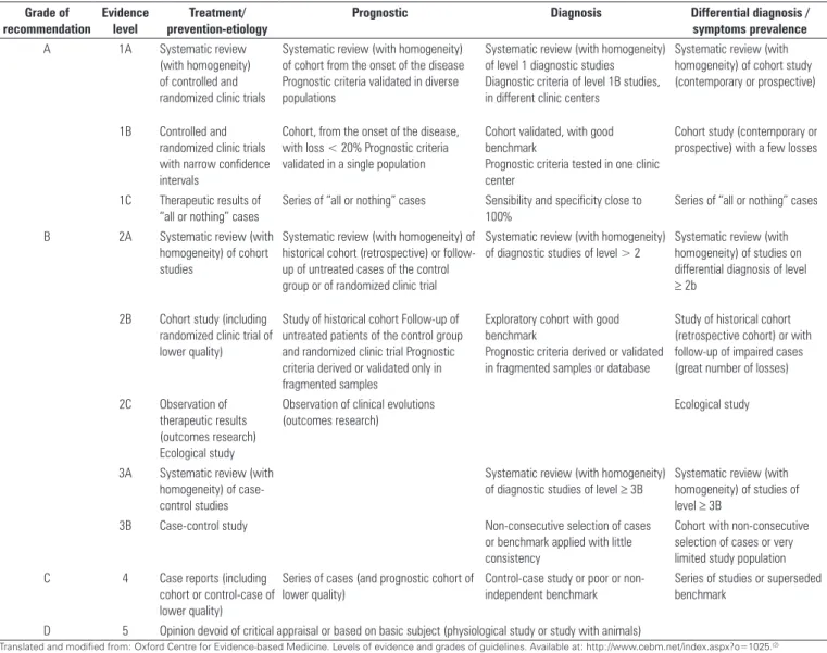
he ive physiotherapists then classiied the information based on grade of recommendation (A, B, C, or D) through the Oxford Centre method (Table 1).(2) he sixth physiotherapist
Table 1 – Level of scientific evidence by study type
Grade of recommendation
Evidence level
Treatment/ prevention-etiology
Prognostic Diagnosis Differential diagnosis /
symptoms prevalence
A 1A Systematic review (with homogeneity) of controlled and randomized clinic trials
Systematic review (with homogeneity) of cohort from the onset of the disease Prognostic criteria validated in diverse populations
Systematic review (with homogeneity) of level 1 diagnostic studies Diagnostic criteria of level 1B studies, in different clinic centers
Systematic review (with homogeneity) of cohort study (contemporary or prospective)
1B Controlled and randomized clinic trials with narrow confidence intervals
Cohort, from the onset of the disease, with loss < 20% Prognostic criteria validated in a single population
Cohort validated, with good benchmark
Prognostic criteria tested in one clinic center
Cohort study (contemporary or prospective) with a few losses
1C Therapeutic results of “all or nothing” cases
Series of “all or nothing” cases Sensibility and specificity close to 100%
Series of “all or nothing” cases
B 2A Systematic review (with homogeneity) of cohort studies
Systematic review (with homogeneity) of historical cohort (retrospective) or follow-up of untreated cases of the control group or of randomized clinic trial
Systematic review (with homogeneity) of diagnostic studies of level > 2
Systematic review (with homogeneity) of studies on differential diagnosis of level
≥ 2b
2B Cohort study (including randomized clinic trial of lower quality)
Study of historical cohort Follow-up of untreated patients of the control group and randomized clinic trial Prognostic criteria derived or validated only in fragmented samples
Exploratory cohort with good benchmark
Prognostic criteria derived or validated in fragmented samples or database
Study of historical cohort (retrospective cohort) or with follow-up of impaired cases (great number of losses)
2C Observation of therapeutic results (outcomes research) Ecological study
Observation of clinical evolutions (outcomes research)
Ecological study
3A Systematic review (with homogeneity) of case-control studies
Systematic review (with homogeneity) of diagnostic studies of level ≥ 3B
Systematic review (with homogeneity) of studies of level ≥ 3B
3B Case-control study Non-consecutive selection of cases or benchmark applied with little consistency
Cohort with non-consecutive selection of cases or very limited study population C 4 Case reports (including
cohort or control-case of lower quality)
Series of cases (and prognostic cohort of lower quality)
Control-case study or poor or non-independent benchmark
Series of studies or superseded benchmark
D 5 Opinion devoid of critical appraisal or based on basic subject (physiological study or study with animals)
reviewed the included and excluded articles and the grades of recommendation and edited the inal document.
Inclusion criteria for articles were clinical trials that were retrospective, prospective, controlled, or non-controlled randomized performed with NBs, infants, children, and/or teenagers in ICUs during MPV or within 12 hours following extubation. We included articles that addressed at least one of the six central themes of these guidelines: airway clearance, lung re-expansion, bed positioning, airway aspiration, inhalation therapy, and assisted coughing.
he following were excluded for the drafting of guidelines: editorials, simple literature reviews, systematic reviews, meta-analyses, experimental studies with animals, and case reports. Literature reviews and some articles excluded from the guidelines were used in the introduction, in the drafting of the rationale.
GUIDELINES FOR AIRWAY CLEARANCE TECHNIQUES
Based on the literature review, the following factors were included in the guidelines for airway clearance tech-niques: 1) assessment, 2) expiratory low increase (EFI), 3) manual hyperinsulation (MH), and 4) chest percussion, as well as combinations of these four categories of respira-tory physiotherapy techniques.
1. What should be assessed in NBs and children before, during, and after the airway clearance techniques?
For the safety of the patients and to ensure the efectiveness of these techniques, it is recommended to assess at least three of the following parameters before, during, and after their application: demographic characteristics (A),(3-5) vital signs (heart rate, respiratory
rate, and pulse oximetry - SpO2) (A),(3,4) noninvasive
systemic blood pressure and arterial blood gas (A),(6)
alveolar pressure and its derived indices (D),(7) dynamic
compliance and airway resistance (B),(8) inspiratory
and expiratory tidal volume (C),(9,10) forced expiratory
volume (B),(8) peak expiratory low (PEF) and its ratio
to peak inspiratory low (PIF) (B),(10) and maximum
inspiratory pressure and MPV parameters (B).(7,8,10)
Rationale
Respiratory physiotherapy techniques that are inten-ded to dislodge and/or remove secretions from airways are called “airway clearance techniques.” hey can be ad-vised or applied by physiotherapists in the neonatal and pediatric age groups,(7) in clinical situations with increased
secretion in the airways,(8,9) and for the prevention of com-plications related to MPV.(7)
Assessment of the patient allows identifying, relating, and prioritizing the problems, thereby assisting in the diagnosis and enhancing the beneits of interventions, especially considering that in most cases, airway clearance techniques are indicated in clinical situations with obstruction of airways caused by secretions and acute respiratory failure.(7)
2. In which situations and for what diseases can the EFI technique be applied?
EFI is recommended to be applied slowly to NBs and slowly or rapidly to infants with a diagnosis of severe acute bronchiolitis; this intervention is suggested to be performed least once a day (C).(9)
Rationale
During MPV, NBs and infants (aged from 26 to 41 weeks) with a diagnosis of severe acute bronchiolitis who developed the illness around the ninth week of life had increased SpO2 and tidal volume when EFI was applied 5 to 10 minutes once a day.(7)
3. Do the MH technique with a self-inlating bag and its combination with other techniques favor the mobilization and displacement of airway secretions?
MH is recommended, with or without vibrocompression, for the mobilization and removal of airway secretions in pediatrics and neonatology (A).(6)
Rationale
MH is one of the techniques used routinely in ICUs. he slow insulation of the self-inlating balloon and the inspiratory plateau recruit collapsed lung areas, while the quick release of the balloon promotes a rapid expiration, thereby increasing the expiratory low rate and helping to mobilize secretions.(11,12)
he mobilization of lower airway secretions is determined by the speed of the airlow in the airways. During the MH technique, the PEF must be greater than the PIF to displace the secretions to proximal airways, and the suitable ratio for this to occur is PIF/PEF ≤ 0.9.(10)
MH combined with vibrocompression during MPV of NBs and children (between 0 to 16 years) with a diagnosis of pulmonary consolidation or atelectasis increases the PEF, thereby increasing the volume of expired air and optimizing the clearance of lower airways.(6)
During MH, it is possible to afect the PIP and the tidal volume (e.g., by the use of one hand or both to compress the balloon, or by the use of a positive end-expiratory pressure (PEEP) valve), which inluences the pressures and volumes ofered to the patient and makes it necessary to use a pressure gauge while applying the technique to ensure safety and avoid barotrauma and/or volutrauma.(13)
he use of a pressure gauge to monitor the peak inspiratory pressure (PIP) during MH is suggested (it should not exceed 20 cmH2O and 30 cmH2O in NBs and children, respectively).
4. Which techniques are not recommended for airway clearance?
horacic percussion applied in NBs immediately after extubation is not recommended (A).(4)
We do not recommend airway desobstruction techniques such as postural drainage and/or chest vibration associated or not with MH with aspiration in cystic ibrosis children undergone endotracheal intubation in the perioperative period (B).(8)
Rationale
horacic percussion can increase intrathoracic pressure and hypoxemia. he latter efect is negligible when the technique is used for less than 30 seconds.(14) horacic per-cussion performed routinely with a duration of one to two minutes in NBs (28 to 37 weeks gestational age) immedia-tely after extubation may collapse small airways.(4)
A randomized study including 18 patients with cystic ibrosis (age varing from 3 to 15 years) undergoing anesthesia in the preoperative period compared desobstruction techniques (postural drainage and/or chest vibration, associated or not with MG and airway aspiration) with aspiration only and showed an increase in airway resistance and a decrease in pulmonary compliance. here was not standardization of the technique applied or the duration of the intervention.(8)
5. What are the main beneits, contraindications, and possible adverse efects of applying airway clearance techniques?
hese techniques improve the SpO2 of children with chronic or acute ventilatory failure(D)(7) in the short
term and increase tidal volume in children with acute bronchiolitis (C).(9)
he major contraindications of these techniques include NBs with extremely low birth weight (A)(4) and
cases of gastroesophageal relux disease (C).(9)
Possible adverse efects include reduced arterial oxygen pressure (PaO2) (D),(7) increased respiratory
rate, reduced respiratory time, and decreased lung elastic recoil pressure during MH (A).(6)
Rationale
Airway clearance techniques prevent and/or treat ai-rway obstructions caused by the presence of secretions, thereby contributing to the reduction of MPV ventilatory parameters and helping to avoid postoperative complica-tions (e.g., atelectasis) and pulmonary infeccomplica-tions. Howe-ver, the lability of the central nervous system, the weight and gestational age of NBs, and the respiratory mechanics of these techniques should be considered during their in-dication and application.(8)
GUIDELINES FOR PULMONARY RE-EXPANSION TECHINIQUES
Based on the literature review, the following pulmo-nary re-expansion techniques were included in these gui-delines: 1) MH and its combination with other techni-ques,(6,10,12,15) 2) intrapulmonary percussive ventilation,(16) and 3) thoracic compressions followed by slow and com-plete release of the rib cage (lung squeezing).(17,18)
6. What should be assessed in NBs and children before, during, and after pulmonary re-expansion techniques?
For the safety of the patients and efectiveness of these techniques, it is recommended to assess at least three of the following parameters before, during, and after their application: demographic characteristics
(A),(6,10,19-21) pulmonary auscultation (A),(20) vital signs
(heart rate, respiratory rate, and SpO2) (A),(4,12,16,17)
noninvasive systemic arterial pressure (A),(15-17) arterial
blood gas (A),(6) chest X-ray (A),(16-19) MPV time (A),(19)
dynamic and static pulmonary compliance (A),(6,10,16,18)
airway resistance (A),(6,18) fraction of inspired oxygen
(A),(17) plateau pressure (A),(16) average airway pressure
(A),(17) inspiratory (B)(10,12) and expiratory tidal
volume (A),(6,10,16,18) PIF/PEF ratio (B),(10) maximum
inspiratory pressure (A)(10,17) with or without manual
during the MH technique by the physiotherapist (D),(12) frequency and amplitude of oscillations during
manual chest vibration (D),(12) and duration of the
application of the technique(s) (A).(12,15,17)
Rationale
Respiratory physiotherapy is also intended to main-tain and/or increase pulmonary volumes (pulmonary re- expansion), and it includes a variety of techniques and resources to prevent or treat pulmonary collapses (atelec-tasis), thereby optimizing gaseous exchange and reducing respiratory efort.(19,21) Re-expansion techniques are used to increase pulmonary volumes by increasing the trans-pulmonary pressure gradient via reducing pleural pressure or increasing intra-alveolar pressure.(21)
he increased oxygenation that occurs after the application of MH results from all of the physiological efects of the technique: removal of secretions combined with alveolar recruitment. he tidal volume applied during MH reaches mainly the regions with greatest pulmonary compliance. Collapsed alveoli are re-expanded through collateral channels and the interdependence phenomenon; this efect explains the need for the evaluation of respiratory and MPV parameters and of respiratory mechanics.(22)
7. Are the MH technique with a self-inlating balloon and its combination with other techniques recommended for pulmonary re-expansion?
he application of MH with a self-inlating bag and its combination with other techniques are recommended in neonatology and pediatrics ifor pulmonary reexpansion (A).(6,10,12,15)
Rationale
During MPV with endotracheal tubes ≤ 3 mm diame-ter, pediatric patients have increased spontaneous expired tidal volume after endotracheal aspiration followed by MH with a low of 10 L/min, PIP of 30 cmH2O, and an inspired oxygen fraction of 100%.(6)
MH combined with manual chest vibration increases PEF and the volume of inspired air; increased PIP increases the lung elastic recoil, favoring the mobilization of secretions throughout the respiratory system and contributing to the re-expansion of collapsed pulmonary areas.(12) Children aged 6 months to 6 years undergoing MH with a PIP of 40 cmH2O and PEEP of 15 cmH2O during 10 cycles/minute showed a lower frequency of atelectasis compared with a continuous positive airway pressure (CPAP) of 5 cmH2O and the control group.(3,15)
MH combined with vibrocompression increases PEF by 4% for each 10% increase in the volume of inspiratory air. A 4% increase in the PEF/PIF ratio is related to a manual force of 10 N (force in Newtons).(10)
It is necessary to provide a protocol for MH according to each clinical situation to determine air volume, peak pressure to be obtained, PIF and PEF, duration and frequency of application, and the need to use a PEEP valve. he application of MH is inadvisable in patients with PEEP ≥ 10 cmH2O.
8. Is intrapulmonary percussive ventilation recommended for pulmonary re-expansion?
Intrapulmonary percussive ventilation is recommended for pulmonary re-expansion in children during MPV in a supine position over conventional respiratory physiotherapy (B).(16)
Rationale
he utilization of intrapulmonary percussive ventilation for pulmonary re-expansion in children during MPV in a supine position improves the atelectasis score and reduces its time of resolution compared to tapping and vibration.(16) Intrapulmonary percussive ventilation was used with a PIP of 15 to 30 cmH2O (or the same PIP used for MPV) and a respiratory rate of 180 to 220 cycles per minute with a mean duration of 10 to 15 minutes every 4 hours.(16)
9. Are chest compressions followed by the slow and complete release of the rib cage recommended for pulmonary re-expansion?
Performing chest compressions followed by a slow and complete release of the rib cage is recommended in preterm NBs (A).(17,18)
Rationale
Chest compressions followed by a slow and complete release of the chest/rib cage decreases the time of MPV, oxygen supplementation, and hospitalization.(17,18)
10. What are the contraindications and possible adverse efects, and which techniques of pulmonary re-expansion are not recommended in NBs, infants, and children?
situations with a reduction in pulmonary volumes, a need to increase ventilatory parameters, and/or a deterioration of blood gases (D).(12)
Chest percussion, with or without a mask, after extubation is not recommended in NBs because it can cause the collapse of small airways (A).(4)
Techniques of pulmonary re-expansion are contraindicated in NBs with extremely low birth weight (A),(4) in thrombocytopenic children with
osteopenia or osteoporosis and clinical instability (D),(12) in children at risk of intra-periventricular
hemorrhage and/or with increased intracranial pressure and metabolic bone disease (B),(10) and if
air leakage through the endotracheal tube is > 20%
(B).(10,12) Other contraindications include persistent
pulmonary hypertension, meconium aspiration syndrome, congenital heart disease with pneumonia (generalized consolidation), immediate postoperative cardiac complication, increased intracranial pressure, hemodynamic instability within the previous 24 hours (20% change in blood pressure, heart rate or in SpO2), SpO2 < 85%, pneumothorax, immediate postoperative complication following thoracoabdominal surgery, extreme prematurity or small for gestational age (B),(17) and clinical evidence
of foreign body aspiration (C).(19)
Possible adverse efects when MH is applied alone in pediatric patients include an increased respiratory rate, decreased lung elastic recoil pressure, and short-term reduction in expiratory time (A).(6)
Children (weight < 3 kg) with atelectasis can present with hypotension during intrapulmonary percussive ventilation (C).(16)
Rationale
Alveolar collapse causes a loss in pulmonary volume with a consequent decrease in functional residual capaci-ty and lung compliance, especially in gravicapaci-ty-dependent pulmonary regions. If not reversed, this can unbalance the ventilation/perfusion (V/Q) ratio with functional con-sequences, including hypoxemia, hypercapnia, increased pulmonary vascular resistance, excessive distention of ad-jacent alveolar units, risk of infection (nosocomial pneu-monia), and pulmonary injury.(18,19)
GUIDELINES FOR BED PLACEMENT
Based on the analyzed articles, considerations related to bed placement are included in these guidelines as an adjuvant to respiratory physiotherapy for airway clearance
and pulmonary re-expansion in chronically ill NBs, in-fants, and children during MPV after thoracoabdominal surgeries and during the process of MPV removal.
11. Which positions can be used for infants and children during MPV?
It is recommended to place chronically ill infants and children (cancer and neurological diseases) receiving MPV and with severe respiratory disease (PaO2/FiO2 < 200) in an elevated prone position with gel pads at the shoulders and hips (B).(20,21)
Rationale
During MPV, infants and children with respiratory disease have increased PaO2 and a reduced oxygenation index when in an elevated prone position, with cushions placed under the hips and shoulders and with the abdomen free.(20) During MPV, chronically ill children (cancer and neurological disea-ses) with severe respiratory disease (PaO2/FiO2 < 200) have an increase in PaO2/FiO2 of roughly 20% when switched from a supine to prone position for 8 hours, with a reverse efect when switched from a prone to supine position.(21)
Children with respiratory disease and a high oxygenation index exhibit an improvement in oxygenation when placed in a prone position. his result starts within the irst 2 hours after the placement and is maintained during the subsequent 12 hours.(21,22)
In NBs, the response time to the prone position, in terms of oxygenation, is variable, and non-response on the irst attempt does not mean a lack of response; however, the initial response can predict subsequent responses.(23)
12. Which positions can be used during MPV in NBs and children following thoracoabdominal surgery?
After thoracoabdominal surgeries, we recommend placing infants and children in an elevated prone position with gel pads at the shoulders and hips, as long as precautions are taken to guard the operatory wound (B).(24,25)
Rationale
Patients in the postoperative period following thoraco-abdominal surgery present with increased functional resi-dual capacities when in an elevated prone position with gel pads at the shoulders and hips.(24)
A prone position is not recommended as a routine procedure during weaning from MPV in NBs, infants, and children (B).(23,25)
Rationale
here was no signiicant diference in the weaning du-ration between the prone and supine positions. SpO2, res-piratory rate, heart rate, and incidence of atelectasis after extubation did not difer between the prone and supine positions.(23,24) NBs, infants, and children in a prone posi-tion during MPV should be monitored to avoid unplan-ned extubation or displacement of the endotracheal tube, catheters, and gastric or bladder tubes.(26,27) he prone po-sition does not alter mortality rates nor the duration of MPV in infants and children.(28,29)
GUIDELINES FOR AIRWAY ASPIRATION
Based on the literature review, guidelines are included for aspiration systems (open and closed), the need for analgesia (for pharmacological and non-pharmacological reasons), and for an increase in sedation before, during, and after the procedure of endotracheal aspiration. We also made some relevant considerations regarding the procedure’s efects on respiratory mechanics and measures to prevent the adverse efects of endotracheal aspiration in NBs, infants, and children.
14. What are the physiological efects of endotracheal aspiration systems (open versus closed) in neonatology and pediatrics?
We recommend the utilization of the closed endotracheal aspiration system to prevent a decrease of SpO2 and bradycardia in NBs under conventional MPV and for extremely preterm NBs(B).(30-32)
Rationale
When the physiological efects of open versus closed endotracheal aspiration systems were compared in NBs under conventional MPV, the results were similar(33) or fa-vored the closed aspiration system, with moderate clinical relevance regarding its efect on SpO2.(30,31)
In NBs under high frequency ventilation, there were no signiicant alterations in SpO2 when both systems were compared.(34,35) In NBs under MPV, pulmonary volume was not inluenced by the aspiration method.(36) When closed and open aspiration methods were compared in extremely preterm NBs under partial ventilation, the closed system provided a greater stability of SpO2 and heart rate.(36)
15. What are the efects of analgesia and sedation on stress reactions provoked by endotracheal aspiration in neonatology and pediatrics?
It is recommended that children under MPV with proper sedation not receive sedation prior to aspiration (B).(30)
In NBs, prior sedation should be carefully judged, and it does not change pain scores (C).(37)
Multisensory stimulation does not alter pain scores after endotracheal aspiration in NBs (C).(38)
Rationale
When the sedation score was evaluated during endo-tracheal aspiration in children under MPV, it was found that this procedure does not alter the level of sedation previously used.(32) In NBs under MPV, the use of seda-tion prior to the procedure did not inluence pain scores as measured using the Bernese Pain Scale for Neonates (BPSN), Premature Infant Pain Proile (PIPP), and Visual Analogue Scale (VAS).(37) Multisensory stimulation after the procedure did not alter pain scores in NBs.(38)
16. Do endotracheal aspiration techniques inluence the respiratory mechanics in neonatology and pediatrics?
here are indications that a reduction in pulmonary volume combined with a worsening of pulmonary ventilation and a decrease in SpO2 may occur in children undergoing conventional MPV after endotracheal aspiration (C).(39)
A transient decrease of pulmonary volume occurs in NBs under high frequency ventilation after endotracheal aspiration using both closed and open aspiration systems(C).(34,35)
Alveolar recruitment through a self-inlatable balloon immediately following endotracheal aspiration is not recommended to improve dynamic compliance and expiratory tidal volume of children under MPV (B).(39)
Rationale
17. Should interventions be undertaken to prevent adverse efects of endotracheal aspiration in NBs, infants, and children during MPV?
Hyperoxia (10% increase over baseline fraction of inspired oxygen) is recommended in preterm NBs to avoid hypoxemia during and after endotracheal aspiration to maintain SpO2 between 88 and 92% (C).(38)
Postural restraint maneuvers are recommended during aspiration procedures in preterm NBs (C).(37)
Rationale
In NBs, hyperoxia (a 10% increase over baseline frac-tion of inspired oxygen) led to favorable efects on the reduction of transient hypoxemia resulting from endotra-cheal aspiration by an open system.(38)
Manual restraint maneuvers, through the placement of the hands on the head and feet of NBs in lexed posture, reduced pain scores during the procedure in preterm NBs.(37)
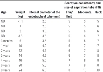
he criteria in table 2 are suggested to enhance the safety of the procedure, and they indicate that airway aspiration of intubated NBs, infants, and children be performed for a maximum of 10 seconds (to prevent ventilatory and hemodynamic alterations inherent to disconnecting the patient from the MPV apparatus) and that the vacuum suction pressure should not exceed 360 mmHg.(40)
Table 2 - Guidelines for airway aspiration in newborns, infants, and children
Secretion consistency and size of aspiration tube (FG)
Age Weight
(kg)
Internal diameter of the endotracheal tube (mm)
Thin/ fluid
Moderate Thick
NB < 1 2.0 5 5 5
NB 1 2.5 5 5 6
NB 2 3.0 5 6 6
NB 3.5 3.5 5 6 7
3 months 6 3.5 5 6 7
1 year 10 4.0 6 7 7
2 years 12 4.5 6 7 8
3 years 14 4.5 6 7 8
4 years 16 5.0 7 8 8
6 years 20 5.5 7 8 8
8 years 24 6.0 8 10 10
NB - newborn; FG - French gauge. Modified from: Morrow BM, Futter MJ, Argent AC. Endotracheal suctioning: from principles to practice. Intensive Care Med. 2004;30(6):1167-74.(40)
To facilitate the appraisal of the multiprofessional team, it is suggested to classify the aspirated secretions as thin or luid (the aspiration tube becomes free of secretions after aspiration using only the vacuum), moderate (when secretions adhere to the tube wall after aspiration but are removed by saline
solution), or thick (when secretions adhere to the tube wall after aspiration even after the use of saline solution).
GUIDELINES FOR INHALATION THERAPY
Based on the analyzed articles, hypertonic saline solu-tion (HS) at 3% and dornase alfa (rhDNA) were included.
18. In which situations can a 3% HS solution be used in NBs and children?
he use of a 3% HS solution is recommended for infants with viral bronchiolitis for the reduction of the disease symptoms and hospitalization time (A)(41) and
for infants with mild to moderate bronchiolitis for the resolution of atelectasis (C).(42)
he use of a 3% HS solution is recommended for NBs with persistent atelectasis unresponsive to conventional treatment (C).(43)
Rationale
Aerosol administration allows the direct and immedia-te availability of drugs in the airways. Drug use through inhalation is an adjuvant to respiratory physiotherapy du-ring techniques for removing secretions from airways and for pulmonary re-expansion.(44) his technique can only be applied with a medical prescription.
A controlled, randomized, double-blind study(41) assessed the use of inhaled 3% HS solution compared with 0.9% saline solution, both in conjunction with epinephrine (1.5 mg), in 52 hospitalized children with acute viral bronchiolitis. he group that used the 3% HS solution exhibited a decrease in symptoms and time of hospitalization.
One study(41) of children with moderate to severe acute bronchiolitis showed a reduction in hospitalization time and faster relief of symptoms in the group using nebulization with the 3% HS solution compared to groups using the 0.9% saline solution not combined with other drugs. Nebulization with 3% HS is safe, inexpensive, efective, well-tolerated, and has no adverse efects.
A controlled and randomized study(42) of 93 infants with mild to moderate bronchiolitis compared the efectiveness and safety of 3% HS solution, salbutamol with 0.9% isotonic saline (SS), and salbutamol on bronchoconstriction, signs of discomfort, and hospitalization time. he group treated with 3% HS solution had a shorter remission time of the bronchoconstriction, remission time of the cough, and hospitalization time.
radiographic scores and SpO2 and reduces the resolution time of the atelectasis.(44) In infants with viral bronchiolitis, HS reduces the disease symptoms and hospitalization time;(44) in infants with mild to moderate bronchiolitis, HS can improve the bronchospasm and signs of respiratory distress and reduce the resolution time of the atelectasis.(42)
19. In which situations is rhDNA recommended?
rhDNA administered by inhalation or via the endotracheal tube is recommended for non-ibrocystic children with persistent atelectasis (C).(45)
Inhalation is recommended for NBs who do not respond to conventional treatment for atelectasis (D);(46) for children during MPV in the postoperative
period of cardiac surgery subjected to direct instillation in the endotracheal tube; and for infants and children (age <2 years) with acute bronchiolitis submitted to daily inhalation for 5 consecutive days for pulmonary re-expansion (B).(47)
It is recommended for the resolution of pulmonary atelectasis in children during prolonged MPV (C).(47-49)
Rationale
Drugs called mucolytics break up secretions and cau-se the lysis of mucoibrils; they are indicated in cacau-ses of hypersecretion with increased mucus viscosity. Infection and inlammation of airways as a result from any con-dition cause signiicant quantities of extracellular DNA, which is a product of the degeneration of leukocytes and epithelial debris. he DNA increases the viscosity and adhesion of the mucus in the airways. In pulmonary in-fections aggravated by atelectasis, high concentrations of DNA can be found, thus making the indication of dorna-se alfa application possible.(45,48)
rhDNA administered through inhalation is used for NBs who did not respond to conventional treatment for atelectasis, aiming to improve radiological scores and respiratory parameters;(46) for children during MPV in the postoperative period of cardiac surgery with luid restriction submitted to direct instillation in the endotracheal tube (dose: 0.2 mg/kg in patients with weight < 5 kg, 0.1 mg/kg for weight > 5 kg; every 12 hours) because it reduces the incidence of atelectasis, the average time of MPV, the average ICU stay, and average costs;(47) and for infants and children (age < 2 years) with acute bronchiolitis, submitted to daily inhalation for ive consecutive days because it yields an improvement in atelectasis score.(50,51) It can only be applied after medical prescription.
GUIDELINES FOR ASSISTED COUGH
Several interventions of respiratory physiotherapy can be indicated for airway clearance to facilitate secretion removal. Among them, mechanically (mechanical insulation-exsu-lation - MIE) and manually assisted coughing have been indicated for children with impaired efectiveness of the cou-gh because it facilitates the expectoration of secretions from airways when applied alone or combined with other manual or mechanical respiratory physiotherapy techniques.(52)
20. In which situations is assisted cough indicated and how it can be performed?
It is recommended for children with neuromuscular diseases and acute or chronic respiratory diseases who present with excessive secretions in airways of diicult expectoration and/or pulmonary atelectasis and/or PEF < 270 L/min (B).(52-54)
Assisted cough can be manually or mechanically performed (B).(52,55)
Rationale
Cough is the most common sign and symptom of di-seases of the respiratory system.(56) his relex is part of the defense mechanism of the airways and can be reproduced and controlled voluntarily or mechanically. Its presence can be correlated to several diseases (e.g., lu, bronchioli-tis, tracheibronchioli-tis, and asthma) and can be acute or chronic.(56) Children with neuromuscular diseases hospitalized in ICUs with an etiology of respiratory disease have a 90% greater risk of morbidity and mortality due to the impossibility of maintaining alveolar ventilation and eliminating secretions from airways because the cough, impaired by the primary disease, becomes less efective in the presence of respiratory diseases and increased volume and consistency of the mucus.(57)
Manually assisted cough through chest or abdomen compression synchronized with the cough performed by the patient increases the PEF, thereby aiding expectoration in cases of mild to moderate cough alterations. Cough assisted combined with MH can increase the efectiveness of the technique.(55)
21. What are the possible adverse efects and contraindications of the assisted cough?
he main adverse efects found were abdominal distention, increased gastroesophageal relux, hemoptysis, chest and/or abdominal discomfort, acute cardiovascular alterations (such as bradycardia), and pneumothorax. he application of these techniques is not recommended in children previously presenting with the clinical conditions mentioned above (B).(52)
Rationale
he main role of the cough is the detachment and displa-cement of the material within the airways during the expulsi-ve phase. he detachment of the secretions occurs as a func-tion of the viscosity, elasticity, and thickness of the mucus and its level of adhesion to the airway wall. he lexibility of the bronchial wall facilitates the transmission of an undulation or transient pressure undulation that, when produced by the cough, rapidly displaces the mucus to the mouth.(58)
CLOSING REMARKS
In this article, we have presented the guidelines for several respiratory physiotherapy interventions for patients in neo-natal and pediatric ICUs under MPV and within 12 hours following extubation, based on exiting evidence. Several te-chniques applied during respiratory physiotherapy were not
included in these guidelines due to lack of scientiic evidence. he role of the physiotherapist in these areas is broader, requiring continuous development of other guidelines for clinical practice with the aim to improve patient safety.
RESUMO
Recomendações para a atuação do isioterapeuta em unidade de terapia intensiva pediátrica e neonatal são fundamentais, pois esses proissionais são responsáveis pela reabilitação de pacientes graves. A reabilitação inclui desde a avaliação e prevenção de altera-ções cinético funcionais às intervenaltera-ções de tratamento (isioterapia respiratória e/ou motora), controle e aplicação de gases medicinais, cuidados da ventilação pulmonar mecânica invasiva e não invasi-va, protocolos de desmame e extubação, insulação traqueal de gás, protocolo de insulação/desinsulação do balonete intratraqueal, aplicação de surfactante, entre outros. Com o objetivo de propiciar a recuperação do doente e seu retorno às atividades funcionais.
Nesse contexto, essas recomendações têm o objetivo de orientar os isioterapeutas sobre algumas intervenções de pre-venção/tratamento de isioterapia respiratória (desobstrução das vias aéreas; reexpansão pulmonar; posicionamento no lei-to; aspiração das vias aéreas; inaloterapia; tosse assistida), que auxiliam no processo de reabilitação de pacientes pediátricos e neonatais em unidade de terapia intensiva em ventilação pul-monar mecânica e até 12 horas após a extubação.
Descritores: Reabilitação; Terapia respiratória; Modalidades de isioterapia; Terapia intensiva neonatal; Respiração artiicial; Criança
REFERENCES
1. Brasil. Ministério da Saúde. Agência Nacional de Vigilância Sanitária. Resolução-RDC nº 7, de 24 de fevereiro de 2010. Dispõe sobre os requisitos mínimos para funcionamento de Unidades de Terapia Intensiva e dá outras providências. Disponível em: http://brasilsus.com.br/legislacoes/ rdc/102985-7.html
2. Oxford Centre for Evidence-based Medicine. Levels of evidence and grades of recommendations. Disponível em: http://www.cebm.net/index. aspx?o=1025.
3. Bailleux S, Lopes D, Geoffroy A, Josse N, Labrune P, Gajdos V. [What evidence for chest physiotherapy in infants hospitalized for acute viral bronchiolitis?]. Arch Pediatr. 2011;18(4):472-5. French.
4. Bagley CE, Gray PH, Tudehope DI, Flenady V, Shearman AD, Lamont A. Routine neonatal postextubation chest physiotherapy: a randomized controlled trial. J Paediatr Child Health. 2005;41(11):592-7.
5. Knight DB, Bevan CJ, Harding JE, Teele RL, Kuschel CA, Battin MR, et al. Chest physiotherapy and porencephalic brain lesions in very preterm infants. J Paediatr Child Health. 2001;37(6):554-8.
6. Morrow B, Futter M, Argent A. A recruitment manoeuvre performed after endotracheal suction does not increase dynamic compliance in ventilated paediatric patients: a randomised controlled trial. Aust J Physiother. 2007;53(3):163-9.
7. Almeida CC, Ribeiro JD, Almeida-Júnior AA, Zeferino AM. Effect of expiratory flow increase technique on pulmonary function of infants on mechanical
ventilation. Physiother Res Int. 2005;10(4):213-21.
8. Tannenbaum E, Prasad SA, Dinwiddie R, Main E. Chest physiotherapy during anesthesia for children with cystic fibrosis: effects on respiratory function. Pediatr Pulmonol. 2007;42(12):1152-8.
9. Bernard-Narbonne F, Daoud P, Castaing H, Rousset A. [Effectiveness of chest physiotherapy in ventilated children with acute bronchiolitis]. Arch Pediatr. 2003;10(12):1043-7. French.
10. Gregson RK, Shannon H, Stocks J, Cole TJ, Peters MJ, Main E. The unique contribution of manual chest compression-vibrations to airflow during physiotherapy in sedated, fully ventilated children. Pediatr Crit Care Med. 2012;13(2):e97-e102.
11. Clini E, Ambrosino N. Early physiotherapy in the respiratory intensive care unit. Respir Med. 2005;99(9):1096-104.
12. Gregson RK, Stocks J, Petley GW, Shannon H, Warner JO, Jagannathan R, et al. Simultaneous measurement of force and respiratory profiles during chest physiotherapy in ventilated children. Physiol Meas. 2007;28(9):1017-28. 13. Redfern J, Ellis E, Holmes W. The use of a pressure manometer enhances
student physiotherapists’ performance during manual hyperinflation. Aust J Physiother. 2001;47(2):121-31.
14. Lamari NM, Martins ALQ, Oliveira JV, Marino LC, Valério N. Bronquiectasia e fisioterapia desobstrutiva: ênfase em drenagem postural e percussão. Braz J Cardiovasc Surg. 2006;21(2):206-10.
16. Deakins K, Chatburn RL. A comparison of intrapulmonary percussive ventilation and conventional chest physiotherapy for the treatment of atelectasis in the pediatric patient. Respir Care. 2002;47(10):1162-7. 17. Wong I, Fok TF. Randomized comparison of two physiotherapy regimens
for correcting atelectasis in ventilated pre-term neonates. Hong Kong Physiother J. 2003;21(1):43-50.
18. Wong I, Fok TF. Effects of lung squeezing technique on lung mechanics in mechanically-ventilated preterm infants with respiratory distress syndrome. Hong Kong Physiother J. 2006;24(1):39-46.
19. Bilan N, Galehgolab BA, Shoaran M. Medical treatment of lung collapse in children. Pak J Biol Sci. 2009;12(5):467-9.
20. Nightlinger K. Developmentally supportive care in the neonatal intensive care unit: an occupational therapist’s role. Neonatal Netw. 2011;30(4):243-8. 21. Unoki T, Mizutani T, Toyooka H. Effects of expiratory rib cage compression
and/or prone position on oxygenation and ventilation in mechanically ventilated rabbits with induced atelectasis. Respir Care. 2003;48(8):754-62. 22. Relvas MS, Silver PC, Sagy M. Prone positioning of pediatric patients with
ARDS results in improvement in oxygenation if maintained > 12 h daily. Chest. 2003;124(1):269-74.
23. Antunes LC, Rugolo LM, Crocci AJ. Efeito da posição do prematuro no desmame da ventilação mecânica. J Pediatr (Rio J). 2003;79(3):239-44. 24. von Ungern-Sternberg BS, Hammer J, Frei FJ, Jordi Ritz EM, Schibler A,
Erb TO. Prone equals prone? Impact of positioning techniques on respiratory function in anesthetized and paralyzed healthy children. Intensive Care Med. 2007;33(10):1771-7.
25. Fineman LD, LaBrecque MA, Shih MC, Curley MA. Prone positioning can be safely performed in critically ill infants and children. Pediatr Crit Care Med. 2006;7(5):413-22.
26. Curley MA, Thompson JE, Arnold JH. The effects of early and repeated prone positioning in pediatric patients with acute lung injury. Chest. 2000;118(1):156-63.
27. Curley MA, Hibberd PL, Fineman LD, Wypij D, Shih MC, Thompson JE, et al. Effect of prone positioning on clinical outcomes in children with acute lung injury: a randomized controlled trial. JAMA. 2005;294(2):229-37.
28. Casado-Flores J, Martínez de Azagra A, Ruiz-López MJ, Ruiz M, Serrano A. Pediatric ARDS: effect of supine-prone postural changes on oxygenation. Intensive Care Med. 2002;28(12):1792-6.
29. Kornecki A, Frndova H, Coates AL, Shemie SD. 4A randomized trial of prolonged prone positioning in children with acute respiratory failure. Chest. 2001;119(1):211-8.
30. Paula LCS, Ceccon MEJ. Randomized comparative analysis between two tracheal suction systems in neonates. Rev Assoc Med Bras. 2010;56(4):434-9. 31. Taheri P, Asgari N, Mohammadizadeh M, Golchin M. The effect of open and
closed endotracheal tube suctioning system on respiratory parameters of infants undergoing mechanical ventilation. Iran J Nurs Midwifery Res. 2012;17(1):33-7.
32. Hoellering AB, Copnell B, Dargaville PA, Mills JF, Morley CJ, Tingay DG. Lung volume and cardiorespiratory changes during open and closed endotracheal suction in ventilated newborn infants. Arch Dis Child Fetal Neonat Ed. 2008;93(6):F436-41.
33. Avena MJ, Carvalho WB, Beppu OS. Avaliação da mecânica respiratória e da oxigenação pré e pós-aspiração de secreção em crianças submetidas à ventilação pulmonar mecânica. Rev Assoc Med Bras. 2003;49(2):156-61. 34. van Veenendaal MB, Miedema M, de Jongh FH, van der Lee JH, Frerichs
I, van Kaam AH. Effect of closed endotracheal suction in high-frequency ventilated premature infants measured with electrical impedance tomography. Intensive Care Med. 2009;35(12):2130-4.
35. Tingay DG, Copnell B, Mills JF, Moreley CJ, Dargaville PA. Effects of open endotracheal suction on lung volume in infants receiving HFOV. Intensive Care Med. 2007;33(4):689-93.
36. Tan AM, Gomez JM, Mathews J, Williams M, Paratz J, Rajadurai VS. Closed versus partially ventilated endotracheal suction in extremely preterm neonates: physiologic consequences. Intensive Crit Care Nurs. 2005;21(4):234-42. 37. Ward-Larson C, Horn RA, Gosnell F. The efficacy of facilitated tucking for
relieving procedural pain of endotracheal suctioning in very low birthweight
infants. MCN Am J Matern Child Nurs. 2004;29(3):151-6; quiz 157-8. 38. Cabello H, Furuya ME, Vargas MH, Tudón H, Garduño J,
González-Ayala J. Evaluation of antihypoxemic maneuvers before tracheal aspiration in mechanically ventilated newborns. Pediatr Pulmonol. 2005;39(1):46-50. 39. Morrow B, Futter M, Argent A. Effect of endotracheal suction on lung
dynamics in mechanically-ventilated paediatric patients. Aust J Physiother. 2006;52(2):121-6.
40. Morrow BM, Futter MJ, Argent AC. Endotracheal suctioning: from principles to practice. Intensive Care Med. 2004;30(6):1167-74.
41. Mandelberg A, Tal G, Witzling M, Someck E, Houri S, Balin A, et al. Nebulized 3% hypertonic saline solution treatment in hospitalized infants with viral bronchiolitis. Chest. 2003;123(2):481-7.
42. Luo Z, Liu E, Luo J, Li S, Zeng F, Yang X, et al. Nebulized hypertonic saline/ salbutamol solution treatment in hospitalized children with mild to moderate bronchiolitis. Pediatr Int. 2010;52(2):199-202.
43. Erdeve O, Uras N, Atasay B, Arsan S. Efficacy and safety of nebulized recombinant human DNase as rescue treatment for persistent atelectasis in newborns: case-series. Croat Med J. 2007;48(2):234-9.
44. De Luca D, Cogo P, Zecca E, Piastra M, Pietrini D, Tridente A, et al. Intrapulmonary drug administration in neonatal and paediatric critical care: a comprehensive review. Eur Respir J. 2011;37(3):678-89
45. Hendriks T, de Hoog M, Lequin MH, Devos AS, Merkus PJ. DNase and atelectasis in non-cystic fibrosis pediatric patients. Crit Care. 2005;9(4):R351-6.
46. Dilmen U, Karagol BS, Oguz SS. Nebulized hypertonic saline and recombinant human DNase in the treatment of pulmonary atelectasis in newborns. Pediatr Int. 2011;53(3):328-31.
47. Prodhan P, Greenberg B, Bhutta AT, Hyde C, Vankatesan A, Imamura M, et al. Recombinant human deoxyribonuclease improves atelectasis in mechanically ventilated children with cardiac disease. Congenit Heart Dis. 2009;4(3):166-73.
48. Riethmueller J, Borth-Bruhns T, Kumpf M, Vonthein R, Wiskirchen J, Stern M, et al. Recombinant human deoxyribonuclease shortens ventilation time in young, mechanically ventilated children. Pediatr Pulmonol. 2006;41(1):61-6. Erratum in Pediatr Pulmonol. 2006;41(4):388.
49. Riethmueller J, Kumpf M, Borth-Bruhns T, Brehm W, Wiskirchen J, Sieverding L, et al. Clinical and in vitro effect of dornase alfa in mechanically ventilated pediatric non-cystic fibrosis patients with atelectases. Cell Physiol Biochem. 2009;23(1-3):205-10.
50. Nasr SZ, Strouse PJ, Soskolne E, Maxvold NJ, Garver KA, Rubin BK, et al. Efficacy of recombinant human deoxyribonuclease I in the hospital management of respiratory syncytial virus bronchiolitis. Chest. 2001;120(1):203-8.
51. Kuzik BA, Al-Qadhi SA, Kent S, Flavin MP, Hopman W, Hotte S, et al. Nebulized hypertonic saline in the treatment of viral bronchiolitis in infants. J Pediatr. 2007;151(3):266-70, 270.e1.
52. Yates K, Festa M, Gillis J, Waters K, North K. Outcome of children with neuromuscular disease admitted to paediatric intensive care. Arch Dis Child. 2004;89(2):170-5.
53. Panitch HB. Airway clearance in children with neuromuscular weakness. Curr Opin Pediatr. 2006;18(3):277-81.
54. Sivasothy P, Brown L, Smith IE, Shneerson JM. Effect of manually assisted cough and mechanical insufflation on cough flow of normal subjects, patients with chronic obstructive pulmonary disease (COPD), and patients with respiratory muscle weakness. Thorax. 2001;56(6):438-44.
55. Gauld LM, Boynton A. Relationship between peak cough flow and spirometry in Duchenne muscular dystrophy. Pediatr Pulmonol. 2005;39(5):457-60. 56. Chatwin M, Ross E, Hart N, Nickol AH, Polkey MI, Simonds AK. Cough
augmentation with mechanical insufflation/exsufflation in patients with neuromuscular weakness. Eur Respir J. 2003;21(3):502-8.
57. O’Connell F, Thomas VE, Pride NB. Adaptation of cough reflex with different types of stimulation. Eur Respir J. 1992;5(10):1296-7.
58. Morice AH, Fontana GA, Belvisi MG, Birring SS, Chung KF, Dicpinigaitis PV, Kastelik JA, McGarvey LP, Smith JA, Tatar M, Widdicombe J; European Respiratory Society (ERS). ERS guidelines on the assessment of cough