SUMMARY
Objective: To characterize the indications of pregnant women who sought the Fetal Medicine Services of the Hospital das Clínicas, at the Medical School of the Universidade de São Paulo for performing invasive diagnostic procedures, and to evaluate the results of fetal karyotypes and their pregnancies. Methods: A retrospective and observational study on pregnant women who underwent chorionic villus sampling (CVS), amniocen-tesis, and cordocentesis in the period from February, 2005 to December, 2009. Other diagnostic or therapeutic procedures were not included. he result of pregnancy was ob-tained by consulting patient electronic records, medical records, and/or telephone call.
Results: 713 procedures were performed (113 CVS, 340 amniocenteses, and 260 cordo-centeses). he main indication for performing invasive procedures was the presence of structural changes in fetuses, followed by increased values of nuchal translucency, and advanced maternal age. Fetal karyotype was altered in 186 cases (26.1%). he18 trisomy was the commonest aneuploidy followed by the 21 trisomy, X monosomy, and 13 tri-somy. here were 4.9% cases of miscarriage, 25.7% cases of stillborn infants, and 13% cases of neonatal deaths. Eight pregnant women opted for legally induced abortion. 99% of pregnant women whose fetuses did not present abnormalities and presented normal fetal karyotype had infants who were born alive.
Uniterms: Karyotype; chorionic villus sampling; fetal blood; fetus.
©2012 Elsevier Editora Ltda. All rights reserved.
Study conducted at the Medical School of the Universidade de São Paulo, São Paulo, SP, Brazil
Submitted on: 05/23/2012
Approved on: 06/30/2012
Correspondence to:
Mário Henrique Burlacchini de Carvalho Department of Obstetrics and
Gynecology Medical School Universidade de São Paulo Av. Dr. Enéas de Carvalho Aguiar, 255 Instituto Central, 10º andar São Paulo, SP, Brazil CEP: 05403-000 Tel: +55 11 2661-6209 [email protected]
Conlict of interest: None.
Analysis of fetal and maternal results from fetal genetic invasive
procedures: an exploratory study at a University Hospital
MARIO KOHATSU1, MÁRIO HENRIQUE BURLACCHINIDE CARVALHO2, ROSSANA PULCINELI VIEIRA FRANCISCO2, ANTÔNIO GOMESDE AMORIM FILHO3, MARCELO ZUGAIB4
1 MSc in Sciences, Department of Obstetrics and Gynecology, Faculdade de Medicina da Universidade de São Paulo (FMUSP), São Paulo, SP, Brazil 2 Professors, Department of Obstetrics and Gynecology, FMUSP, São Paulo, SP, Brazil
INTRODUCTION
Pre-natal screening for aneuploidy started in the 1970s, with maternal age as its main indication1.
he risk of chromosomal abnormalities increases with the maternal age. In a study that evaluated approximately 89,000 classical amniocenteses, pregnant women aged 35 years or over presented more chromosomal changes than women aged less than 35 years. he frequencies of trisomy 21, trisomy 18, and trisomy 13 for women aged over 35 years were1/100, 1/454, and 1/1438, respectively. Con-versely, the frequencies for women aged under 35 years were 1/591, 1/2862, and 1/4651 for the same aneuploidies2. he use of maternal age of 35 years or more as a cut-of point for indicating fetal karyiotype research presents a sensitivity of approximately 30%.
In the 1990s, biochemical screening in the second tri-mester started by evaluating alpha-fetoprotein, beta cho-rionic gonadotropin (β-HCG), and unconjugated estriol in maternal plasma, with a sensitivity of 60% and a false-positive rate of 5% for trisomy 213. With the improvement of ultrasound devices and equipment, screening was ad-vanced to the irst trimester by nuchal translucency evalu-ation. he detection rate for Down syndrome through nuchal translucency is 77%, with a false-positive rate of 5%4. he association of nuchal translucency with protein dosage in maternal blood, maternal serum free-beta-cho-rionic gonadotropin (free β-HCG) and pregnancy-associ-ated plasma protein-A (PAPP-A) raises the sensitivity to 90%, with the same false-positive rate5,6. Another beneit of combined screening is the reduction of cases with in-dication for invasive procedure, reducing the exposure of pregnant women to the risks of such a procedure7.
Morphological ultrasound scan can be also used for screening chromosomal anomalies in the second trimes-ter of gestation. Some abnormalities can also be related to aneuploidy, such as ventriculomegaly, facial clet, cardi-opathy, diaphragmatic hernia, nephrcardi-opathy, omphalocele, shortened limbs, and club-foot8,9.
he deinite diagnosis of chromosomal abnormality in the antenatal period is only possible by performing inva-sive procedures and analysis of the fetal tissue or its compo-nents, such as trophoblast, amniotic luid, and fetal blood10. Chorionic villus sampling (CVS) presents a risk of total fatal loss varying from 2.3% to 3.7%11-13. hose rates take into account the early procedure, and as a consequence, the presence in this group of chromosomally altered fe-tuses that would evolve to spontaneous abortion. he ad-vantage of this examination is the early gestational age of diagnosis. he main disadvantage is conined placental mosaicism, which occurs in approximately 1% of the cases and causes the need to repeat the procedure in another environment (amniotic luid or fetal blood)14,15. Classical amniocentesis ofers a risk of fetal loss of 0.3% to 1.0%16-18.
Cordocentesis is a procedure that allows for the evaluation of fetal karyotype, besides anemia diagnosis, infections, and fetal hemoglobinopathies. his procedure presents a 1.4% risk of fetal loss19.
At the Obstetrics Department of the Hospital das Clínicas of the Medical School at the Universidade de São Paulo (HCFMUSP), the Fetal Medicine Services is a refer-ence for cases of fetuses with abnormalities or increased risk for aneuploidies. he service performs invasive pro-cedures for fetal karyotype studies in cases diagnosed at the service and in cases referred to from external services. However, data regarding fetal procedures, karyotype re-sults, and evolution of pregnant women who underwent an invasive procedure have not yet been published, and national data are scarce. Publishing those data is relevant, as it represents a reference service in the city of São Paulo and provides useful information on advice to future preg-nant women, at local and national level.
he main objective of this study was to characterize the indications of pregnant women who sought the Fetal Med-icine Services of a tertiary center for performing invasive diagnostic procedures for fetal karyotype studies. Second-ary objectives were to evaluate fetal kSecond-aryotype results and their gestations.
METHODS
A retrospective, observational, and cross-sectional study was performed at the Fetal Medicine Services of the Ob-stetric Department at the HCFMUSP; the study was ap-proved by the ethics in research committee of the institu-tion (CaPPesq – HCFMUSP), No. 0600/09.
Pregnant women who underwent invasive procedures (CVS, amniocentesis, and cordocentesis) were selected in the period from February, 2005 to December, 2009.
Pregnant women were referred from other fetal medi-cine services or from this institution. Ater genetic coun-seling and explanation of the risks related to the proce-dure, the pregnant woman or her legal guardian signed an informed consent. Before performing the invasive proce-dure, all pregnant women underwent morphological ul-trasonography for evaluation of fetal structure.
Only pregnant women who underwent invasive diag-nostic procedures for fetal karyotype (CVS, amniocentesis and cordocentesis) were included. CVS was performed between 11 and 14 weeks, six days of gestation; amniocen-tesis, from 14 weeks; and cordocenamniocen-tesis, from 19 weeks.
Procedures with other purposes, such as amniotic luid drainage, placement of drains, and laser or punctures for fetal karyotype collected from other dry secretions were not included.
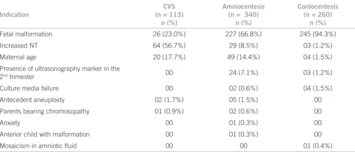
Indication
CVS (n = 113)
n (%)
Amniocentesis (n = 340)
n (%)
Cordocentesis (n = 260)
n (%)
Fetal malformation 26 (23.0%) 227 (66.8%) 245 (94.3%)
Increased NT 64 (56.7%) 29 (8.5%) 03 (1.2%)
Maternal age 20 (17.7%) 49 (14.4%) 04 (1.5%)
Presence of ultrasonography marker in the
2nd trimester 00 24 (7.1%) 03 (1.2%)
Culture media failure 00 02 (0.6%) 04 (1.5%)
Antecedent aneuploidy 02 (1.7%) 05 (1.5%) 00
Parents bearing chromosopathy 01 (0.9%) 02 (0.6%) 00
Anxiety 00 01 (0.3%) 00
Anterior child with malformation 00 01 (0.3%) 00
Mosaicism in amniotic luid 00 00 01 (0.4%)
CVS, chorionic villus sampling; NT, nuchal translucency.
Table 1 – Indications for performing invasive procedures for fetal karyotype research. HCFMUSP, 2005 to 2009 on Obstetrics and Gynecology (Sistema Informatizado de
Laudos em Obstetrícia e Ginecologia — SILOG), which is used by the Fetal Medicine Services for ultrasonography scans report, invasive procedures, and fetal and gestation scan results. Postnatal results were obtained by consulting paper-based medical records in the HCFMUSP and in SI-LOG, and/or by telephone call to patients.
Population features evaluated were: patients’ age; indi-cations for performing invasive procedures; number and type of procedures performed (CVS, amniocentesis, and cordocentesis); results of fetal karyotype; and evolution of gestations (legally and non-legally induced abortion; miscarriage deined as gestation loss up to twenty weeks of pregnancy; stillborn, deined as birth of fetus dead ater twenty weeks of pregnancy; and neonatal deaths, deined as death until 28 days post-birth)20.
Numerical variables were described as maximum, minimum, average and standard deviation, or median. For the categorical variables, simple and relative frequencies were used.
RESULTS
In the period studied, 713 diagnostic invasive procedures were performed as follows: 113 CVS, 340 amniocentesis, and 260 cordocentesis. Table 1 describes indications for diagnostic invasive procedures performed during the pe-riod studied.
Regarding CVS, maternal age for performing the pro-cedure varied from 15 to 45 years, with an average of 32.4 (± 7.7) years. he average gestational age was 13.4 (± 1.3) weeks of pregnancy.
In the group of pregnant women who underwent am-niocentesis, maternal age for performing the procedure var-ied from 14 to 47 years, with an average of 30.7 (± 8.2) years.
he gestational age for performing the procedures varied from 14.3 to 34 weeks, with an average of 20.1 (± 3.6) weeks. Among the cases of amniocen-tesis, a case of maternal death was observed 22 days ater the procedure, in a case of high risk to the mother’s life with a diagnosis of sickle-cell anemia.
Regarding cordocentesis, maternal age varied from 13 to 48 years, with an average of 28 (± 7.5). he average gestational age was of 27.1 (± 3.6) weeks of pregnancy.
From those pregnant women who underwent cordocentesis, 53.5% (139/260 had children born alive, 24.6% (64/260) had stillborn children, 20.8% (54/260) had neonatal death, and 1.6% (3/260) opt-ed for legally inducopt-ed abortion.
Table 2 shows the results of fetal karyotypes from the cases submitted to invasive procedures at the Fetal Medicine Services of the Obstetrics De-partment at the HCFMUSP in the period studied. he result of pregnancies is shown in Table 3.
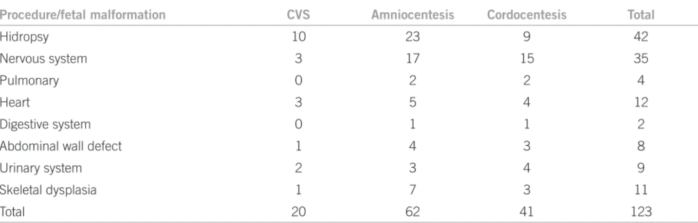
Table 4 describes the main fetal abnormalities in fetuses evolving to intrauterine death.
DISCUSSION
he majority of patients who were referred to this Fetal Medicine Services presented morphological fetal changes, considering this was the main reason for performing fetal karyotype invasive procedures, representing 69.8% of indications.
Procedure/karyotype CVS n (%)
Amniocentesis n (%)
Cordocentesis n (%)
Total n (%)
Normal 77 (68.2%) 214 (63%) 187 (67.3%) 478 (67%)
13 trisomy 03 (2.6%) 12 (3.5%) 12 (4.6%) 27 (3.8%) 18 trisomy 09 (8.0%) 30 (8.8%) 19 (7.3%) 58 (8.1%)
21 trisomy 10 (8.8%) 20 (5.9%) 14 (5.4%) 44 (6.2%) X monosomy 06 (5.3%) 24 (7.1%) 04 (6.2%) 34 (4.8%)
Triploidy 0 04 (1.2%) 01 (0.4%) 05 (0.7%)
Other aneuploidy 01 (0.9%) 08 (2.3%) 10 (3.8%) 19 (2.7%)
Culture media failure 07 (6.2%) 28 (8.2%) 13 (5.0%) 48 (6.7%) CVS, chorionic villus sampling.
Table 2 – Results of fetal karyotypes of the cases submitted to diagnostic invasive procedures.
Procedure/result
CVS 113 n (%)
Amniocentesis 340 n (%)
Cordocentesis 260 n (%)
Total 713 n (%)
Total intrauterine death 39 (34.5%) 125 (36.8%) 67 (25.8%) 231(32.4%)
Total intrauterine death associated with
fetal malformation or abnormal karyotype 39 (34.5%) 124 (36.4%) 67 (25.8%) 230 (32.2%)
Legally induced abortion 01 (0.9%) 04 (1.2%) 03 (1.2%) 8 (1.1%) Neonatal death 03 (2.6%) 36 (10.6%) 52 (20%) 91 (12.8%)
Born alive 71 (62.9%) 179 (52.6%) 141 (54.2%) 391 (54.8%) Total intrauterine death, the sum of intrauterine deaths associated with fetal malformation or abnormal karyotype and legally induced abortion; otal intrauterine death associated with fetal malformation or abnormal karyotype, total intrauterine deaths with malformation or abnormal fetal karyotype including legally induced abortions of altered fetuses; CVS, chorionic villus sampling.
Table 3 – Results of pregnant women who underwent invasive procedures for fetal karyotype study
Procedure/fetal malformation CVS Amniocentesis Cordocentesis Total
Hidropsy 10 23 9 42
Nervous system 3 17 15 35
Pulmonary 0 2 2 4
Heart 3 5 4 12
Digestive system 0 1 1 2
Abdominal wall defect 1 4 3 8
Urinary system 2 3 4 9
Skeletal dysplasia 1 7 3 11
Total 20 62 41 123
CVS, chorionic villus sampling.
Table 4 – Description of malformation by fetal system of the cases evolving to intrauterine death
to factors such as the non-existence of public policy for screening chromosomal abnormalities, diiculty of ac-cess to diagnostic examination services, patients fearful of adverse results ater performing the procedures, and impossibility or limitation to treatment ater certain di-agnoses. In addition, this study was performed in a ter-tiary center where cases of abnormalities are referred to, many times in an advanced stage of pregnancy. Maternal age 35 years or over was, in the past, the main indication
the third commonest indications respectively. he other indications observed (antecedent aneuploidies, parents bearing chromosomopathies, and anterior child bear-ing abnormalities) presented frequencies similar to the medical literature23,24.
In this study, it was also possible to observe a difer-ence in the number of chromosomal abnormalities found (27.1%), a very high rate compared to other studies, which presented chromosomal abnormality rates up to 14%8,14,22. his rate cannot be extended to the general population, since this is a reference service where patients are referred to for possible diagnosis and counseling.
Another diference observed is the culture media fail-ure rate of 6.7% (48/713). he medical literatfail-ure reports rates lower than 1.0%14,15,25,26. his rate may be related to technical collection problems, as this is a teaching hospital, but it also indicates the need to review laboratory routines. Among the cases of culture media failure, in only six cases was a new puncture performed. Five cases of intrauterine death were observed among those cases of culture media failure, occurring in the time interval between the punc-ture and the return of fetal karyotype results. In 41 cases, the failure occurred in analyzing fetal karyotype in amni-otic luid or cordocentesis. he advanced gestational age in these procedures, approximately 20 weeks for amnio-centesis and 27 weeks for cordoamnio-centesis, a period in which pregnant women initially feel fetal movements, may have inluenced the decision to not perform a new puncture, for fear of a greater loss in advanced gestational age.
Adverse results, such as miscarriage, stillborn, and neonatal death also presented high rates when compared to rates in other studies: 4.9%, 25.7%, and 13% respec-tively. his was probably due to the presence of a great proportion of fetuses bearing chromosomal abnormali-ties and anomalies, besides the non-existence of a law for induced abortion in case of malformed fetuses. In 11 Eu-ropean countries, induced abortion by fetal indication is allowed regardless of the gestational age27. In medical liter-ature, miscarriage rates have been described from 0.35% to 2.58%; of stillborns, from 0.35% to 1.0%; and of neonatal death, from 0% to 8.33%14,17,21,28-30. Antsaklis et al.30, in fe-tuses presenting ultrasonography changes who underwent cordocentesis, reported 15.5% of intrauterine deaths and 8.33% neonatal deaths. hese rates are lower than those observed in the present study, even though only fetuses bearing ultrasonography changes were included, probably demonstrating the severe condition in which such infants are referred to the service.
Knowledge of fetal karyotype has allowed counseling to pregnant women. In some cases, pregnant women were allowed to make decisions they deemed more adequate for their gestation, opting for legally induced abortion, obtained by preliminary order in cases of chromosomal
abnormalities incompatible with extra-uterine life. For pregnant women whose fetuses had severe abnormalities incompatible with life, chromosomal changes, or when they presented the need for psychological support, psycho-logical counseling had was ofered. he knowledge of fetal karyotype also allowed for appropriate delivery planning.
In the study, a patient who underwent amniocentesis with 16 weeks and ive days of pregnancy died. he in-dication for performing the procedure was the presence of omphalocele and fetal cardiopathy. he karyotype result was 46, XY. he patient was hospitalized ater 15 days, and discharged ater a week of antibiotic therapy for pneumonia. Death occurred 22 days ater the amnio-centesis procedure. he maternal death was probably not related to the invasive procedure, as the pregnant woman had sickle cell anemia and had been on in other occa-sions during this pregnancy due to algic crisis, needing blood transfusion. Deaths related to invasive procedures take place ater maternal sepsis, starting up to 30 hours ater the procedure31; this fact does not appear to be re-lated to this patient.
Among pregnant women with normal fetal karyotype and no structural changes, the gestational results were superior, presenting a survival rate of 99% (95/96) among all of the cases submitted to invasive procedures.
CONCLUSION
Fetal karyotype is an important diagnostic examination that should be ofered to all patients ater genetic counsel-ing and screencounsel-ing test. his invasive procedure presents a risk of pregnancy loss; however, the present study dem-onstrates that the majority of fetal losses was related to a subjacent fetal condition (presence of fetal abnormalities and aneuploidies). Only one case evolved to intrauterine death with no fetal abnormality or abnormal karyotype. HCFMUSP still considers malformation and abnormali-ties as the main indication for fetal karyotype research, and such cases are referred to in advanced gestational age, which may be responsible for the higher number of cordo-centesis (36.4%) for evaluation of fetal karyotype.
REFERENCES
1. Canadian guidelines for antenatal diagnosis of genetic disease: a joint state-ment. Can Med Assoc J. 1974;111(2):180-3.
2. Forabosco A, Percesepe A, Santucci S. Incidence of non-age-dependent chro-mosomal abnormalities: a population-based study on 88965 amniocenteses. Eur J Hum Genet. 2009;17(7):897-903.
3. Burton BK, Prins GS, Verp MS. A prospective trial of prenatal screening for Down syndrome by means of maternal serum alpha-fetoprotein, human chorionic gonadotropin, and unconjugated estriol. Am J Obstet Gynecol. 1993;169(3):526-30.
4. Snijders RJ, Noble P, Sebire N, Souka A, Nicolaides KH. UK multicentre proj-ect on assessment of risk of trisomy 21 by maternal age and fetal nuchal-trans-lucency thickness at 10-14 weeks of gestation. Fetal Medicine Foundation First Trimester Screening Group. Lancet. 1998;352(9125):343-6.
6. Nicolaides KH, Brizot ML, Snijders RJ. Fetal nuchal translucency: ultrasound screening for fetal trisomy in the irst trimester of pregnancy. Br J Obstet Gyn-aecol. 1994;101(9):782-6.
7. Nicolaides KH. Nuchal translucency and other irst-trimester sono-graphic markers of chromosomal abnormalities. Am J Obstet Gynecol. 2004;191(1):45-67.
8. Nicolaides KH, Snijders RJ, Gosden CM, Berry C, Campbell S. Ultrasono-graphically detectable markers of fetal chromosomal abnormalities. Lancet. 1992;340(8821):704-7.
9. Benacerraf BR, Nadel A, Bromley B. Identiication of second-trimester fetuses with autosomal trisomy by use of a sonographic scoring index. Radiology. 1994;193(1):135-40.
10. Ball RH. Invasive fetal testing. Curr Opin Obstet Gynecol. 2004;16(2):159-62. 11. Jackson LG, Zachary JM, Fowler SE, Desnick RJ, Golbus MS, Ledbetter DH,
et al. A randomized comparison of transcervical and transabdominal chori-onic-villus sampling. he U.S. National Institute of Child Health and Human Development Chorionic-Villus Sampling and Amniocentesis Study Group. N Engl J Med. 1992;327(9):594-8.
12. Brambati B, Lanzani A, Tului L. Transabdominal and transcervical chorionic villus sampling: eiciency and risk evaluation of 2,411 cases. Am J Med Genet. 1990;35(2):160-4.
13. Smidt-Jensen S, Permin M, Philip J, Lundsteen C, Zachary JM, Fowler SE, et al. Randomised comparison of amniocentesis and transabdominal and tran-scervical chorionic villus sampling. Lancet. 1992;340(8830):1237-44. 14. Brambati B, Tului L, Cislaghi C, Alberti E. First 10,000 chorionic villus
sam-plings performed on singleton pregnancies by a single operator. Prenat Diagn. 1998;18(3):255-66.
15. Sundberg K, Bang J, Smidt-Jensen S, Brocks V, Lundsteen C, Parner J, et al. Randomised study of risk of fetal loss related to early amniocentesis versus chorionic villus sampling. Lancet. 1997;350(9079):697-703.
16. Crandall BF, Howard J, Lebherz TB, Rubinstein L, Sample WF, Sarti D. Follow-up of 2000 second-trimester amniocenteses. Obstet Gynecol. 1980;56(5):625-8.
17. Tabor A, Philip J, Madsen M, Bang J, Obel EB, Nørgaard-Pedersen B. Randomised controlled trial of genetic amniocentesis in 4606 low-risk women. Lancet. 1986;1(8493):1287-93.
18. Midtrimester amniocentesis for prenatal diagnosis. Safety and accuracy. JAMA. 1976;236(13):1471-6.
19. Tongsong T, Wanapirak C, Kunavikatikul C, Sirirchotiyakul S, Piyamongkol W, Chanprapaph P. Fetal loss rate associated with cordocentesis at midgestation. Am J Obstet Gynecol. 2001;184(4):719-23.
20. Bittar RE, Miyadahira S, Zugaib M. Obstetrícia: conceito e desaios. In: Zugaib M. Zugaib obstetricia. Barueri: Manole; 2012. p.3-14.
21. Odibo AO, Gray DL, Dicke JM, Stamilio DM, Macones GA, Crane JP. Revisit-ing the fetal loss rate ater second-trimester genetic amniocentesis: a sRevisit-ingle center’s 16-year experience. Obstet Gynecol. 2008;111(3):589-95.
22. Mademont-Soler I, Morales C, Clusellas N, Soler A, Sánchez A, Group of Cytogenetics from Hospital Clínic de Barcelona. Prenatal cytogenetic diag-nosis in Spain: analysis and evaluation of the results obtained from amni-otic luid samples during the last decade. Eur J Obstet Gynecol Reprod Biol. 2011;157(2):156-60.
23. Tabor A, Alirevic Z. Update on procedure-related risks for prenatal diagnosis techniques. Fetal Diagn her. 2010;27(1):1-7.
24. Akolekar R, Bower S, Flack N, Bilardo CM, Nicolaides KH. Prediction of mis-carriage and stillbirth at 11-13 weeks and the contribution of chorionic villus sampling. Prenat Diagn. 2011;31(1):38-45.
25. Sikkema-Raddatz B, Bouman K, Verschuuren-Bemelmans CC, Stoepker M, Mantingh A, Beekhuis JR, et al. Four years’ cytogenetic experience with the culture of chorionic villi. Prenat Diagn. 2000;20(12):950-5.
26. Nagel HT, Vandenbussche FP, Keirse MJ, Oepkes D, Oosterwijk JC, Beverstock G, et al. Amniocentesis before 14 completed weeks as an alterna-tive to transabdominal chorionic villus sampling: a controlled trial with infant follow-up. Prenat Diagn. 1998;18(5):465-75.
27. Gissler M, Fronteira I, Jahn A, Karro H, Moreau C, Oliveira da Silva M, et al. Terminations of pregnancy in the European Union. BJOG. 2012;119(3):324-32.
28. Tabor A, Vestergaard CH, Lidegaard Ø. Fetal loss rate ater chorionic villus sampling and amniocentesis: an 11-year national registry study. Ultrasound Obstet Gynecol. 2009;34(1):19-24.
29. Pitukkijronnakorn S, Promsonthi P, Panburana P, Udomsubpayakul U, Chittacharoen A. Fetal loss associated with second trimester amniocentesis. Arch Gynecol Obstet. 2010 Oct;284(4):793-7.
30. Antsaklis A, Daskalakis G, Papantoniou N, Michalas S. Fetal blood sampling-indication-related losses. Prenat Diagn. 1998;18(9):934-40.