129
Rodrigues LP et al. Madelung’s disease: a case report
Radiol Bras. 2012 Mar/Abr;45(2):129–131
Madelung’s disease: a case report and literature review
*
Doença de Madelung: relato de caso e revisão da literatura
Ligia Persici Rodrigues1, Ernesto Lima Araujo Melo2
Madelung’s disease is characterized by the presence of symmetric masses of adipose tissue on the neck, shoulders, arms and the upper trunk. Computed tomography demonstrates characteristic imaging findings, and it is considered to be the method of choice for diagnosis, pre-operative staging, and postoperative follow-up. The authors report the case of a man with typical tomographic findings of Madelung’s disease.
Keywords: Madelung’s disease; Benign multiple symmetric lipomatosis; Lipomas; Alcoholism.
A doença de Madelung é caracterizada por massas simétricas de tecido adiposo no pescoço, ombros, braços e parte superior do tronco. A tomografia computadorizada demonstra achados de imagem característicos e é considerada o método de escolha para diagnóstico, estadiamento pré-operatório e acompanhamento pós-cirúrgico. Relatamos o caso de um homem com aspectos tomográficos típicos da doença de Madelung.
Unitermos: Doença de Madelung; Lipomatose simétrica múltipla benigna; Lipomas; Alcoolismo.
Abstract
Resumo
* Study developed at the Imaging Center of Hospital Geral Dr. César Cals (HGCC) – SESA, Fortaleza, CE, Brazil.
1. MD, Resident of Radiology, Instituto Dr. José Frota, Forta-leza, CE, Brazil.
2. PhD, MD, Radiologist, Hospital Geral Dr. César Cals (HGCC) – SESA, Fortaleza, CE, Brazil.
Mailing Address: Dra. Ligia Persici Rodrigues. Rua Pedro Ru-fino, 100, 802C, Varjota. Fortaleza, CE, Brazil, 60175-100. E-mail: [email protected]
Received June 19, 2011. Accepted after revision October 29, 2011.
Rodrigues LP, Melo ELA. Madelung’s disease: a case report and literature review. Radiol Bras. 2012 Mar/Abr;45(2):129–131.
0100-3984 © Colégio Brasileiro de Radiologia e Diagnóstico por Imagem CASE REPORT
alcoholism and other systemic diseases has been observed(4).
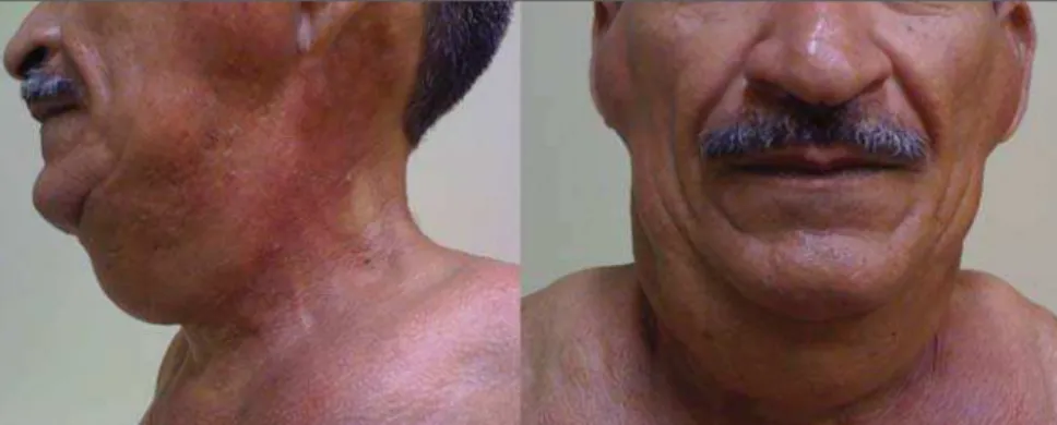
The authors report the case of a 50-year-old man with painless masses in the cervi-cofacial region and in the upper trunk, which increase over a period of three to four years (Figure 1).
CASE REPORT
The patient’s clinical history indicated systemic arterial hypertension effectively controlled with medication. The patient de-nied increase in caloric intake and reported alcohol consumption of 1.0 liter of distilled beverage per day during 28 years. At physi-cal examination, foci of volume increase were observed on the face, neck and upper
INTRODUCTION
Multiple symmetric lipomatosis, also known as Madelung’s disease or Launois-Bensaude syndrome, is a rare entity char-acterized by painless, subcutaneous diffuse deposition of adipose tissue on the neck, upper trunk, arms and legs(1).
Historically, Madelung’s disease has been most frequently observed in men (male-female ratio = 15:1) aged between 30 and 60 years. The disease prevalence is increased among the Mediterranean popu-lation (reported incidence in Italy: 1/25,000 men), and there is a relationship between this disease and excessive consumption of alcohol, particularly red wine(2). Such a condition develops over a period of months to years. The patients complain of their appearance although, in advanced cases, the increase in fat deposits may cause dys-pnea or dysphagia(3). Association of Madelung’s disease with diabetes, gout,
Figure 1. Lateral and frontal views of the patient demonstrating symmetrical enlargement of soft tissues resulting from fat deposition.
130
Rodrigues LP et al. Madelung’s disease: a case report
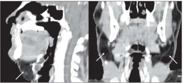
Radiol Bras. 2012 Mar/Abr;45(2):129–131 Figure 3. Contrast-en-hanced axial image dem-onstrating diffuse depo-sition of adipose tissue involving deep structures.
DISCUSSION
Madelung was the first author to exten-sively describe this entity in a series of patients in 1888; hence the name Made-lung’s disease. In 1889, Launois and Ben-saude reported 65 cases, expanding the description of this disease that also has been called as Launois-Bensaude syn-drome. Currently, reports on more than 270 cases are found in the literature(2,5).
Multiple symmetric lipomatosis is clas-sified into two types, as follows: type I, with lipomatous masses in the parotid gion, neck, suprascapular and deltoid re-gions. In this group, deep involvement causing dyspnea and dysphagia may be observed in some patients. In type II, li-pomatosis is diffuse, with clinical appear-ance of simple obesity, and without the symptoms of deep involvement present in type I(6). Typically, the masses do not re-spect cleavage planes, penetrating all lev-els of the tissue, so their surgical exeresis is more difficult(4).
Several types of lipomatosis typically differentiated by anatomic location and varied clinical symptoms have been de-scribed. Such types include multiple sym-metric lipomatosis, congenital infiltrative lipomatosis of the face, encephalocranio-cutaneous lipomatosis, scapular waist pomatosis, adiposis dolorosa, pelvic li-pomatosis and mediastinal lili-pomatosis(5). The clinical diagnosis is based on the rec-ognition of the unique appearance of the patient, but in atypical cases, there may be confusion with lymphoproliferative disor-ders and other conditions(4).
The etiology of Madelung’s disease still remains unknown. Alcohol ingestion of more than 80 g per day during at least ten years – a factor observed in up to 90% of cases described in the literature -, was also observed in the present case. Polyneuropa-thy, described as a sensory, motor and au-tonomic dysfunction, and detected in about 85% of patients with Madelung’s disease, generally develops several years after the onset of lipomas, and was not present in this case(7).
Chest radiography may show mediasti-nal enlargement. At CT, lipomatous, non-encapsulated and homogeneous tissue is observed, possibly in association with deep deposition (submuscular) of adipose tissue (Figure 3). This latter presentation is most frequently observed around the trachea and/or deeply located in the trapezius
muscle. In some cases, calcifications are identified within lipomas(7).
131
Rodrigues LP et al. Madelung’s disease: a case report
Radiol Bras. 2012 Mar/Abr;45(2):129–131 sentation of findings, facilitating the analy-sis of adipose tissues distribution both by radiologists and nonradiologists (Figure 2). The utilization of CT allows the inves-tigator to rule out relevant differential di-agnosis such as lymphoma and metastatic lymph node involvement. Also, it allows the tumor staging in cases where multiple symmetric lipomatosis is associated with malignant disease, particularly, squamous cell carcinoma of upper airways resulting from alcoholism and smoking whose asso-ciation is frequently observed(9).
Sonographic findings of lipomatosis are typical. However, despite the frequent uti-lization of ultrasonography in the investi-gation of neck masses, measurements of density by CT provide a higher diagnostic fidelity(6).
MRI findings usually overlap with CT findings, but it is important to highlight that MRI is a more expensive and generally less available method(9).
Despite the relatively low incidence of Madelung’s disease, radiologists should know the characteristic imaging findings of this condition in order to be able to promptly achieve a correct diagnosis, ruling out over-lap with other conditions and providing appropriate information for the required clinical management of the disease.
REFERENCES
1. Feliciani C, Amerio P. Madelung’s disease: inher-ited from an ancient Mediterranean population? N Engl J Med. 1999;340:1481.
2. Landis MS, Etemad-Rezai R, Shetty K, et al. Case 143: Madelung disease. Radiology. 2009;250: 951–4.
3. Josephson GD, Sclafani AP, Stern J. Benign sym-metric lipomatosis (Madelung’s disease). Oto-laryngol Head Neck Surg. 1996;115:170–1. 4. Ramos GHA, Trevizan GL, Pepe ES. Doença de
Madelung. Rev Bras Otorrinolaringol. 2002;68: 587–90.
5. Murphey MD, Carroll JF, Flemming DJ, et al. From the archives of the AFIP: benign musculoskeletal lipomatous lesions. Radiographics. 2004;24:1433– 66.
6. Vieira MV, Grazziotin RU, Abreu M, et al. Lipo-matose simétrica múltipla (doença de Madelung): relato de um caso. Radiol Bras. 2001;34:119–21. 7. Bulum T, Duvnjak L, Car N, et al. Madelung’s dis-ease: case report and review of the literature. Dia-betologia Croatica. 2007;36:25–30.
8. Vidal MGC, Haygert CJP, Zagoury AR, et al. Doença de Madelung: relato de caso e revisão da literatura. Radiol Bras. 2010;43:275–6. 9. Souza RP, Paes Junior AJO, Rapoport A. Doença