DOI: 10.5935/2359-4802.20170085
ORIGINAL ARTICLE
Mailing Address: Fátima Derlene da Rocha Araújo
Rua Indiana, 789, Ap 301. Postal Code: 30460350, Jardim América, Belo Horizonte, MG – Brazil. E-mail: [email protected], [email protected]
Prognosis of Dilated Cardiomyopathy with Severe Heart Failure according to Functional
Classification Scales in Childhood
Fátima Derlene da Rocha Araújo, Rose Mary Ferreira Lisboa da Silva, Henrique de Assis Fonseca Tonelli, Adriana Furletti M Guimarães, Sandra Regina Tolentino Castilho, Zilda Maria Alves Meira
Hospital das Clínicas da Universidade Federal de Minas Gerais, Belo Horizonte, MG – Brazil
Manuscript received February 14, 2017, revised manuscript April 17, 2017, accepted June 05, 2017
Abstract
Background: Heart failure is the main manifestation of dilated cardiomyopathy in childhood, and the systematic evaluation of signs and symptoms allows monitoring the treatment outcome.
Objective: To evaluate the use of three functional classification scales of heart failure in children and adolescents with dilated cardiomyopathy.
Methods: Longitudinal and observational study including patients from zero to 18 years with dilated cardiomyopathy and severe initial heart failure. All of them were followed up using the New York Heart Association (NYHA), The New York University Pediatric Heart Failure Index (The NYU PHFI) and Ross version 2012 scales. Statistical analyzes were done using Statistical Package for Social Science, version 14.0, with Mann-Whitney test, Chi-Square test or Fisher's test, application of the Operating Characteristic Curve, Wilcoxon test and Kappa coefficient for comparison of scales and Kaplan-Meier curve for survival evaluation. The level of significance adopted was 5%.
Results: A total of 57 patients, aged from 1 to 200 months (mean of 48.7 ± 55.9) and follow-up of 6 to 209 months (mean of 63.6 ± 48.4) were included. There was substantial agreement between the Ross 2012 scales, The NYU PHFI and NYHA (Kappa = 0.71 and 0.82, respectively). Paired analysis by the Wilcoxon test, comparing the scales before and after treatment, was significant (p < 0.0001). The greatest survival was found in patients with class I/II by NYHA or scores lower than 11 points in the others.
Conclusion:The use of functional assessment scales of heart failure proved to be useful in the follow-up and evaluation of the therapeutic response and there was no difference between them. Patients who remained in functional classification III or IV NYHA or scores ≥ 11 in Ross 2012 or The NYU PHFI had worse prognosis. (Int J Cardiovasc Sci. 2018;31(1)12-21)
Keywords: Cardiomyopathy, Dilated; Prognosis; Heart Failure; Scales; Child; Adolescents.
Introduction
Dilated Cardiomyopathy (DCM) is the main type of cardiomyopathy that occurs in childhood, characterized by ventricular dilatation and impaired myocardial
function.1,2 Heart failure (HF) is its main manifestation,
being the initial symptom in approximately 70 to 90% of the cases.3-5 The HF degree is related to the prognosis;1,4,6
however, there are specific characteristics of its clinical spectrum according to each age range, which makes
it difficult to carry out its objective quantification
through scales or scores.7 Additionally, obtaining
Currently, the scales available for assessing HF severity in childhood and adolescence are the New York Heart Association (NYHA) Functional Classification
(FC),8 The New York University Pediatric Heart Failure
Index (The NYU PHFI) score,9 and the Ross scales
(version 199210 modified by Läer et al.11 in 2002 and the
2012 version).12 The NYHA classification, widely used
in adults, is reported to be useful to quantify HF in older children and adolescents. The 1992 version of the Ross scale is an adaptation of the NYHA scale for the pediatric age group, with the 2012 version being the most comprehensive one, which includes data from physical assessment and complementary examinations. Similarly, The NYU PHFI, in addition to including these data, also adds the types of medications used. Both are scores that correspond to the total sum of each scored item.
The aim of the present study was to evaluate the evolution of children and adolescents with severe HF as the initial presentation of DCM, using the HF functional
classification scales (NYHA, The NYU PHFI and Ross
scale, version 2012).
Methods
This was a longitudinal and observational study, which included patients undergoing treatment by the same team
since 1999 and, prospectively, those admitted from January
2010 to December 2015. Children and adolescents under 18 years of age with a diagnosis of DCM|, whose manifestation was severe acute HF (FC III or IV of the NYHA) were included in the study. Patients with myocardial dysfunction due to sepsis, primary pulmonary hypertension, congenital heart diseases, primary arrhythmias, neuromuscular diseases, rheumatic valvulopathy and ischemic processes were excluded.
All patients were admitted to the Intensive Care Unit (ICU) at the diagnosis, due to the HF severity. After hospital discharge, they were referred for outpatient treatment. All of them were followed by the same professionals during the entire treatment period and submitted to a chronic HF treatment strategy, according to data in the literature and service clinical protocol.
The tool used to evaluate treatment response were the three HF FC scales (NYHA, The NYU PHFI and Ross 2012). The scales were applied in the original versions by a researcher blinded to the patients’ clinical evolution. Symptom-related data were reported by parents or guardians in the case of children under 12 years of age and by the patients themselves who were older than
12 years. Laboratory and complementary test results were obtained from direct consultation of patients’ medical records. This study was approved by the institutional Research Ethics Committee.
Statistical Analysis
The Statistical Package for Social Science (SPSS) software, version 14.0, was used for data analysis. Categorical variables were expressed as frequencies or percentages and compared with the Chi-square or Fisher's test, as appropriate. Quantitative variables were described as means ± standard deviation. The Mann-Whitney test was used to compare the variables between the scales (non-parametric distribution), and a normality test was not performed. The Wilcoxon test was applied for the paired analysis of the FC scales of HF before and after treatment.
The Receiver Operating Characteristic (ROC) curve was constructed based on the NYHA FC III in relation to the total scores in the Ross 2012 and The NYU PHFI scales. The area under the curve was calculated and the best cutoff point was defined in the NYU PHFI and Ross scales that corresponded to NYHA FC III. A Kaplan-Meier curve was constructed to evaluate the death-free or transplant-free survival according to the calculated scores, and a log-rank test was used to compare the curves. The level of significance was set at 5%. The Kappa statistic was used to evaluate the agreement between the Ross 2012 and NYHA scores at the diagnosis and after complete therapy of HF. For the interpretation, we considered the classification
according to Landis and Kock,13 namely: 15 < 0, without
agreement; 0-0,19, poor agreement; 0.20-0.39, reasonable agreement; 0.40-0.59, moderate agreement; 0.60-0.79, high agreement; 0.80-0.99, almost perfect agreement; 1, perfect agreement.
To compare the Ross 2012 scale with NYHA, Ross et al.10
categorization was used, which considered FC I (zero to 5 points), FC II (6 to 10 points), FC III (11 to 15 points), and FC IV (16 to 20 points).
Results
Table 1 – Clinical and demographic characteristics of patients with dilated cardiomyopathy who presented with acute heart failure (functional classes III and IV of the New York Heart Association) on admission to the intensive care unit
Characteristics Patients n (%)
Age, months
0-60 38 (66.7)
61-120 11 (19.3)
≥ 121 8 (14.0)
Female gender 34 (60.0)
Skin color/ethnicity
Caucasian 6 (10.5)
Black or mixed-race 51 (89.5)
Inotropic use 44 (77.2)
Endotracheal intubation 27 (47.4)
Gamma globulin use 8 (14.0)
Cardiac arrest event 7 (12.3)
Cardiogenic shock 30 (52.6)
Blood transfusion 21 (36.8)
Consanguinity 5 (8.8)
Previous viral disease report 9 (15.6)
A history of viral disease preceding the diagnosis up to 3 months was reported in nine patients (15.6%), with only one patient reporting the disease in the gastrointestinal tract. Respiratory symptoms were the most commonly reported by family members as the initial clinical picture (88%), and in 80% of infants the diagnosis at hospitalization was bronchopneumonia.
In adolescents, the prevailing initial symptoms were precordial pain, exercise intolerance, fatigue, and abdominal pain. Of the total of 57 patients, only one patient had ascites at the diagnosis, but nine (16%) patients had this symptom during evolution. Ascites was related to some degree of right ventricular dysfunction, and all patients who had ascites died or required heart transplantation.
Evaluation of dilated cardiomyopathy etiology
The cause of DCM was identified in 36 (79%) patients. The most common causes were myocarditis and DCM induced by anthracyclines. The highest frequency of myocarditis was found in children younger than 2 years (76.5%), while those induced
by anthracyclines was observed in children older than 5 years (66.7%). Left ventricular noncompaction cardiomyopathy was identified in five patients (8%) in the echocardiographic or magnetic resonance imaging study. Cardiac catheterization was performed in 14 patients to investigate coronary anomalies, and no coronary abnormalities were found. The distribution of patients, according to the causes and age range of DCM, is shown in Table 2 and Figure 1.
Functional class categorization according to the scales
Table 2 – Distribution of patients with dilated cardiomyopathy by age group according to the etiological diagnosis
Etiology
Age range (months)
0-12 13-60 61-120 ≥ 121
Myocarditis 11 4 2 0
Idiopathic 5 2 3 2
Anthracyclines 0 4 4 4
Ventricular noncompaction 4 1 0 0
Mitochondriopathy 4 0 0 1
Genetic syndromes 3 0 1 1
Familial 0 0 1 1
In the agreement evaluation of the Ross 2012 and NYHA scores, the Kappa coefficient was 0.67 (95%CI: 0.570-0.770; p < 0.0001) for the calculated score at the diagnosis of the patients and 0.71 (95%CI: 0.702-0.79, p < 0.0001) for the score after HF treatment optimization.
Evolution of patients according to the functional classification
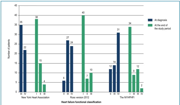
The initial categorization of the patients according to the FC scales of HF carried out at admission to the ICU showed 35 patients (61.4%) in NYHA FC III and 22 patients (38.6%) in FC IV, with scores at the Ross 2012 scale ranging from 10 to 20 points (mean of 14.7 ± 3.05 points) and by the NYU PHFI, ranging from 10 to 24 points (mean of 16 ± 4.5 points).
After treatment optimization for HF, it was observed that 16 patients did not show a satisfactory response and died or were submitted to heart transplantation. Considering evaluation through the scales, it was verified that four (7%) patients did not show FC improvement according to the NYHA, 11 (19.1%) according to Ross 2012 scale and 15 (26.3%) according to The NYU PHFI.
Time until symptom improvement ranged from 1 to 48 months (mean of 10.2 ± 9.8 months). Paired analysis by the Wilcoxon test, comparing scales before and after treatment, was significant (p < 0.0001). Patient evolution, according to the classification by the three scales at the diagnosis and after the treatment optimization, is shown in Figure 3.
An association between HF severity demonstrated by the three scales and patient evolution was also observed (Figure 4). Patients who remained in NYHA FC III or IV or with a score equal to or greater than 11 in the other
scales showed worse evolution, considering death or need for heart transplantation (p < 0.0001).
Similar result was obtained in the Kaplan-Meier survival curve plot (Figure 5). Patients who remained in NYHA FC III or IV or with a score of 11 points or higher in the Ross 2012 and NYU PHFI scales had lower death-free or heart transplantation-free survival (p < 0.0001).
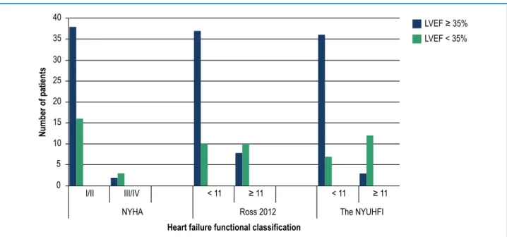
It was observed that 16 (28%) patients, after complete HF treatment, were asymptomatic at rest and at mild and moderate exertion, although they persisted with Left Ventricular Ejection Fraction (LVEF) < 35%. There was association between HF severity and LVEF < 35% (Figure 6) when the Ross 2012 and the NYU PHFI scores were equal to or greater than 11 points (p < 0.0001). However, this association was not observed for the NYHA scale (p = 0.232).
Discussion
The etiology of DCM is diverse, and current diagnostic methods make it possible to improve the etiological diagnosis, but most (about two thirds) remain idiopathic. However, in this study, the probable cause was identified in 79% of the patients. In a study that included 1,426 children, the frequency of identification of DCM etiology
was obtained in only 31%.14 Another, which followed
83 patients after the first hospitalization for DCM, obtained a rate of 39% of idiopathic cardiomyopathy and causal
identification in 61% of the cases.6 These differences can
Myocarditis
Idiopathic
Use of Anthracyclines
Ventricular noncompaction
Mitochondriopathy
Genetic syndromes
Familial
0% 5% 10% 15% 20% 25% 30% 35%
30%
21%
21%
9%
9%
7%
3%
Figure 1 – Frequency distribution of patients with dilated cardiomyopathy, according to the etiological diagnosis.
1.0
1.0 0.8
0.8 0.6
0.6 0.4
0.4 0.2
0.2 0.0
0.0
Specificity
Sensitivity
Ross 2012
(AUC 0.90 (95% CI: 0.829-0.979))
(AUC 0.89 (95% CI: 0.801-0.971)) The NYU PHFI
Figure 2 – ROC curve for evaluation of the functional classification scales of heart failure (Ross version 2012 and The New York University Pediatric Heart Failure Index (NYU PHFI in relation to the New York Heart Association) in initial clinical presentation of dilated cardiomyopathy AUC: area under the curve; 95%CI: 95% confidence interval.
HF is a common event in DCM presentation and, very often, of great severity at the initial presentation. In a study with 142 children, it was observed that 24.5%
and 58.5% were admitted with an initial manifestation of
HF in NYHA FC III and IV, respectively.15 Of these, 30.5%
45
40
35
30
25
20
15
10
5
0
Number of patients
III IV I II III II III IV I II III II III IV I III III IV
New York Heart Association Ross version 2012 The NYHPHFI
Heart failure functional classification
At diagnosis
At the end of the study period 35
38
22
15
4
6 27
24
7 10 40
12 14
31 34
9 12
2
Figure 3 – Distribution of patients according to the heart failure functional classification (Ross scale version 2012 and The New York University Pediatric Heart Failure Index (NYU PHFI) in relation to the New York Heart Association) at the the diagnosis of dilated cardiomyopathy and at the end of the follow-up period.
41 41
39
13 13
3 6 3
10
1
Favorable evolution
Transplant or death 45
40
35
30
25
20
15
10
5
0
Number of patients
I e II III e IV
NYHA
< 11 ≥ 11 < 11 ≥ 11
Ross 2012 The NYHPHFI
Heart failure functional classification
1.0
0.8
0.6
0.4
0.2
0.0 0.0
Survival (%)
50.0 100.0 150.0 200.0 250.0 p = 0.001
Time of follow-up (months)
Classes I and II (NYHA) Classes III and IV (NYHA)
(A)
1.0
0.8
0.6
0.4
0.2
0.0
Survival (%)
0.0 50.0 100.0 150.0 200.0 250.0 p < 0.0.0001
Time of follow-up (months)
Ross < 11 pontos Ross ≥ 11 pontos
(B)
1.0
0.8
0.6
0.4
0.2
0.0
0.0 50.0 100.0 150.0 200.0 250.0 p < 0.0001
Survival (%)
Time of follow-up (months)
The NYUHFI < 11 pontos The NYUHFI ≥ 11 pontos
(C)
Figure 5 – Kaplan-Meier survival curve for the events of death or heart transplantation of patients with dilated cardiomyopathy, according to the Ross 2012 version, The New York University Pediatric Heart Failure Index (NYU PHFI) and the New York Heart Association (NYHA) scales.
the relative risk of death was 2.15 (95%CI: 1.73-2.67). The present study included only those patients with severe initial presentation (FC III or IV). This decision was important, since it homogenized the initial severity, minimizing the evolution classification bias. We are not aware of a similar study in the literature.
Previous studies have shown that the persistence of HF severity during treatment is associated with
poor prognosis in patients with DCM.4,5,15-17 This study
also showed that HF severity detected by any of the three scales was associated with death or need for heart transplantation.
HF symptoms vary according to the age group.7 In
young infants, symptoms may be nonspecific, such as sweating and dyspnea on feeding, tachypnea, tachycardia, irritability and low weight gain. In older children, dyspnea, orthopnea, paroxysmal nocturnal dyspnea, ascites, peripheral edema and exercise intolerance are the most frequent symptoms. Symptoms such as nausea, vomiting, anorexia and postprandial abdominal pain can also be present at any age and are explained by mesenteric ischemia
secondary to low systemic output.1,6 This diversity and
40
35
30
25
20
15
10
5
0
I/II III/IV < 11 ≥ 11 < 11 ≥ 11
NYHA Ross 2012 The NYUHFI
LVEF ≥ 35%
LVEF < 35%
Heart failure functional classification
Number of patients
Figure 6 – Distribution of children and adolescents with dilated cardiomyopathy, according to left ventricular ejection fraction and heart failure functional class, at the end of the follow-up period. NYHA: New York Heart Association; NYU PHFI: The New York University Pediatric Heart Failure Index.
make it difficult to systematize the evaluation, which is more easily applicable to the adult population.
The estimate of children’s and adolescents’ functional capacity is crucial for the evaluation of both the therapeutic response and the prognosis. However, the objective categorization, through the ergospirometry tests with oxygen consumption measurement, is not always available and accessible to all age groups.
Reference values are available for the adult population,18
but they are not well established for the pediatric population. On the other hand, a study showed that the objective values of the cardiopulmonary test were not correlated with the functional class assessed by the
medical team.19 Similarly, in adults, cardiopulmonary
exercise testing is a part of the decision-making process for heart transplantation waiting lists. However, the experience using cardiopulmonary exercise testing as a prognostic tool in children is still limited.
The study by Guimarães et al.20 provided evidence to
support the use of cardiopulmonary exercise testing to stratify DCM risk in older children. Similar results were
obtained by Giardini et al.,21 when they demonstrated that
these tests were feasible in outpatient children with DCM and height greater than 120 cm and when they correlated it with the outcome of death or heart transplantation.
Therefore, the scales, despite being subjective assessment methods, based on patient or family
information, are still of universal use and easy to apply. Cardiopulmonary tests are reserved for older children and are available at referral centers.
The Ross scale, of which first version dates from 1992, is described as the most adequate one for use in younger children and represents the adaptation of
the NYHA scale data to the symptoms in children.9
Subsequently, this Ross classification was modified
to be adapted to the peculiarities of each age group.10
The latest version of the Ross scale, published in
2012, is wide-ranging and difficult to use routinely.12
It assigns scores according to the classification of clinical findings, physical examination, laboratory and complementary exams, and can add up from zero to 20 points. Similar to the NYHA scale, the Ross 2012 scale can also be categorized into classes from I to IV, according to the score ranges. A similar methodology is also used in the The NYHPHFI scale, which results in a score from zero to 30. Clinical indicators, findings of complementary tests, therapeutic regimens and the type of ventricular pathophysiology are scored, not considering the age group distinction.
used. These modifications make it difficult to use these scales in daily practice. The reality of the care of these patients in our environment still does not allow the use of such difficult tools.
It is noteworthy that NYHA FC, in addition to being the best-known scale, is also the one that is easier to
apply. Tavares et al.22 developed a version of the NYHA
classification for the symptoms in the pediatric range and created a graphical representation of the four functional classes. Through this visual method, the perception of the children and their caregivers can be obtained, in addition to the categorization carried out by the medical team.
Although several parameters of the echocardiographic
analysis,23-25 especially LVEF, are associated with
prognosis, this study found that almost one third of the patients (16 patients/28%) showed HF symptom improvement after treatment, although they persisted
with LVEF ≤ 35%. For this group of patients, the Ross 2012
and The NYU PHFI scales continued to show high scores, as they add points for medications and echocardiographic findings, such as LVEF. For this group, the NYHA scale correlated better with functional capacity.
The present study demonstrated that the three scales used to classify HF in children were useful for HF severity evolution assessment in this population. There were small differences between the scales, probably due to the parameters used in the score, but there was no statistical significance. The NYHA scale considers only the clinical symptoms. The Ross scale version 2012 associates symptom data, physical and complementary examinations. The NYU PHFI scale includes scores for the type of drug used, as well as the presence of signs and symptoms.
Even though DCM is not very prevalent in children, it has a worse prognosis in relation to the adult
population.26 Studies of methods that allow DCM
evolution follow-up and its response to treatment contribute to a greater knowledge of the disease, as well as to the increased survival of these patients. There are no other studies in the literature describing the use of scales for the evolution assessment of children and adolescents with DCM. This study is a pioneer in demonstrating the behavior of a high-risk population due to the initial disease severity, by using HF functional class assessment scales. It collaborates by verifying that both the more complex scales (Ross 2012 and The NYU PHFI) and the best known (NYHA) scale can be used for patient evolution assessment. It is recommended that
patients who remain symptomatic after HF treatment optimization should also be assessed regarding the scores on the other scales. Thus, a more complete prognosis estimate can be attained.
Conclusion
All assessed scales were adequate for follow-up, evaluation of therapeutic response and prognostic estimate of patients with dilated cardiomyopathy and heart failure undergoing treatment. Patients who, after optimized treatment for heart failure, remained in NYHA functional class III or IV, or with a score equal to or greater than 11 at Ross 2012 or The NYU PHFI, had a worse prognosis.
Study limitations
The limitations are related to sample size, disease etiological diversity, wide-ranging affected age group and inclusion of retrospective data. These reflect the difficulties previously reported by other authors regarding the follow-up of cohorts of patients with severe and low-prevalence diseases.
Author contributions
Conception and design of the research: Meira ZMA, Araujo FDR. Acquisition of data: Araujo FDR, Tonelli HAF, Guimaraes AFM, Castilho SRT. Analysis and interpretation of the data: Araujo FDR, Silva RMFL, Meira ZMA. Statistical analysis: Silva RMFL, Araujo FDR. Writing of the manuscript: Araujo FDR, Meira ZMA. Critical revision of the manuscript for intellectual content: Araujo FDR, Meira ZMA. Supervision / as the major investigador: Meira ZMA.
Potential Conflict of Interest
No potential conflict of interest relevant to this article was reported.
Sources of Funding
There were no external funding sources for this study.
Study Association
1. Hsu DT, Canter CE. Dilated cardiomyopathy and heart failure in children. Heart Fail Clin. 2010;6(4):415-32. doi: 10.1016/j.hfc.2010.05.003.
2. Maron BJ, Towbin JA, Thiene G, Antzelevitch C, Corrado D, Arnett
D, et al; American Heart Association; Council on Clinical Cardiology, Heart Failure and Transplantation Committee; Quality of Care and Outcomes Research and Functional Genomics and Translational Biology Interdisciplinary Working Groups; Council on Epidemiology and Prevention. Contemporary definitions and classification of the cardiomyopathies: an American Heart Association Scientific statement from the Council on Clinical Cardiology, Heart Failure and Transplantation Committee; Quality of Care and Outcomes Research and Function. Circulation. 2006;113(14):1807-16. doi: 10.1161/ CIRCULATIONAHA.106.174287.
3. Carvalho JS. Cardiomyopathies. In: Anderson RH, Baker EJ, MaCartney FJ, Rigby ML, Shinebourne EA, Tynan M. Paediatric cardiology. 2nd ed.
New York: Churchill-Livingstone; 2002. p. 1595-643.
4. Hollander SA, Berstein D, Yeh J, Dao D, Sun HY, Rosenthal D.
Outcomes of children following a first hospitalization for dilated cardiomyopathy. Circ Heart Fail. 2012;5(4):437-43. doi: 10.1161/ CIRCHEARTFAILURE.111.964510.
5. Lipshultz SE, Sleeper LA, Towbin JA, Lowe AM, Orav EJ, Cox GF, et al.
The incidence of pediatric cardiomyopathy in two regions of the United
States. N Engl J Med. 2003;348(17):1647-55. doi: 10.1056/NEJMoa021715.
6. Rupp S, Apitz C, Tholen L, Latus H, Ostermayer SH, Schmidt D, et al. Upgraded heart failure therapy leads to an improved outcome of dilated cardiomyopathy in infants and toddlers. Cardiol Young. 2015;25(7):1300-5. doi: 10.1017/S1047951114002406.
7. Azeka E, Jatene MB, Jatene IB, Horowitz ES, Branco KC, Souza Neto JD, et
al; Sociedade Brasileira de Cardiologia. [I Guidelines of heart failure and heart transplantation in the fetus, in children and adults with congenital cardiopathy, The Brazilian Society of Cardiology]. Arq Bras Cardiol. 2014;103(6 Suppl 2):1-126. doi: 10.5935/abc.2014S005.
8. The Criteria Committee of the New York Heart Association. Nomenclature and criteria for diagnosis of diseases of the heart and great vessels. 9th ed. Boston: Little, Brown & Co; 1994. p. 253-6.
9. Connolly D, Rutkowski M, Auslender M, Artman M. The New York University Pediatric Heart Failure Index: a new method of quantifying chronic heart failure severity in children. J Pediatr. 2001;138(5):644-8. doi: 10.1067/mpd.2001.114020.
10. Ross RD, Bollinger RO, Pinsky WW. Grading the severity of congestive heart failure in infants. Pediatr Cardiol. 1992;13(2):72-5. doi: 10.1007/ BF00798207.
11. Läer S, Mir TS, Behn F, Eiselt M, Ssholz H, Venzke A, et al. Carvedilol therapy in pediatric patients with congestive heart failure: a study
investigating clinical and pharmacokinetic parameters. Am Heart J.
2002;143(5):916-22. PMID: 12040358.
12. Ross RD. The Ross Classification for heart failure in children after 25 years: a review and an age-stratified revision. Pediatr Cardiol.
2012;33(8):1295-300. doi: 10.1007/s00246-012-0306-8.
13. Landis JR, Koch GG. The mensurement of observer agrément for
categorical data. Biometrics. 1977;33(1):159-74. PMID: 843571.
14. Towbin JA, Lowe AM, Colan SD, Sleeper LA, Orav EJ, Clunie S, et al.
Incidence, causes, and outcomes of dilated cardiomyopathy in children.
JAMA. 2006;296(15):1867-76. doi: 10.1001/jama.296.15.1867.
15. Azevedo VM, Santos MA, Abanezi Filho M, Castier MB, Tura BR, Amino
JG. Outcome factors of idiopathic dilated cardiomyopathy in children
- a long-term follow-up review. Cardiol Young. 2007;17(2):175-84. doi: 10.1017/S1047951107000170.
16. Alexander PM, Daubeney PE, Nugent AW, Lee KJ, Turner C, Colan SD, et al;
National Australian Childhood Cardiomyopathy Study. Long-term outcomes of dilated cardiomyopathy diagnosed during childhood: results from a national population-based study of childhood cardiomyopathy. Circulation. 2013;128(18):2039-46. doi: 10.1161/CIRCULATIONAHA.113.002767.
17. Everitt MD, Sleeper LA, Lu M, Canter CE, Pahl E, Wilkinson JD,
et al; Pediatric Cardiomyopathy Registry Investigators. Recovery of echocardiographic function in children with idiopathic dilated cardiomyopathy: results from the pediatric cardiomyopathy
registry. J Am Coll Cardiol. 2014;63(14):1405-13. doi: 10.1016/j.
jacc.2013.11.059.
18. Weber KT, Kinasewitz GT, Janicki JS, Fishman AP. Oxygen utilization
and ventilation during exercise in patients with chronic cardiac failure. Circulation. 1982;65(6):1213-23. PMID: 6804111.
19. Guimarães GV, Bellotti G, Mocelin AO, Camargo PR, Bocchi EA. Cardiopulmonary exercise testing in children with heart failure secondary to idiopathic dilated cardiomyopathy. Chest. 2001;120(3):816-24. PMID: 11555515.
20. Guimarães GV, d'Avila VM, Camargo PR, Moreira LF, Lanz JR, Bocchi
EA. Prognostic value of cardiopulmonary exercise testing in children with heart failure secondary to idiopathic dilated cardiomyopathy in a
non-beta-blocker therapy setting. Eur J Heart Fail. 2008;10(6):560-5. doi:
10.1016/j.ejheart.2008.04.009.
21. Giardini A, Fenton M, Andrews RE, Derrick G, Burch M. Peak oxygen uptake correlates with survival without clinical deterioration in ambulatory children with dilated cardiomyopathy. Circulation. 2011;124(16):1713-8. doi: 10.1161/CIRCULATIONAHA.111.035956.
22. Tavares AC, Bocchi EA, Guimarães GV. Functional class in children with idiopathic dilated cardiomyopathy. a pilot study. Arq Bras Cardiol. 2016;106(6):502-9. doi: 10.5935/abc.20160066.
23. Morhy SS. [Dilated cardiomyopathy in children--is there an ecocardiographic prognostic index?]. Arq Bras Cardiol. 2004;82(6):501-2. doi: /S0066-782X2004000600001.
24. Azevedo VM, Albanesi Filho FM, Santos MA, Castier MB, Tura BR. How can the echocardiogram be useful for predicting death in children with idiopathic dilated cardiomyopathy? Arq Bras Cardiol 2004;82(6):505-14. doi: /S0066-782X2004000600003.
25. Lewis AB. Prognostic value of echocardiography in children with
idiopathic dilated cardiomyopathy. Am Heart J. 1994;128(1):133-6.
PMID: 8017266.
26. Puggia I, Merlo M, Barbati G, Rowland TJ, Stolfo D, Gigli M, et al. Natural history of dilated cardiomyopathy in children. J Am Heart Assoc. 2016; 5(7):pii: e003450. doi: 10.1161/JAHA.116.003450.
References
This is an open-access article distributed under the terms of the Creative Commons Attribution License
Ethics approval and consent to participate
This study was approved by the Ethics Committee of the Federal University of Minas Gerais under the protocol number CAAE - 11280413300005149. All the