Key words:
Antioxidants; Calcium
Oxalate; Cocos; Phytotherapy; Urolithiasis
Int Braz J Urol. 2013; 39: 108-17
__________________
Submitted for publication: February 05, 2012
__________________
Accepted after revision: November 30, 2012 Purpose: Many medicinal plants have been employed during ages to treat urinary stones
though the rationale behind their use is not well established. Thus, the present study was proposed to evaluate the effect of coconut water as a prophylactic agent in experimen-tally induced nephrolithiasis in a rat model.
Materials and Methods: The male Wistar rats were divided randomly into three groups. Animals of group I (control) were fed standard rat diet. In group II, the animals were ad-ministrated 0.75% ethylene glycol in drinking water for the induction of nephrolithiasis. Group III animals were administrated coconut water in addition to ethylene glycol. All the treatments were continued for a total duration of seven weeks.
Results and Conclusion: Treatment with coconut water inhibited crystal deposition in renal tissue as well as reduced the number of crystals in urine. Furthermore, coconut water also protected against impaired renal function and development of oxidative stress in the kidneys. The results indicate that coconut water could be a potential candidate for phytotherapy against urolithiasis.
INTRODUCTION
Urolithiasis is a common ocurrence, affec-ting up to 10%-15% of the population at some point during their lifetime (1). Increased incidence of kidney stones in the industrialized world is as-sociated with improved standards of living, race, ethnicity and geographical area (2). Studies by Trinchieri et al. (3) reported recurrence rates of 50 - 75% after 10 and 20 years, respectively. Kidney sto-nes composed of CaOx, either alone or mixed with calcium phosphate, are hitherto the most common stones accounting for more than 80% of them (4). Earlier studies have shown that tubular cell injury facilitates calcium oxalate (CaOx) crystal formation and deposition in the renal tubules (5). Similarly
various authors demonstrated that calcium oxala-te crystals increased lipid peroxidation that further led to renal epithelial injury (6,7).
Although during recent years development of modern techniques such as extracorporeal sho-ck wave lithotripsy and percutaneous nephrolitho-tomy has revolutionized the surgical management of the problem, yet not much progress has been made towards the medical management of uroli-thiasis. Many medicinal plants have been employed during ages to treat urinary stones though their mechanism of action is not well established throu-gh systematic and pharmacological studies, except for some composite herbal drugs and plants (8-10).
Coconut water (Cocos nucifera L.) is the most nutritious wholesome beverage in all the
Prophylactic effect of coconut water (Cocos nucifera L.)
on ethylene glycol induced nephrocalcinosis in male
wistar rat
_______________________________________________
M. Gandhi, M. Aggarwal, S. Puri, S.K. Singla
Department of Biochemistry (MG, MA, SKS), Department of Biotechnology (SP), University Institute of Engineering and Technology, Panjab University, Chandigarh, India
ABSTRACT
ARTICLE
INFO
coconut producing countries. Coconut water, the liquid endosperm of coconut, contains sugars, vi-tamins, minerals, proteins, free amino acids and growth promoting factors. It is a natural isotonic beverage and is acclaimed in the tropics for its numerous medicinal properties. Coconut water is an essential dietary ingredient of South India where the incidence of urolithiasis is very low (11). However, so far no systematic study has been re-ported regarding the antiurolithiatic property of coconut water. So the present study was designed to evaluate the efficacy of coconut water on ethy-lene glycol induced nephrolithiasis.
MATERIALS AND METHODS
Collection of coconut water
Fresh coconuts were purchased from the market, broken carefully, liquid endosperm were collected and used for each day experiment. Whi-le purchasing coconuts from the market, the nuts stored in cold conditions and from the same stock were procured.
Animals
Male Wistar rats weighing 150-170 g were housed in polypropylene cages in a room maintai-ned at 25 ± 1º C with alternate exposure to light and dark for 12 hours. The animals were acclima-tized for one month in polypropylene cages under hygienic conditions. All procedures were done in accordance with ethical guidelines for care & use of laboratory animals and were approved by the local experimental animal ethical committee. The animals were provided standard animal feed and water ad libitum. The standard rat diet was acqui-red from Aashirwad Company (Ludhiana, Punjab, India), and the composition of the diet is given in Table-1. To induce urolithiasis in animals, they were exposed to 0.75% ethylene glycol in their drinking water for 7 weeks. The protocol to induce urolithiasis was adapted from Huang et al. (12), where the authors showed evidence that the admi-nistration of “0.75 % ethylene glycol” in drinking water can induce “urolithiasis” in male wistar rats.
Rats were divided into three groups of six rats each and fed the following diet:
Group 1: Normal rat diet (control).
Group 2: Normal rat diet + 0.75% ethylene glycol (EG) mixed with tap water for 7 weeks ad libitum (12).
Group 3: Normal rat diet + 0.75% EG + 10% Coconut water for 7 weeks ad libitum.
Methods
A 10% of kidney homogenate was prepared in 0.1 mM tris buffer (pH 7.4) and was used for assaying lipid peroxidation (13), antioxidant enzy-mes superoxide dismutase (14) and catalase (15).
All the animals were kept in individual metabolic cages and urine samples were collec-ted throughout 24hours, one day before sacrifi-cing the animals. Blood was collected from orbi-tal sinus under mild anesthetic conditions, using diethyl ether as an anesthetic agent and animals were sacrificed by cervical decapitation. Serum was separated by centrifugation at 3,000 x g for 15 minutes, analyzed for creatinine (code no. FRE-BCERM0082) and urea (code no. FRCER0034) by Erba Manheim diagnostic kits.
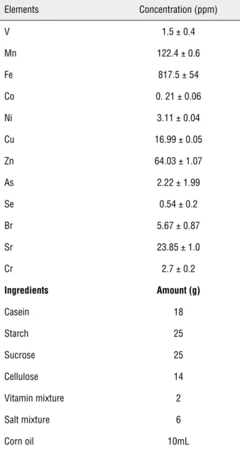
Table 1 - Composition of standard animal diet.
Elements Concentration (ppm)
V 1.5 ± 0.4
Mn 122.4 ± 0.6
Fe 817.5 ± 54
Co 0. 21 ± 0.06
Ni 3.11 ± 0.04
Cu 16.99 ± 0.05
Zn 64.03 ± 1.07
As 2.22 ± 1.99
Se 0.54 ± 0.2
Br 5.67 ± 0.87
Sr 23.85 ± 1.0
Cr 2.7 ± 0.2
Ingredients Amount (g)
Casein 18
Starch 25
Sucrose 25
Cellulose 14
Vitamin mixture 2
Salt mixture 6
Corn oil 10mL
and 30 cycles for CAT), each cycle consisting of denaturation for 45 sec at 94º C followed by an-nealing for 45 sec at 55.5º C and extension for 1 minutes at 68º C.
Though ideally, urine chemistry and the pH should have been noted; based on our earlier observation and those of others (21-23) it was as-sumed that the urine chemistry & pH did not differ significantly in the 3 groups of rats. A 24hours urine collection was done for polarization micros-copy to compare calcium oxalate crystallization in the 3 groups. For analysis of crystalluria a drop of
urine sample was spread on a glass slide and visu-alized under polarized light using Leica DM3000 light microscope (24).
For histopathological studies, the kidneys were removed and their transverse sections were fixed in 10% buffered formalin solution (pH 7). The tissues were dehydrated and embedded in pa-raffin wax (68º C). The papa-raffin sections were then cut and finally stained with Delafield’s Hemato-xylin and Eosin staining (H&E staining).
Data analysis
The results were expressed as mean ± standard deviation (S.D) for six animals in each group. Kolmogorov Smirnov test was applied to check the parametric distribution of the data. Di-fferences between groups were assessed by one way analysis of variance (ANOVA) using the SPSS software package for Windows. Post hoc test was performed for inter-group comparisons using the least significance difference (LSD) test; significan-ce at P-values < 0.001, < 0.01, < 0.05 have been given respective symbols in the tables and figures.
RESULTS
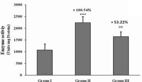
of coconut water decreased the enzyme activity of the SOD and CAT in the group III compared to group II (Figures 3a and 3b) but decrease in the expression level of catalase in the group III com-pared to group II was not as that much as in case of SOD (Figures 1, 2a and 2b).
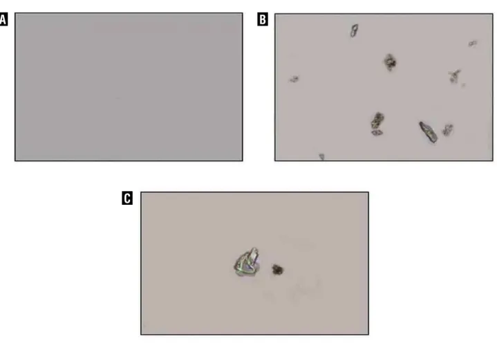
On observing urine samples under pola-rization microscope, no crystal deposition was observed in control animals (Figure-4a), whereas group II rats revealed presence of abundant CaOx crystals deposition (Figure-4b). In group III, a dras-tic decrease in the number of urinary crystals was observed (Figure-4c). The histopathological obser-vation of renal tissue under light microscope sho-wed normal architectural and intactness without
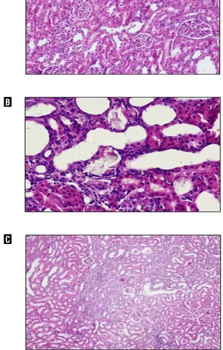
any apparent damage in control rats. However, the urolithiatic rats showed tubulae containing crys-talline particles, mostly distal segments with con-sequent dilation of distal tubules, thin loop and collecting ducts. In group III, no depositions of crystalline particles were observed (Figure-5c).
DISCUSSION
Coconut is grown and consumed largely in South India where incidence of urolithiasis is low. It is therefore pertinent to investigate the effect of coconut water in nephrocalcinosis model of male Wistar rats. Some investigators (21,25) have studied various natural therapeutic agents in vivo
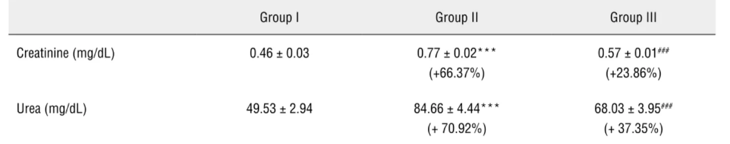
Table 2 - Effect of various treatments on serum creatinine and urea level in experimental rats.
Group I Group II Group III
Creatinine (mg/dL) 0.46 ± 0.03 0.77 ± 0.02***
(+66.37%)
0.57 ± 0.01###
(+23.86%)
Urea (mg/dL) 49.53 ± 2.94 84.66 ± 4.44***
(+ 70.92%)
68.03 ± 3.95###
(+ 37.35%)
Values in brackets are percentage increase (+) or percentage decrease (−) compared with control (group I); *p < 0.05 **p < 0.01, ***p < 0.001 indicates significant change in comparison with control group I; #p < 0.05, ##p < 0.01, ###p < 0.001 indicates significant change in comparison with group II.
Table 3 - Effect of coconut water on Lipid peroxidation status in the kidney.
Animal group Lipid peroxidation (mol MDA/mg protein/15 min.)
Group I 13.36 ± 3.36
Group II 38.99 ± 3.86*** (+191.84 %)
Group III 27.68 ± 2.45### (+107.18%)
Values in brackets are percentage increase (+) or percentage decrease (−) compared with control (group I); *p < 0.05, **p < 0.01, ***p < 0.001 indica-tes significant change in comparison with control group I; #p < 0.05, ##p < 0.01, ###p < 0.001 indicates significant change in comparison with group II.
Figure 1 - Effect of various treatment on the expression level of different antioxidant enzymes ( SOD, CAT and GAPDH).
Ladder GpI GpII GpIII
SOD (446)
CAT (652)
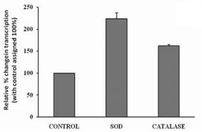
Figure 2 - Densitometry scanning of antioxidant enzymes following RT PCR program using scion image software in different groups. (A) Urolithiatic group II vs group I(control), (B) coconut water treated vs group I (control).
A
B
animal models. In the present study, efficacy of coconut water was evaluated on ethylene glycol induced nephrocalcinosis in male Wistar rats. Stone formation in ethylene glycol fed animals is caused by hyperoxaluria, which causes incre-ased renal retention and excretion of oxalate (26). Under hyperoxaluric condition, oxalate has been reported to induce lipid peroxidation and to cause renal damage by reacting with polyunsa-turated fatty acids in the cell membrane (27). In
Figure 3 - Effect of coconut water administration on Superoxide dismutase (A) and Catalase activity (B) in kidney tissue of animals in different groups.
Values are expressed as mean ± SD and percentage increase (+) or percentage decrease (−) compared with control (group I); *p < 0.05, **p < 0.01, ***p < 0.001 indicates significant change in comparison with control group I, #p < 0.05, ##p < 0.01, ###p < 0.001 indicates significant change in comparison with group II.
A
Figure 4 - Polarization micrograph of rat’s urine sample.
(A) Urine of control rats (group I), devoid of any crystals. (B) Urine of urolithiatic rats (group II) showing CaOx crystals. (C) Urine of coconut water treated rats showing reduced crystals deposition. Original magnifications of 100x.
increased antioxidant enzymes SOD and CAT acti-vity and their expression at mRNA level in kidney tissues (Figures 1, 2a, 2b, 3a and 3b). The increase in SOD activity as observed in our experimenta-tion could be an adaptive response of this enzyme to increased production of superoxide ions follo-wing activation of NAD(P)H oxidase via cytokine TGF-b1 (transforming growth factor b1) induction (28). The increase in SOD activity will further lead to high production of hydrogen peroxide (H2O2). Catalase is responsible for the decomposition of hydrogen peroxide, therefore after ethylene glycol
exposure an increase in catalase activity was also observed. The rebalancing of elevated antioxidant enzyme’s activity and their expression by coco-nut water post treatment further substantiated the protective nature of coconut water against free ra-dical induced oxidative stress. Furthermore, ethy-lene glycol administration increased the level of serum creatinine and urea (Table-2) indicating re-nal dysfunction due to urolithiatic condition. Here again rebalancing of serum urea further unveils the potential effect of coconut water on maintai-ning renal functiomaintai-ning.
A
C
Figure 5 - Representative microscopic examination under light microscope of renal tissue.
(A) Renal tissue of control (group1) rats showing no sign of crystallization. (B) Renal tissue of urolithiatic rats (group 2) showing Crystals deposition mainly located in renal tubules (200×). (C) Renal tissue of group 3 treated rats showing no crystal formation.
A
B
In the present study, administration of co-conut water reduced the number of crystals in the urine as compared to urolithiatic rats (Figure-4c). Our findings are consistent with the studies by Itoh et al. (29) who showed that green tea reduces CaOx crystal deposits in the kidneys of rats made hype-roxaluric by the administration of ethylene glycol. The present histopathological studies sho-wed EG induced crystal deposition in the renal cells (Figure-5b) and most of crystal deposition took place in the renal tubules, which corroborates the results of other studies reporting that crystals depo-sition mainly occur in tubules (30). Administration of coconut water to urolithiatic animals prevented supersaturation of calcium oxalate and thus decre-ased their deposition in renal tubules (Figure-5c).
We are aware of some limitations of the study such as absence of liquid ingestion control by the animals, yet the study does suggest bene-ficial effect of coconut water in nephrocalcinosis.
CONCLUSIONS
In conclusion, coconut water has potential to inhibit the genes of oxidative stress to push the activity of these enzymes towards normal. The re-balancing of elevated antioxidant enzyme gene expression by coconut water treatment, reduced mineral deposits in kidney tissue further substan-tiated the prophylactic nature of coconut water in nephrolithiasis.
ACKNOWLEDGEMENT
The Senior Research Fellowship to Manish Gandhi (Grant no. 45/10/2010-BMS) by the Indian Council of Medical Research, New Delhi is grate-fully acknowledged.
CONFLICT OF INTEREST
None declared.
REFERENCES
1. xLong LO, Park S: Update on nephrolithiasis management. Minerva Urol Nefrol. 2007; 59: 317-25.
2. Stamatelou KK, Francis ME, Jones CA, Nyberg LM, Curhan GC: Time trends in reported prevalence of kidney stones in the United States: 1976-1994. Kidney Int. 2003; 63: 1817-23. 3. Trinchieri A, Ostini F, Nespoli R, Rovera F, Montanari E,
Zanetti G: A prospective study of recurrence rate and risk factors for recurrence after a first renal stone. J Urol. 1999; 162: 27-30.
4. Kaufman DW, Kelly JP, Curhan GC, Anderson TE, Dretler SP, Preminger GM, et al.: Oxalobacter formigenes may re-duce the risk of calcium oxalate kidney stones. J Am Soc Nephrol. 2008; 19: 1197-203.
5. Khan SR, Hackett RL: Retention of calcium oxalate crystals in renal tubules. Scanning Microsc. 1991; 5: 707-11; dis-cussion 711-2.
6. Thamilselvan S, Khan SR: Oxalate and calcium oxalate crystals are injurious to renal epithelial cells: results of in vivo and in vitro studies. J Nephrol. 1998; 11(Suppl 1): 66-9.
7. Bashir S, Gilani AH: Antiurolithic effect of Bergenia ligulata rhizome: an explanation of the underlying mechanisms. J Ethnopharmacol. 2009; 122: 106-16.
8. Miyaoka R, Monga M: Use of traditional Chinese medicine in the management of urinary stone disease. Int Braz J Urol. 2009; 35: 396-405.
9. Atmani F, Sadki C, Aziz M, Mimouni M, Hacht B: Cynodon dactylon extract as a preventive and curative agent in ex-perimentally induced nephrolithiasis. Urol Res. 2009; 37: 75-82.
10. Aggarwal A, Singla SK, Gandhi M, Tandon C: Preventive and curative effects of Achyranthes aspera Linn. extract in experimentally induced nephrolithiasis. Indian J Exp Biol. 2012; 50: 201-8.
11. Marickar YM, Joseph D. Abraham PA: Clinical study of 192 urinary stones in Kerala. Ind J Surg. 1977; 39: 144-50. 12. Huang HS, Chen CF, Chien CT, Chen J: Possible biphasic
changes of free radicals in ethylene glycol-induced nephro-lithiasis in rats. BJU Int. 2000; 85: 1143-9.
13. Buege JA, Aust SD: Microsomal lipid peroxidation. Meth-ods Enzymol. 1978; 52: 302-10.
14. Kono Y: Generation of superoxide radical during autoxida-tion of hydroxylamine and an assay for superoxide dis-mutase. Arch Biochem Biophys. 1978; 186: 189-95. 15. Luck H: Catalase. In: Bergmeyer HU (ed.), Methods of
En-zymatic Analysis. Academic Press, New York 1971; pp. 885-93.
16. Williams K, Frayne J, Hall L: Expression of extracellular glutathione peroxidase type 5 (GPX5) in the rat male repro-ductive tract. Mol Hum Reprod. 1998; 4: 841-8.
18. Cederberg J, Galli J, Luthman H, Eriksson UJ: Increased mRNA levels of Mn-SOD and catalase in embryos of dia-betic rats from a malformation-resistant strain. Diabetes. 2000; 49: 101-7.
19. Park EJ, Park K: Gene expression profiles of cultured rat cardiomyocytes (H 9C2 cells) in response to arsenic triox-ide at subcytotoxic level and oxidative stress. J Health Sci. 2006; 52: 512-21.
20. Liu J, Lei D, Waalkes MP, Beliles RP, Morgan DL: Genomic analysis of the rat lung following elemental mercury vapor exposure. Toxicol Sci. 2003; 74: 174-81.
21. Freitas AM, Schor N, Boim MA: The effect of Phyllanthus niruri on urinary inhibitors of calcium oxalate crystalliza-tion and other factors associated with renal stone forma-tion. BJU Int. 2002; 89: 829-34.
22. Grases F, March JG, Ramis M, Costa-Bauza A: The influ-ence of Zea mays on urinary risk factors for kidney stones in rats. Phytother Res. 1993; 7: 146-49.
23. Grases F, Ramis M, Costa-Bauzá A, March JG: Effect of Herniaria hirsuta and Agropyron repens on calcium oxalate urolithiasis risk in rats. J Ethnopharmacol. 1995; 45: 211-4. 24. Herrmann U, Schwille PO, Kuch P: Crystalluria deter-mined by polarization microscopy. Technique and results in healthy control subjects and patients with idiopathic re-current calcium urolithiasis classified in accordance with calciuria. Urol Res. 1991; 19: 151-8.
25. Bashir S, Gilani AH, Siddiqui AA, Pervez S, Khan SR, Sar-faraz NJ, et al.: Berberis vulgaris root bark extract prevents hyperoxaluria induced urolithiasis in rats. Phytother Res. 2010; 24: 1250-5.
26. Selvam R, Kalaiselvi P, Govindaraj A, Bala Murugan V, Sath-ish Kumar AS: Effect of A. lanata leaf extract and Vediuppu chunnam on the urinary risk factors of calcium oxalate urolithiasis during experimental hyperoxaluria. Pharmacol Res. 2001; 43: 89-93.
27. Ernester LN: Oxidation and phosphorylation. In: Ronald WE MEe (ed.), Methods in Enzymology. Academic Press, New York. 1967; pp; 574-80.
28. Rashed T, Menon M, Thamilselvan S: Molecular mecha-nism of oxalate-induced free radical production and glu-tathione redox imbalance in renal epithelial cells: effect of antioxidants. Am J Nephrol. 2004; 24: 557-68.
29. Itoh Y, Yasui T, Okada A, Tozawa K, Hayashi Y, Kohri K: Preventive effects of green tea on renal stone formation and the role of oxidative stress in nephrolithiasis. J Urol. 2005; 173: 271-5.
30. Khan SR, Glenton PA, Byer KJ: Modeling of hyperoxaluric calcium oxalate nephrolithiasis: experimental induction of hyperoxaluria by hydroxy-L-proline. Kidney Int. 2006; 70: 914-23.