Key words:
Epidemiology; etiology; Prevalence; Penile diseases
Int Braz J Urol. 2013; 39: 55-62
__________________
Submitted for publication: January 16, 2012
__________________
Accepted after revision: July 19, 2012
Purpose: To report the prevalence and risk factors of penile lesions/anomalies in a Metropolitan Brazilian city.
Materials and Methods: All participants undergoing prostate cancer screening in the city of Curitiba were systematically examined to identify penile lesions including cuta-neous mycosis, sexually transmitted diseases, penile cancer, meatal stenosis, hypos-padias, and Peyronie’s disease. Outcomes of interest included the prevalence and the relative risk and 95% confidence intervals of the lesions/anomalies according to age, school level, race, personal history of diabetes, arterial hypertension, nonspecific ure-thritis, and vasectomy.
Results: Balanoposthitis occurred in 11.8% of all participants, with an increased risk in those with diabetes (RR = 1.73), or past history of nonspecific urethritis (RR = 1.58); tinea of the penis was present in 0.2%; condyloma acuminata in 0.5%; herpes virus infection in 0.4%; urethral discharge in 0.2%; genital vitiligo in 0.7%, with an increa-sed prevalence in non-white men (RR = 4.43), and in subjects with lower school level (RR = 7.24); phimosis in 0.5%, with a nearly 7-fold increased risk in diabetics; lichen sclerosus in 0.3%; stenosis of the external urethral meatus in 0.7%, with a higher pre-valence in subjects with lichen sclerosus (RR = 214.9), and in those older than 60 years of age (RR = 3.57); hypospadia in 0.6%; fibrosis suggestive of Peyronie’s disease in 0.9%, especially in men older than 60 years (RR = 4.59) and with diabetes (RR = 3.91); and penile cancer in 0.06%.
Conclusion: We estimated the prevalence and risk factors of commonly seen penile diseases in an adult cohort of Brazilian men.
INTRODUCTION
Genital lesions/anomalies are commonly seen in the office practice. Although these lesions are frequently referred to urologists, they are often discovered incidentally during physical examina-tion by various other specialists including general physicians and surgeons.
The prevalence of genital lesion/anomalies is difficult to estimate. Results differ according to age, gender, racial/ethnic influences, geographic location, comorbidities, and socioeconomic status of the patient. The setting in which the study is conducted (based in the population/community or hospital/clinic setting), the type of study (retros-pective or pros(retros-pective), and the type of diagnostic
Prevalence and risk factors for penile lesions/anomalies
in a cohort of Brazilian men
≥
40 years of age
_______________________________________________
Frederico R. Romero, Antonio W. Romero, Rui Manuel S. de Almeida, Fernando Cesar de Oliveira
Jr., Renato Tambara Filho
Hospital Policlínica Cascavel, (FRR, AWR); Faculdade Assis Gurgacz (FAG) (FRR, RMSA), Cascavel; Instituto Curitiba de Saúde (FRR, FCOJR) and Hospital de Clínicas da Universidade Federal do Paraná (FRR, RTF) , Curitiba, PR, Brazil
ABSTRACT
ARTICLE
INFO
assessment (clinical, laboratorial, or by imaging studies) also influence prevalence levels.
Epidemiological studies are important be-cause they contribute to the appropriate approach of the conditions, improving awareness, promo-ting educational practices and preventive mea-sures, and expediting treatment. They may also allow for intra- and inter-country comparisons, temporal variations between different ages and time periods, and to guide future research eva-luating pathogenesis, etiology and risk factors of these diseases.
Prospective epidemiological studies about common genital diseases are limited worldwide, with only scant reports from Brazil. The objective of this manuscript is to report the prevalence and risk factors of penile lesions/anomalies collected prospectively in a cohort of participants in the Metropolitan Brazilian City of Curitiba.
MATERIALS AND METHODS
Between December 2006 and April 2011, 1731 subjects were included in this research. Par-ticipants were men aged 40 years or older under-going outpatient urologic evaluation in the City of Curitiba (PR) as part of a free prostate cancer scre-ening program conducted by the City employees’ Health Care System. The study protocol was re-viewed and approved by the Institutional Ethics Committee on Human Research (registry number 2253.147/2010-06).
During evaluation, participants were clas-sified by a single examiner as white, or non-white (including brown or black) race; they answered a general questionnaire including age, school level, personal history of diabetes or arterial hyperten-sion, and past history of nonspecific urethritis or vasectomy (Table-1); and were offered a complete genital-pelvic examination.
Urological examination was standardized as follows, and it was performed in all participants in the supine position by a single examiner. Penile inspection with prepuce retraction was performed to identify cutaneous lesions including balano-posthitis, sexually transmitted diseases (STDs), penile cancer, and other infectious/inflammatory, hypochromic or hyperchromic lesions. Lesions/
anomalies of the urethral meatus such as meatal stenosis and hypospadia were also registered, and the penile shaft was palpated for areas of thicke-ning or fibrosis suggestive of Peyronie’s disease.
Outcomes of interest included the preva-lence of penile lesions/anomalies, and the relative risk (RR) and 95% confidence intervals (95% CI) of the lesions/anomalies according to age (≥ 60 vs. < 60 years), school level (elementary school or lo-wer vs. high-school or higher), race (non-white vs. white), personal history of diabetes, arterial hyper-tension, nonspecific urethritis, and vasectomy (yes vs. no, to all). Statistics were calculated using the Fisher´s Exact Test or Pearson´s Chi-square Test, whichever appropriate, and statistical significance was set when p < 0.05 or when the 95% CI did not include the null hypothesis (RR = 1.00).
RESULTS
Cutaneous lesions of the penis were iden-tified in 15.2% (263/1731) of participants. Bala-noposthitis was responsible for most of these le-sions (77.6%, 204/263), corresponding to 11.8% (204/1731) of all participants, and it was associa-ted with tinea cruris in 28.9% (59/204) of them. Balanoposthitis occurred more commonly in par-ticipants with diabetes (RR = 1.73, p < 0.05), and past history of nonspecific urethritis (RR = 1.58, p < 0.05) (Table-2). Tinea of the penis was present in 1.5% (4/263) of skin lesions and 0.2% (4/1731) overall, 75% (3/4) of which had associated bala-noposthitis or tinea cruris. The risk-adjusted pre-valence of balanoposthitis and tinea of the penis are summarized in Table-2.
presented an increased prevalence of herpes virus infection than those individuals with lower school level (0.7% vs. 0.0%, p < 0.05) (Table-2).
Other infectious/inflammatory lesions en-countered included phimosis, responsible for 3.0% (8/263) of penile skin lesions and 0.5% (8/1731) overall; lichen sclerosus in five men (1.9% [5/263]
and 0.3% [5/1731]); and psoriasis in one (0.4% [1/263] and 0.06% [1/1731]). Risk-adjusted preva-lence of these lesions demonstrated that phimo-sis was more common in subjects with history of diabetes (RR = 6.88, p < 0.05). The prevalence of lichen sclerosus was non-significantly increased in men with lower education level, and in those with a history of diabetes (Table-2).
Hypochromic or hyperchromic lesions were identified in 10.3% (27/263) of all skin le-sions on the penis. Genital vitiligo was responsible for 4.6% (12/263) and 0.7% (12/1731) of the cuta-neous lesions and the complete sample, respec-tively. Vitiligo was more prevalent in non-white men than in white men (RR = 4.43, p < 0.05), as well as in subjects with lower school level (RR = 7.24, p < 0.05) (Table-2).
Traumatic lesions including post-coital excoriation, superficial hemorrhagic effusion, and keloid were identified in one participant each (0.4% [1/263] and 0.06% [1/1731]). A median ra-phe cyst of the penis was found in one subject (0.4% [1/263] and 0.06% [1/1731]), and biopsy--confirmed penile cancer was also diagnosed in one man (0.4% [1/263] and 0.06% [1/1731]).
Twenty-two (1.3%, 22/1731) participants presented with lesions at the urethral meatus. Ste-nosis of the external urethral meatus was identified in 0.7% (12/1731) of all participants. Meatal steno-sis was more frequent in men with lichen sclerosus compared to those without it (25.0% vs. 0.1%, RR = 214.9, 95% CI 39.38-1172.40, p < 0.05), and in men aged 60 years or more in comparison with those < 60 years (RR = 3.57, p < 0.05). Subjects with a past history of nonspecific urethritis had a nonsig-nificant increased risk of meatal stenosis (Table-2). Hypospadia was detected in 0.6% (10/1731) of the participants, eight of which (80%, 8/10) were situa-ted on the glans penis, and two (20%, 2/10) on the coronal sulcus. Meatal stenosis was not detected in any of these subjects. The risk-adjusted prevalence of hypospadias is summarized in Table-2.
Palpation of the penile shaft revealed a circumscribed area of fibrosis suggestive of Peyronie’s disease in 0.9% (15/1731) of the parti-cipants. A higher risk was identified in men > 60 years (RR = 4.59, p < 0.05), and in diabetics (RR = 3.91, p < 0.05) (Table-2).
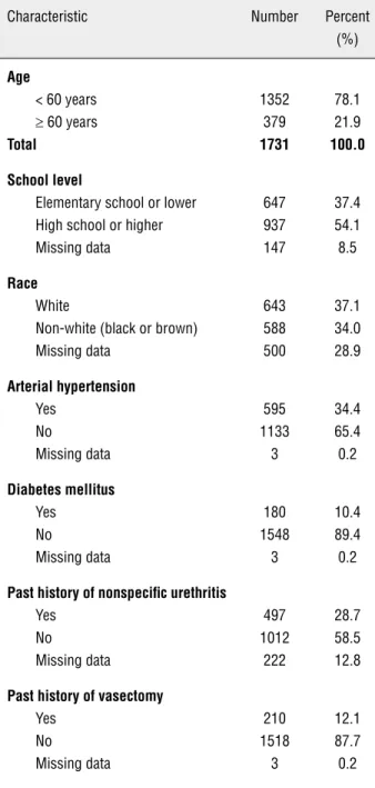
Table 1 - Demographic and clinical characteristics of study population.
Characteristic Number Percent (%)
Age
< 60 years ≥ 60 years
Total
1352 379
1731
78.1 21.9
100.0
School level
Elementary school or lower High school or higher Missing data
647 937 147
37.4 54.1 8.5
Race
White
Non-white (black or brown) Missing data
643 588 500
37.1 34.0 28.9
Arterial hypertension
Yes No
Missing data
595 1133
3
34.4 65.4 0.2
Diabetes mellitus
Yes No
Missing data
180 1548
3
10.4 89.4 0.2
Past history of nonspecific urethritis
Yes No
Missing data
497 1012
222
28.7 58.5 12.8
Past history of vasectomy
Yes No
Missing data
210 1518
3
Table 2 - Prevalence of penile diseases, and relative risks (RR) and 95% confidence intervals (CI) of the lesions/anomalies ac-cording to age, school level, race, and history of diabetes mellitus, arterial hypertension, nonspecific urethritis, and vasectomy.
Age ≥ 60 vs.
<60 RR 95% CI
School-level Elementary School
vs. High-School or higher RR 95% CI
Race Non-white
vs. White RR 95% CI
Diabetes Yes vs. No RR 95% CI
Arterial hypertension
Yes vs. No RR 95% CI
Past history of nonspecific
urethritis Yes vs. No RR 95% CI
Past history of vasectomy Yes vs. No RR 95% CI
Balanoposthitis
12.1 vs. 11.7 1.04 0.76-1.41
11.4 vs. 11.6 0.98 0.74-1.30
11.1 vs. 10.4 1.06 0.77-1.46
18.9 vs. 10.9 (*) 1.73 1.24-2.42
13.4 vs. 10.9 1.24 0.95-1.61
14.7 vs. 9.3 (*) 1.58 1.19-2.11
12.4 vs. 11.7 1.06 0.72-1.56
Tinea of the Penis
0.5 vs. 0.1 3.57 0.50-25.24
0.0 vs. 0.4 0.00 0.00-NaN
0.0 vs. 0.3 0.00 0.00-NaN
0.0 vs. 0.3 0.00 0.00-NaN
0.2 vs. 0.3 0.63 0.07-6.09
0.6 vs. 0.1 6.11 0.64-58.58
0.5 vs. 0.2 2.41 0.25-23.06
Sexually Transmitted Diseases
0.5 vs. 1.8 (*) 0.30 0.07-1.24
1.1 vs. 2.0 0.53 0.23-1.26
1.2 vs. 1.6 0.77 0.29-2.00
0.6 vs. 1.0 0.53 0.07-4.00
1.0 vs. 1.9 0.54 0.22-1.34
1.8 vs. 1.3 1.41 0.61-3.28
1.0 vs. 1.6 0.58 0.14-2.42
Condyloma Acuminata
0.0 vs. 0.7 0.00 0.00-NaN
0.6 vs. 0.4 1.45 0.36-5.77
0.7 vs. 0.3 2.19 0.40-11.90
0.0 vs. 0.6 0.00 0.00-NaN
0.7 vs. 0.4 1.52 0.41-5.65
0.6 vs. 0.5 1.22 0.29-5.09
0.0 vs. 0.6 0.00 0.00-NaN
Herpesvirus Infection
0.0 vs. 0.5 0.00 0.00-NaN
0.0 vs. 0.7 (*) 0.00 0.00-NaN
0.2 vs. 0.5 0.36 0.04-3.49
0.0 vs. 0.5 0.00 0.00-NaN
0.2 vs. 0.5 0.31 0.04-2.63
0.6 vs. 0.3 2.04 0.41-10.05
0.5 vs. 0.4 1.20 0.15-9.96
Phimosis
1.1 vs. 0.4 2.85 0.77-10.58
0.6 vs. 0.3 1.93 0.43-8.60
0.3 vs. 0.3 1.09 0.20-23.80
2.2 vs. 0.3 (*) 6.88 1.86-25.39
0.8 vs. 0.4 2.38 0.64-8.83
0.0 vs. 0.7 0.00 0.00-NaN
0.0 vs. 0.6 0.00 0.00-NaN
Lichen Sclerosus
0.3 vs. 0.3 0.89 0.10-7.96
0.5 vs. 0.1 4.34 0.45-41.68
0.3 vs. 0.2 2.19 0.20-24.06
0.6 vs. 0.3 2.15 0.24-19.13
0.0 vs. 0.4 0.00 0.00-NaN
0.2 vs. 0.3 0.68 0.07-6.51
0.0 vs. 0.3 0.00 0.00-NaN
Genital Vitiligo
0.8 vs. 0.7 1.19 0.32-4.37
1.5 vs. 0.2 (*) 7.24 1.59-32.94
1.4 vs. 0.3 (*) 4.43 0.94-20.76
0.0 vs. 0.8 0.00 0.00-NaN
0.5 vs. 0.8 0.63 0.17-2.34
0.8 vs. 0.6 1.36 0.38-4.79
1.4 vs. 0.6 2.41 0.66-8.83
Meatal Stenosis
1.6 vs. 0.4 (*) 3.57 1.16-11.00
0.6 vs. 0.4 1.45 0.36-5.77
0.3 vs. 0.6 0.55 0.10-2.97
1.1 vs. 0.6 1.72 0.38-7.79
0.3 vs. 0.9 0.39 0.09-1.76
0.8 vs. 0.3 2.71 0.61-12.08
0.5 vs. 0.7 0.66 0.09-5.06
Hypospadia
1.6 vs. 1.0 1.53 0.59-3.95
1.2 vs. 1.1 1.16 0.46-2.92
0.9 vs. 0.8 1.09 0.32-3.76
1.1 vs. 1.2 0.96 0.22-4.08
0.7 vs. 1.4 0.48 0.16-142
1.6 vs. 0.9 1.82 0.70-4.68
1.0 vs. 1.2 0.80 0.19-3.44
Fibrosis (Peyronie)
2.4 vs. 0.5 (*) 4.59 1.72-12.23
0.9 vs. 0.6 1.45 0.47-4.47
0.5 vs. 0.5 1.09 0.22-5.40
2.8 vs. 0.7 (*) 3.91 1.37-11.12
1.3 vs. 0.7 1.90 0.72-5.05
0.6 vs. 0.7 0.87 0.23-3.36
1.4 vs. 0.9 1.67 0.48-5.81
DISCUSSION
Penile mycosis
In the present study, penile mycosis was identified in 11.9% of all participants: balanopos-thitis in 99.0%, and tinea of the penis in 1.9%.
Balanoposthitis is the inflammation of the foreskin/glans penis caused by multiple infec-tious and noninfecinfec-tious agents. It occurs at any age, especially in uncircumcised men, accounting for 11%-13% of them (1,2). It is more common in diabetics (3), in whom it is frequently chronic or recurrent, with an increased risk of 73% compared to non-diabetics in the present research. We also found a higher prevalence of balanoposthitis in men with past history of nonspecific urethritis.
Tinea of the penis is a relatively uncom-mon mycotic infection of the penile shaft (4,5), with an incidence of 1.2% of men with derma-tophytosis (4). It is frequently associated to bala-noposthitis or other dermatophytosis (4-6). In our study, the prevalence of penile tinea was increased by 257% in men > 60 years, and by more than 6-fold in participants with a past history of nons-pecific urethritis. However, the small number of participants with penile tinea prevented a signifi-cant correlation when controlling the analysis for potential risk factors.
Sexually Transmitted Diseases
The prevalence of STDs varies according to the etiologic agent, and the age, gender, so-cioeconomic factors, and sexual behavior of the patient. In Brazil, the prevalence of STDs in men > 20 years-old is 7.1%, diminishing progressively with the increasing of age, with a RR = 6.5 in the group of 20-30 years-old, compared to those > 70 years-old (7). In our study, STDs were identified in 1.8% of men between 40-60 years of age, and 0.5% of those > 60 years-old.
Condiloma acuminata (genital warts) are caused by human papillomavirus (HPV), the most common viral STD in the world (8,9). The preva-lence of genital warts is 1.1% in Brazilian men be-tween 20-49 years-old (7) and it was 0.5% in the participants aged > 40 years-old evaluated in our study. However, visible genital warts are detecta-ble in only a small percentage of HPV carriers (9).
In a study evaluating men with clinical suspicion of HPV infection through DNA testing, the preva-lence of HPV was 0.4% at the ages of 61-70 years, 3.1% at 51-60 years, 8.3% at 41-50 years, 19.8% at 31-40 years, and 50.3% at 21-30 years (9).
In our study, the risk-adjusted prevalence of all STDs was similar between non-white and white participants, similarly to several studies (10,11), although others show an increased risk of some STDs among blacks, compared to white in-dividuals (12).
Genital herpes virus (HSV) is a frequently under-recognized and underestimated STD becau-se infection is often subclinical (13). In the US, although the estimated seroprevalence of HSV is about 25%-28% of the population (14,15), 88.4% of people with laboratory evidence of HSV are unaware of their diagnosis (15). In our cohort, the overall prevalence of HSV identified through a group of blisters and/or ulcers was 0.4% of all participants.
The annual prevalence of Chlamydia and Gonorrhea (nonspecific urethritis) in the sexually active population in Brazil is estimated collecti-vely as 60.8% of all STDs, followed by syphilis in 16.2%, HPV in 11.9%, and HSV in 11.1% (16). In the present study, the prevalence of urethral dis-charge was only 13.6% of all STDs, and there were no laboratory-confirmed cases of syphilis. The prevalence of HPV and HSV, on the other hand, reached respectively 40.9% and 31.8% of all STDs.
The low rates of nonspecific urethritis in this series may be explained by the frequently asymptomatic or oligosymptomatic clinical mani-festations of urethritis caused by Chlamydia. Ho-wever, although nonspecific urethritis and syphilis are estimated as the most prevalent STDs in all age-groups in Brazil (16), HPV and HSV may be more prevalent in older age groups because they are chronic, frequently recurrent, and up to this day noncurable diseases.
Phimosis
gradually separated by intermittent penile erec-tions and accumulation of epithelial debris under the prepuce (17). The prevalence of phimosis is 58% at 1 year of life, 10%-35% at 3 years, 8% at 6 years, and less than 1% by 17 years of age (18). In the present study, phimosis was observed in 0.5% of men > 40 years, more commonly in diabetics, that had an increased risk of nearly 7-fold compa-red to non-diabetics.
Lichen sclerosus
Lichen sclerosus (balanitis xerotica oblite-rans), which commonly appears as white plaques on the glans, often with involvement of the prepuce that becomes thickened and non-retractile, occurs at any age. The underlying cause is unknown (19), but it has been frequently associated with phimosis, either as a cause or as a consequence (17,20).
The prevalence of lichen sclerosus in the general population is estimated to be 0.1%-0.3% (21). In our study, although it reached a 334% hi-gher prevalence in men with low school level, and it was 115% increased among men with a history of diabetes; statistical analysis was non-signifi-cant, probably due to the low prevalence of lichen sclerosus.
Genital vitiligo
Vitiligo is an acquired disorder of skin depigmentation that affects 0.5%-2% of the po-pulation, and it is limited to the genitalia in less than 0.3% of men (22,23). It is particularly more noticeable in darker-skinned individuals (23), with a prevalence 4.4-fold higher in non-whites than in whites in our cohort, as well as a 7.2-fold incre-ased risk in men with lower school level than in those with higher education.
Meatal stenosis
Urethral meatal stenosis is a narrowing of the urethra at the external meatus. One of the most common causes of meatal stenosis is lichen sclerosus, but it also occurs in adults after inflam-mation, specific or nonspecific urethral infections, and urethral instrumentation or surgery (21). The prevalence of meatal stenosis in our study was 0.7%, with an increased risk in men with lichen sclerosus, and in those > 60 years. The risk of
me-atal stenosis in participants with past history of nonspecific urethritis was increased by 171%, but it did not reach statistical significance.
Hypospadia
Hypospadia is an abnormal ventral ope-ning of the urethral meatus anywhere from the ventral aspect of the glans penis to the perineum. In the US, the prevalence of hypospadias is up to 0.8% of live male births, 87% of which are glan-dular or coronal (24). In Brazil, the prevalence of hypospadias is approximately 1.8%-4.1% of live male births (25,26).
Hypospadia in the adult is uncommon and frequently overlooked because most severe cases are treated during childhood, and the remaining cases are clinically insignificant or merely unaes-thetic. In our cohort of men > 40 years-old, hy-pospadias were present in 0.6%, all of which were located on the glans penis or the coronal sulcus, and none were associated with meatal stenosis.
Penile cancer
Cancer of the penis is a rare neoplasm, with a prevalence that varies according to different ge-ographic regions between countries, and within a single country. In Brazil, penile cancer accounts for 2.1% of male malignancies, with the highest incidence in the Northeast region (5.3%), and the lowest in the Southern region (1.2%), where the present study was conducted (27).
Penile cancer occurs more frequently in the sixth decade of life. Risk-factors for the deve-lopment of penile cancer include phimosis, STDs, lichen sclerosus, low socioeconomic level, and poor personal hygiene (27-29).
Peyronie’s disease
po-tential risk factor for Peyronie’s disease, with a 3 to 4-fold increased risk of the disease (30).
Strengths and limitations of the study
Although this survey is strengthened by a prospective and systematic collection of data per-formed by a single examiner in a medium-sized co-hort of subjects, it has several limitations. First, it involves exclusively men > 40 years of age from an established private Health Care System, and therefo-re should be extrapolated with caution. However, it approaches two specific age-range of adults (40-60 years, and > 60 years of age), allowing important insights about the prevalence and risk factors of se-veral diseases more or less common to these age groups. Second, with the exception of cancer, we did not routinely use biopsy or laboratory tests to confirm the clinical diagnosis of the lesions encoun-tered. This practice is commonly used and clinically recommended for most lesions/anomalies because complementary examination will not modify treat-ment, but it is not adequate for research purposes because it may result in false positive/negative bias. Additionally, the prevalence of lesions/anomalies in our cohort did not include conditions previously treated (e.g. history of circumcision) or in clinical remission (e.g. history of herpes virus infection). One of the most interesting aspects of our cohort, however, is the establishment of several epidemio-logical risk factors poorly evaluated in the literature for penile lesions/anomalies. Future studies should validate the consistency of these associations.
CONCLUSIONS
Penile lesions/anomalies are frequently found in the adult population. They may have mul-tiple causes, including infectious, inflammatory, traumatic, congenital, or idiopathic. We estimated the prevalence and risk factors of penile diseases commonly seen in the office in an adult cohort of Brazilian men.
ACKNOWLEDGEMENTS
This article is part of a doctoral dissertation in development by the main author at the Federal University of Paraná, Brazil.
CONFLICT OF INTEREST
None declared.
REFERENCES
1. Edwards S: Balanitis and balanoposthitis: a review. Genito-urin Med. 1996; 72: 155-9.
2. Fakjian N, Hunter S, Cole GW, Miller J: An argument for circumcision. Prevention of balanitis in the adult. Arch Der-matol. 1990; 126: 1046-7.
3. Romano C, Ghilardi A, Papini M: Nine male cases of tinea genitalis.Mycoses. 2005; 48: 202-4.
4. Das JK, Sengupta S, Gangopadhyay A: Dermatophyte in-fection of the male genitalia. Indian J Dermatol. 2009; 54: 21-3.
5. Pielop J, Rosen T: Penile dermatophytosis. J Am Acad Der-matol. 2001; 44: 864-7.
6. Vora SN, Mukhopadhyay A. Incidence of dermatophytosis of the penis and scrotum. Indian J Dermatol Venereol Lep-rol. 1994; 60: 89-91.
7. Carret ML, Fassa AG, da Silveira DS, Bertoldi AD, Hallal PC: Sexually transmitted diseases symptoms in adults: preva-lence and risk factors. Rev Saude Publica. 2004; 38:76-84. 8. Fleischer AB Jr, Parrish CA, Glenn R, Feldman SR: Con-dylomata acuminata (genital warts): patient demographics and treating physicians. Sex Transm Dis. 2001; 28: 643-7. 9. Carestiato FN, Silva KC, Dimetz T, Oliveira LH, Cavalcanti
SM: Prevalence of human papillomavirus infection in the genital tract determined by hybrid capture assay. Braz J Infect Dis. 2006; 10: 331-6.
10. Tortolero-Luna G: Epidemiology of genital human papillo-mavirus. Hematol Oncol Clin North Am. 1999; 13: 245-57. 11. Brill JR: Diagnosis and treatment of urethritis in men. Am
Fam Physician. 2010; 81: 873-8.
12. Dinh TH, Sternberg M, Dunne EF, Markowitz LE: Genital warts among 18- to 59-year-olds in the United States, national health and nutrition examination survey, 1999--2004. Sex Transm Dis. 2008; 35: 357-60. Erratum in: Sex Transm Dis. 2008; 35: 772-3.
13. Benedetti J, Corey L, Ashley R: Recurrence rates in genital herpes after symptomatic first-episode infection. Ann In-tern Med. 1994; 121: 847-54.
14. Stanberry L, Cunningham A, Mertz G, Mindel A, Peters B, Reitano M, et al.: New developments in the epidemiology, natural history and management of genital herpes. Antiviral Res. 1999; 42: 1-14.
16. Brasil. Ministério da Saúde: DST no Brasil. Available in: <http://www.aids.gov.br/pagina/dst-no-brasil>. Last ac-cessed in: 05/11/12.
17. El Achkar ME, Machado AB, Pereima MJ, Bastos JC: Clini-cal analysis and anatomopathologic research on patient prepuces referred to postectomy. An Bras Dermatol. 2004; 79: 29-37.
18. Elder JS: Abnormalities of the genitalia in boys and their surgical management. In: Campbell-Walsh Urology. 9th Ed., Edited by: Wein AJ, Kavoussi LR, Novick AC, Partin AW, Peters CA, Philadelphia: WB Saunders Elsevier. 2007; chap 126, pp. 3746.
19. Powell JJ, Wojnarowska F: Lichen sclerosus. Lancet. 1999; 353: 1777-83.
20. Edmonds EV, Hunt S, Hawkins D, Dinneen M, Francis N, Bunker CB: Clinical parameters in male genital lichen scle-rosus: a case series of 329 patients. J Eur Acad Dermatol Venereol. 2012; 26: 730-7.
21. Navalón Verdejo P, Pallás Costa Y, Juan Escudero J, Fabuel Deltoro M, Ordoño Domínguez F, Monllor Peidro E, et al.: Dorsal meatoplasty for the treatment of meatal stenosis in patients with balanitis xerotica obliteran. Arch Esp Urol. 2007; 60: 1.156-60.
22. Moss TR, Stevenson CJ: Incidence of male genital vitiligo. Report of a screening programme. Br J Vener Dis. 1981; 57: 145-6.
23. Link RE: Cutaneous diseases of the external genitalia. In: Campbell-Walsh Urology. 9th Ed., Edited by: Wein AJ, Ka-voussi LR, Novick AC, Partin AW, Peters CA, Philadelphia: WB Saunders Elsevier. 2007; chap 13, pp. 433.
24. Borer JG, Retik AB: Hypospadias. In: Campbell-Walsh Urol-ogy. 9th Ed., Edited by: Wein AJ, Kavoussi LR, Novick AC, Partin AW, Peters CA, Philadelphia: WB Saunders Elsevier. 2007; chap 13, pp. 3708.
25. Ronchetti MH: Características anatômicas da genitália ex-terna no recém-nascido. In: http://www.lume.ufrgs.br/bit-stream/handle/10183/11368/000611747.pdf?sequence=1 26. Monteleone Neto R, Castilla EE, Paz JE Hypospadias: an
epidemiological study in Latin America. Am J Med Genet. 1981; 10: 5-19.
27. Favorito LA, Nardi AC, Ronalsa M, Zequi SC, Sampaio FJ, Glina S: Epidemiologic study on penile cancer in Brazil. Int Braz J Urol. 2008; 34: 587-91; discussion 591-3.
28. Guimaraes GC, Rocha RM, Zequi SC, Cunha IW, Soares FA: Penile cancer: epidemiology and treatment. Curr Oncol Rep. 2011; 13: 231-9.
29. Zequi SD, Guimaraes GC, da Fonseca FP, Ferreira U, de Matheus WE, Reis LO, et al.: Sex with Animals (SWA): Behavioral Characteristics and Possible Association with Penile Cancer. A Multicenter Study. J Sex Med. 2012; 9: 1860-7.
30. Sommer F, Schwarzer U, Wassmer G, Bloch W, Braun M, Klotz T: Epidemiology of Peyronie’s disease. Int J Impot Res. 2002; 14: 379-83.
31. Lindsay MB, Schain DM, Grambsch P, Benson RC, Beard CM, Kurland LT: The incidence of Peyronie’s disease in Rochester, Minnesota, 1950 through 1984. J Urol. 1991; 146: 1007-9.
_____________________
Correspondence address: