Authors
Aline Felicio Bueno 1 Fernando de Aguiar Lemos 1 Matheus Elias Ferrareze 1
William Antonio Martins dos Santos 1
Francisco Veríssimo Veronese 2
Alexandre Simões Dias 3
1 Universidade Federal do Rio Grande do Sul, Porto Alegre - RS, Brazil. 2 Universidade Federal do Rio Grande do Sul, Hospital de Clínicas de Porto Alegre, Serviço de Nefrologia, Porto Alegre - RS, Brazil.
3 Hospital de Clínicas de Porto Alegre, Serviço de Fisioterapia, Programas de Pós Graduação em Ciências Pneumológicas e em Ciências do Movimento Humano, Porto Alegre - RS, Brazil.
Submitted on: 07/06/2016. Approved on: 09/22/2016.
Correspondence to: Aline Felicio Bueno.
Fundo de Incentivo à Pesquisa do Hospital de Clínicas de Porto Algre - FIPE HCPA. E-mail: alinefeliciobueno@ gmail.com
Muscle thickness of the pectoralis major and rectus
abdomi-nis and level of physical activity in chronic hemodialysis
patients
Espessura muscular do
pectoralis major
e do
rectus abdominis
e
nível de atividade física em pacientes em hemodiálise crônica
Introdução: Pacientes que realizam hemodiálise crônica tendem a perder massa magra e ter comportamento sed-entário. Objetivo: Comparar o nível de atividade física e morfologia dos múscu-los peitoral maior e reto do abdômen de pacientes que realizam hemodiálise com indivíduos saudáveis. Métodos: Foram estudados 17 pacientes e 17 indivíduos saudáveis. As espessuras musculares foram avaliadas por meio de ultrassono-grafia, e o nível de atividade física pelo Questionário Internacional de Atividade Física versão longa (IPAQ). Resultados: Os pacientes apresentaram menores es-pessuras do peitoral maior (5,92 ± 0,35 mm vs. 8,35 ± 0,62 mm, p < 0,001) e de reto abdominal (0,96 ± 0,10 mm vs. 2,21 ± 0,40 mm, p < 0,001) compara-dos aos sujeitos saudáveis. Os pacientes foram fisicamente menos ativos que os indivíduos saudáveis: 1502.55(788.19-2513.00) MET-minutos/semana vs. 2268.0(1680.0-4490,8) MET-minutos/ semana (p = 0,006); o gasto calórico se-manal dos pacientes também foi menor: 1384,0(480,7-2253.7) kcal/kg/semana
vs. 1680,0(1677,4-4950,0) kcal/kg/ semana (p = 0,010). O tempo médio gasto sentado por semana dos pacien-tes foi maior que dos sujeitos saudáveis (394,0 ± 33,1 min/dia vs. 293,0 ± 38,6,
p = 0,009), assim como o tempo mé-dio gasto sentado durante o fim de se-mana (460,0 ± 40,1 vs. 201,0 ± 10,7,
p = 0,003). Conclusão: Pacientes renais crônicos em hemodiálise apresentam comportamento sedentário e menores espessuras musculares do tronco.
R
ESUMOPalavras-chave: atividade motora; diálise renal; insuficiência renal crônica.
Introduction: Patients on chronic he-modialysis tend to lose lean body mass and have sedentary behavior. Objecti-ve: To compare the level of physical activity and the morphology of the muscles pectoralis major and rectus abdominis of patients on hemodialy-sis with healthy subjects. Methods: We studied 17 patients and 17 heal-thy individuals. Muscle thickness were evaluated by ultrasound, and the level of physical activity by the Inter-national Physical Activity Question-naire (IPAQ), long version. Results: The patients had lower thicknesses of the pectoralis major (5.92 ± 0.35 mm vs. 8.35 ± 0.62 mm, p < 0.001) and rectus abdominis (0.96 ± 0.10 mm vs. 2 21 ± 0.40 mm, p < 0.001) compared to healthy subjects. Pa-tients were physically less active than healthy individuals: 1502.55(788.19-2513.00) MET-minutes/week vs. 2268.0(1680.0-4490,8) MET-minu-tes/week (p = 0.006); the weekly ca-loric expenditure of patients was also lower: 1384.0(480,7-2253.7) kcal/ kg/week vs. 1680.0(1677.4-4950.0) kcal/kg/week (p = 0.010). The avera-ge time spent sitting per week of the patients was higher than in healthy subjects (394.0 ± 33.1 min/day vs. 293.0 ± 38.6, p = 0.009) as well as the average time spent sitting during weekend (460.0 ± 40.1 vs. 201.0 ± 10.7, p = 0.003). Conclusion: Chronic renal failure patients on hemodialysis have sedentary behavior and lower muscle thickness of the trunk.
A
BSTRACTI
NTRODUCTIONChronic kidney disease (CKD) is a common disease in Brazil and in the world, and because of its high prevalence is considered a public health problem.1,2 The hemodialysis (HD) is recommended in the ter-minal stage of CKD. The HD treatment, however, is associated with decreased physical activity and is accompanied by a number of comorbidities, such as protein-energy malnutrition, reduced lean body mass and muscle strength.3-6
So often a generalized weakness state is present in these patients, the clinical condition research is required as well as the structure and function of the muscular system.7-9 The most studied are the antigravity musculature of the lower limbs. This may be related to the fact that neuropathies and uremic myopathies have a higher incidence in the lower limbs in relation to the upper.10,11 However, the muscles of the trunk, such as the pectoralis ma-jor, has an important role in the multiaxial shoul-der joint. The atrophy in the muscles can inhibit movements such as flexion, extension, adduction and horizontal shoulder flexion preventing many activities of daily living.
The abdominal muscles have a stabilizing ac-tion of the lumbar spine, to load external loads or to maintain the posture in various positions of the activities of daily living. Furthermore, during HD patients remain about 4 hours three times a week in a lying or sitting position, which may lead to a reduction in the use state, generating atrophies and reducing the range of motion of the trunk and the upper limbs.4,12,13
However, it was not found in the literature stud-ies evaluating the morphology of these musculature. One way to evaluate these muscles is through ultra-sound that tells about the muscle thickness, which is directly associated with the amount of contractile tissue and muscle strength production capacity.4,14,15 In this context, the objective of this study is to evalu-ate the thickness of the muscles pectoral muscles and abdominal rectus through muscle ultrasound of pa-tients undergoing chronic hemodialysis compared to healthy subjects. Such information can assist in the organization of preventive protocols of muscle func-tion of maintaining the trunk of these patients.
M
ETHODSThe sample was chosen intentionally, consisting of 17 patients with terminal CKD diagnosis on hemo-dialysis in Porto Alegre Clinical Hospital (HCPA), followed at the Nephrology Service and Exercise Pathophysiology Laboratory. Seventeen healthy sub-jects were included as a control group.
The sample size was calculated by G * Power 3.1.3 software (FrauzFaurUniversität Kiel, Germany), where the “EffectSize” adopted was 0.69, the alpha of 0.05 and power of the study to 0.80, using Student’s t test for independent sam-ples as statistical test for comparison of variable.16 This study was approved by the Research Ethics Committee of the Porto Alegre Clinical Hospital (CAAE 36473714.1.0000.5327). All participants read and signed the consent form.
Were used as inclusion patients criteria aged above 18 years, terminal CKD treated with he-modialysis for at least three months, regardless of gender, age and severity of disease, and to provide clinically stable, without acute complica-tions (ex., infeccomplica-tions) in the last three months. The subjects in the control group were matched for sex, age, total body weight, height and body mass index.
The study excluded individuals who had: a) co-morbidities unrelated to the pathological pro-cess of origin; b) absolute contraindications or for holding the tests; c) difficulty in understand-ing the procedures proposed by researchers; d) were in a period of exacerbation of the disease, and) did not agree to participate and f) patients with neuromuscular diseases who presented mo-tor deficits resulting from stroke (stroke), mul-tiple sclerosis, amyotrophic lateral sclerosis or
Guillain-Barré.
INTERNATIONAL PHYSICAL ACTIVITY QUESTIONNAIRE
LONGVERSION (IPAQ)
Among the outcome measures of the International Physical Activity Questionnaire (IPAQ), long ver-sion, were total physical activity, expressed in MET-minutes per day and minutes reported in vigorous-intensity activity, moderate-intensity ac-tivity, and hiking. Moderate intensity was assigned as 4 METs (Metabolic Equivalent of Task), vigor-ous intensity as 8 METs, and hiking equivalent to 3.3 METs. The metabolic equivalent/minute (MET-min) was calculated by multiplying METs per minute participation in physical activities of vigorous intensity, moderate and hiking, as the formula for MET-minutes calculation considered as follows:
• Walking MET-minutes/week = 3.3 * hiked minutes * days of trekking;
• Moderate MET minutes/week = 4.0 * minutes of moderate intensity activity * days of mod-erate or vigorous activities;
• Vigorous MET-minutes/week = 8.0 * minutes of vigorous intensity activities * day activities with vigorous intensity.
The total physical activity MET-min/week was calculated as the sum of the scores Hikinh + MET-min/week moderate + vigorous. Low activity level was considered when checking to values below 600 MET-minutes/week. For moderate activity, consid-ered 5 or more days of any combination of walking, moderate intensity or vigorous intensity activities achieving a minimum of at least 600 MET-minutes/ week. And how high physical activity, considered 7 days or more of any combination of walking, mod-erate or vigorous intensity activities accumulating at least 3,000 MET-minutes/week.
The caloric expenditure in MET minutes/week was measured by multiplying the value of MET activity held by its weekly frequency and duration. The value ob-tained was multiplied by the weight and divided by 60 minutes to transform into kilocalories (kcal/min).
EVALUATIONOFMUSCLETHICKNESS
For the evaluation of transverse muscle thickness, was used an ultrasound machine and a linear array trans-ducer B-mode ultrasonography (HD7.XE®, Philips
with a frequency of 7.5 MHz. The transducer was soaked in gel water soluble transmission, promoting acoustic contact, not depress the skin surface.
To ensure that subsequent images were made in the exact anatomical location markings bony prominences were considered.17,18 After demarca-tions, a transverse image to which it was possible to view the musculature was obtained. Thus, for evaluation of cross muscle thickness measure-ments were performed by ultrasound by means of an ultrasound device, considering the following regions: the upper inner edge of the aponeurosis of higher and lower abdominal and pectoral both bilateral.
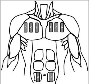
For evaluation of the pectoralis major probe was positioned parallel to the sternum and perpendicu-lar to the clavicle in the second and third rib in three points: 1) Proximal to the sternum; 2) Midpoint of the muscle belly; 3) Distal to the sternum.18 For the evaluation of the abdominal muscles the probe was positioned longitudinally to the muscle, brain-caudal direction. Two images were placed, one to two centi-meters to the right and one to two centicenti-meters to the left of the umbilicus.19 All images were made bilater-ally (Figure 1).
All examinations were performed by the Figure 1. Probe positioning to collect the thickness of the pectoralis major and rectus abdominis.
Patients were classified as moderately active: 1502.55 (788.19-2513.00) MET-minutes/week, while the control subjects were classified as highly active: 2268.0 (1680.0-4490,8) MET-minutes/ week (p = 0.006); the weekly caloric expenditure of patients was also lower: 1384.0 (480,7-2253.7) measurements of muscle thickness (MT) was used Intra-class correlation coefficient (ICC). Six ran-dom images between groups and measured five times by the same person on different days were chosen. The values of reliability coefficients range from zero (0) to one (1) where values closer to 1 indicate higher reliability. The following catego-ries for reliability levels were applied: greater than 0.75 = high reliability; between 0.4 and 0.75 = reasonable reliability, and lower than 0.4 = poor reliability.19
The test Shapiro-Wilk and Levene were used to verify the normality and homogeneity of data. In the descriptive analysis we used mean and stan-dard deviation or median and interquartile range. The independent t test was used to compare the linkage variables (age, weight, height) and mor-phological variables (transverse muscle thickness) between the groups. The Mann-Whitney test was used to assess the measure of central tendency be-tween physical activity levels bebe-tween the groups. All analyzes were performed using SPSS 20.0 for Windows (Chicago, IL, USA). The level of signifi-cance was p < 0.05.
R
ESULTSThe sample consisted of 17 patients with CKD, 10 men and 7 women, the same distribution occurred in the control group. There was no statistically signifi-cant difference between patients and controls with re-spect to age, total body weight, height and body mass index, as shown in Table 1.
Patients Control p
Age (years) 54.1 ± 14.1 48.3 ± 15.2 0.33 Stature (cm) 161.3 ± 8.1 166.0 ± 12.0 0.87 Body mass (Kg) 64.2 ± 11.8 73.0 ± 20.6 0.41 BMI (kg/m²) 24.5 ± 3.1 26.4 ± 5.0 0,83
TABLE 1 DEMOGRAPHICANDANTHROPOMETRICDATAOF HEMODIALYSISPATIENTSANDHEALTHYSUBJECTS
Cm: centimeters; kg: kilogram; kg/m2: kilogram per square meter; BMI = body mass index; Significance level p < 0,05.
kcal/kg/week vs. 1680.0 (1677.4 to 4950.0) kcal /kg/week (p = 0.010). The weekly energy expen-diture of patients was significantly lower than controls: 1384.0 (480,7-2253.7) kcal/kg/week vs.
1680.0 (1677.4 to 4950.0) kcal/kg/week, respec-tively (p = 0.010).
The average time spent sitting week the patients were significantly higher than those of healthy sub-jects (394.0 ± 33.1 min/day 293.0 ± 38.6; p = 0.009). The same was observed in relation to the average time spent sitting during the weekend, the values were 460.0 ± 0.1 for patients and 201.0 ± 10.7 for the con-trols (p = 0.003).
The intraclass correlation test and retest the as-sessment of muscle thickness of the pectoralis major was r = 0.971 and the rectus abdominis of r = 0.984. Regarding muscle morphology, the groups showed significant difference in the thickness of the pecto-ralis major (p = 0.001) and abdominal (p = 0.001). Patients have a thickness average value of the pecto-ralis major of 5.92 ± 0.35 mm and the control group an average of 8.35 ± 0.62 mm. For abdominal muscle, patients have a mean value of 0.96 ± 0.10 mm, where-as in the control group this value wwhere-as 2.21 ± 0.40 mm (Figure 2).
D
ISCUSSIONThe main finding of this study was that chronic re-nal failure patients on hemodialysis had lower muscle thickness of the pectoralis major and rectus abdomi-nis. Compared to healthy subjects, patients had a lower level of physical activity, caloric expenditure and metabolic equivalent, and more time sitting in the week and the weekend.
The maintenance treatment of CKD by hemodi-alysis makes these patients staying for long periods estate throughout the week, a fact that is not balanced with physical activity, can promote muscle weakness, fatigue and increased hospitalization rate.13,20,21 The results of the study of Medina et al.,23 as well as the present study show that the population of chronic re-nal HD is insufficiently active, contributing to sed-entary lifestyles and changes in muscle structure as atrophy and changes in its architecture.3
The study Sakkas et al.10 evaluated through muscle biopsy the rectus abdominis muscle of patients who underwent peritoneal dialysis, compared to healthy subjects. The authors found muscle atrophy of the tis-sue sample of patients compared to control subjects. The findings of this study correlate with these data, as were observed less muscle thickness values of the rec-tus abdominis, although the assessment was by ultra-sound and not by microscopic analysis of the tissue.
Few studies have evaluated the loss of muscle mass musculature of the trunk, such as the rectus abdominis. This muscle has an important stabiliz-ing role of the spine durstabiliz-ing movement, moreover, it can also be affected when there is disuse and atrophy of skeletal muscles of the lower limbs.9,23 Furthermore, the smallest thickness of the rectus abdominis of the patient may indicate a general-ized muscular atrophy.10,11
Regarding the pectoralis major, it was also found a smaller thickness in patients in the con-trol group. This study is the first to evaluate the muscle thickness pectoralis by ultrasound in pa-tients with CKD who perform HD. Two other studies using muscle biopsy also showed atrophy of the deltoid muscle.24,25 These findings indicate a decrease of contractile tissue in the tissue sample, predominantly of type II fibers.
The importance of evaluating the pectoralis ma-jor is in preventing problems that its atrophy may induce such as loss of acessories inspiratory
muscu-and difficulty in performing activities of daily living that require movements performed with upper limbs. In addition, patients with lower limb strength reduc-tion can constantly order the pectoralis major to sit or stand up with the help of hand support due to its water main function bilaterally which assists in trunk stabilization.27
The pectoralis major assists directly movements that perform internal rotation of the arm, as is the cam function to slow shoulder extension movements, horizontal abduction and external27 rotation, which can often be traced body support moves during basic activities such as bathing and bedtime. In such cases, it is important that the pectoralis major is preserved to prevent injuries such as breaks, if it is used too much force during extension and external shoulder rotation.28
It has been shown that proximal musculature of the lower limbs of kidney patients suffer more from the loss tecidual.29,30 In advanced cases of high urea, strength reduction level of the lower limbs due to myopathies and/or polineuromyopa-thies is very high.31,32 Among the mechanisms in-volving the loss of lean body mass are involved aspects such as the development of insulin resis-tance and inflammation and activation of muscle proteolysis by ubiquitin-proteassoma complex.33 In addition, patients with CKD have high risks in their nutritional status due to uremia, a restricted diet, low physical activity, chronic inflammation, comorbidities, and metabolic disorders. These fac-tors affect energy expenditure and affect the body composition of these patients.34
Evidence suggests that in patients with sarcopenia CKD complex condition mediated by an imbalance between anabolism and catabolism of muscle pro-tein.35 Still, there is a gap in the literature regarding the different methods for the evaluation of muscle mass in these patients, as well as studies to perform a combination of these methods with lean mass loss markers.
functional limitations of upper limb muscles, chest and abdominal wall.
As a limitation of this study, the presence and as-sociation of other lean mass conventional markers with muscle thickness parameter assessed by ultra-sound has not been evaluated.
C
ONCLUSIONThis study showed that patients with terminal CKD treated with HD feature: 1) Lower level of physical activity; 2) Increased time spent sitting during the week and the weekend; and 3) Minor muscle thickness of the largest and rectus abdomi-nis muscles chest.
R
EFERENCES1. Bastos MG, Bregman R, Kirsztajn M. Doença renal crônica: frequente e grave, mas também prevenível e tratável. RevAs-socMed Bras 2010;56:248-53.
2. Pinho NA, Silva GV, Pierin AMG. Prevalência e fatores asso-ciados à doença renal crônica em pacientes internados em um hospital universitário na cidade de São Paulo, SP, Brasil. J Bras-Nefrol 2015;37:91-7.
3. Sesso RC, Lopes AA, Thomé FS, Lugon JR, Watanabe Y, Santos DR. Relatório do Censo Brasileiro de Diálise Crôni-ca 2012. J Bras Nefrol 2014;36:48-53. DOI: http://dx.doi. org/10.5935/0101-2800.20140009
4. Johansen KL, Shubert T, Doyle J, Soher B, Sakkas GK, Kent-Braun JA. Muscleatrophy in patientsreceivinghemodialysis: ef-fects on musclestrength, musclequality, and physicalfunction. Kidney Int 2003;63:291-7. DOI: http://dx.doi.org/10.1046/ j.1523-1755.2003.00704.x
5. Toyama K, Sugiyama S, Oka H, Sumida H, Ogawa H. Exer-cisetherapycorrelates with improvingrenalfunction through modifyinglipidmetabolism in patients with cardiovasculardis-ease and chronickidneydiscardiovasculardis-ease. J Cardiol 2010;56:142-6. DOI: http://dx.doi.org/10.1016/j.jjcc.2010.06.007
6. Domański M, Ciechanowski K. Sarcopenia: a majorchallenge in elderlypatients with end-stagerenaldisease. J Aging Res 2012;2012:754739.DOI: 10.1155/2012/754739. DOI: http:// dx.doi.org/10.1155/2012/754739
7. Ikizler TA, Himmelfarb J. Musclewasting in kidneydisease: Let’sgetphysical. J Am Soc Nephrol 2006;17:2097-8.
8. Fahal IH. Uraemicsarcopenia: aetiology and implications.
Nephrol Dial Transplant 2014;29:1655-65. DOI: http://dx.doi. org/10.1093/ndt/gft070
9. Souza VA, Oliveira D, Mansur HN, Fernandes NMS, Bastos MG. Sarcopenia na Doença Renal Crônica. J Bras Nefrol 2015;37:98-105. 10. Sakkas GK, Ball D, Mercer TH, Sargeant AJ, Tolfrey K, Na-ish PF. Atrophy of non-locomotormuscle in patients with end-stagerenalfailure. Nephrol Dial Transplant 2003;18:2074-81. DOI: http://dx.doi.org/10.1093/ndt/gfg325
11. Cotton JR, Woodard T, Carter NW, Knochel JP. Restingskel-etalmusclemembranepotential as an index of uremictoxicity. A proposed new method to assess adequacy of hemodialysis. J Clin Invest 1979;63:501-6. PMID: 429569 DOI: http://dx.doi. org/10.1172/JCI109328
12. Campistol JM. Uremic myopathy. Kidney Int 2002;62:1901-13. PMID: 12371997 DOI: http://dx.doi.org/10.1046/j.1523-1755.2002.00614.x
13. Hall YN, Larive B, Painter P, Kaysen GA, Lindsay RM, Nis-senson AR,et al.; FrequentHemodialysisNetwork Trial Group. Effects of sixversusthreetimesperweekhemodialysis on physi-calperformance, health, and functioning: FrequentHemo-dialysisNetwork (FHN) randomizedtrials. Clin J Am Soc Nephrol2012;7:782-94.
14. Capitanini A, Galligani C, Lange S, Cupisti A. Upper-limbdisability in hemodialysispatients: evaluation of con-tributingfactorsaside from amyloidosis. Ther Apher Dial 2012;16:242-7. DOI: http://dx.doi.org/10.1111/j.1744-9987.2011.01056.x
15. Herzog W, ter Keurs HE. Force-length relation of in-vivo hu-man rectus femoris muscles. Pflugers Arch 1988;411:642-7. DOI: http://dx.doi.org/10.1007/BF00580860
16. Radaelli R, Wilhelm Neto EN, Marques MFB, Pinto RS. Espes-sura e qualidade musculares medidas a partir de ultrassono-grafia: influência de diferentes locais de mensuração. RevBras-Cineantropom Desempenho Hum2011;13:87-91.
17. Lindenau JDR, Guimarães LSP. Calculando o tamanho do efeito no SPSS. Rev HCPA 2012;32:363-81.
18. Sumerling TJ, Quant SP. Measurements of the human anterior chest wall by ultrasound and estimates of chest wall thickness for use in determination of transuranic nuclides in the lung. Radiat Prot Dosimetry 1983;3:203-10.
19. Bradley M, Donnell P. Atlas of Musculoskeletal Ultrasound Anatomy. London: Greenwich Medical Media; 2002.
20. Dancey CP, Reidy J. Estatística sem matemática para psicologia usando SPSS para windows.3ª ed.Porto Alegre: Artmed; 2008. 21. Almeida LAB, Pitanga FJG, Freitas MM, Pitanga CPS, Dan-tas EHM, Beck CC. Caloric expenditure of different domains of physical activity as predictors of the absence of diabetes in adults. Rev Bras Med Esporte 2012;18:17-21. DOI: http:// dx.doi.org/10.1590/S1517-86922012000100003
22. Kopple JD, Kim JC, Shapiro BB, Zhang M, Li Y, Porszasz J, et al. Factorsaffectingdailyphysicalactivity and physicalperfor-mance in maintenancedialysispatients. J Ren Nut 2015;25:217-22. DOI: http://dx.doi.org/10.1053/j.jrn.2014.10.017 23. Medina LAR, Vanderlei FM, Vanderlei LCM,Torres DB,
Pa-dulla AT, Freitas AEA, et al. Atividade física e qualidade de vida em pacientes com doença renal crônica submetidos à he-modiálise.ConScientiae Saúde 2010;9:212-9.
24. Hodges PW, Richardson CA. Contraction of the abdominal musclesassociated with movement of the lower limb. Phys Ther 1997;77:132-42. DOI: http://dx.doi.org/10.1093/ptj/77.2.132 25. do Prado LB, do Prado GF, Oliveira AS, Schmidt B, Carvalhaes
JT. Histochemicalstudy of the skeletalmuscle in children with chronicrenalfailure in dialysistreatment. Arq Neuropsiquiatr 1998;56:381-7.
26. Bautista J, Gil-Necija E, Castilla J, Chinchon I, Rafel E. Dialysis-myopathy. Report of 13cases. Acta Neuropathol 1983;61:71-5. DOI: http://dx.doi.org/10.1007/BF00688389
27. Cunha MS, Andrade V, Guedes CAV, Meneghetti CHZ, Agu-iar AP, Cardoso AL. Avaliação da capacidade funcional e da qualidade de vida em pacientes renais crônicos submetidos a tratamento hemodialítico. Fisioter Pesqui2009;16:155-60. 28. Floyd RT. Manual de cinesiologia estrutural. 16ª ed. Barueri:
Manole; 2011.
29. Oliveira FC, Alves MDS, Bezerra AP. Co-morbidades e mortal-idade de pacientes com doença renal: atendimento terceirizado de nefrologia. Acta Paul Enferm 2009;22:476-80. DOI: http:// dx.doi.org/10.1590/S0103-21002009000800003
30. Bohanon RW. Sit-to-stand test for measuring performance of lower extremity muscles. Percept Mot Skills 1995;80:163-6. DOI: http://dx.doi.org/10.2466/pms.1995.80.1.163
32. Mert T, Gunes Y, Guven M, Gunay I, Ozcengiz D. Effects of calcium and magnesium on peripheral nerveconduction. Pol JPharmacol 2003;55:25-30. PMID: 12856822
33. Mitch WE, Remuzzi G. Diets for patients with chronic kid-ney disease, should we reconsider? BMC Nephrol2016;17:80. PMID: 27401192
34. Koefoed M, Kromann CB, Juliussen SR, Hvidtfeldt D, Ekelund B, Frandsen NE, et al. NutritionalStatus of MaintenanceDialysisPa-tients: LowLeanBodyMassIndex and Obesity Are Common, Pro-tein-EnergyWasting Is Uncommon. PLoS One 2016;11:e0150012. DOI: http://dx.doi.org/10.1371/journal.pone.0150012