Relationship between Fetuin A, Vascular
Calcification and Fracture Risk in Dialysis
Patients
Hung Yuan Chen1,2, Yen Ling Chiu1,2, Shih Ping Hsu1,2, Mei Fen Pai1,2, Ju Yeh Yang1,2, Yu Sen Peng1,2*
1Division of Nephrology, Department of Internal Medicine, Far Eastern Memorial Hospital, New Taipei City, Taiwan,2Division of Nephrology, Department of Internal Medicine, National Taiwan University Hospital, National Taiwan University College of Medicine, Taipei, Taiwan
Abstract
Background
Fractures are a common morbidity that lead to worse outcomes in dialysis patients. Fetuin A inhibits vascular calcification (VC), potentially promotes bone mineralization and its level positively correlates with bone mineral density in the general population. On the other hand, the presence of VC is associated with low bone volume in dialysis patients. Whether the fetuin A level and VC can predict the occurrence of fractures in dialysis patients remains unknown.
Methods
We performed this prospective, observational cohort study including 685 dialysis patients (629 hemodialysis and 56 peritoneal dialysis) from a single center in Taiwan for a median follow-up period of 3.4 years. The baseline fetuin A level and status of presence of aortic arch calcification (VC) and incidence of major fractures (hip, pelvis, humerus, proximal fore-arm, lower leg or vertebrae) were assessed using adjusted Cox proportional hazards mod-els, recursive partitioning analysis and competing risk models.
Results
Overall, 177 of the patients had major fractures. The incidence rate of major fractures was 3.29 per 100 person-years. In adjusted analyses, the patients with higher baseline fetuin A levels had a lower incidence of fractures (adjusted hazard ratio (HR), 0.3; 95% CI, 0.18-0.5, fetuin A tertile 3vs. tertile 1 and HR, 0.52; 95% CI, 0.34-0.78, tertile 2vs. tertile 1). The
pres-ence of aortic arch calcification (VC) independently predicted the occurrpres-ence of fractures (adjusted HR, 1.95; 95% CI, 1.34-2.84) as well. When accounting for death as an event in competing risk models, the patients with higher baseline fetuin A levels remained to have a lower incidence of fractures (SHR, 0.31; 95% CI, 0.17-0.56, fetuin A tertile 3vs. tertile 1 and
0.51; 95% CI, 0.32-0.81, tertile 2vs. tertile 1).
a11111
OPEN ACCESS
Citation:Chen HY, Chiu YL, Hsu SP, Pai MF, Yang JY, Peng YS (2016) Relationship between Fetuin A, Vascular Calcification and Fracture Risk in Dialysis Patients. PLoS ONE 11(7): e0158789. doi:10.1371/ journal.pone.0158789
Editor:Martijn van Griensven, Klinikum rechts der Isar—Technical University Munich—TUM,
GERMANY
Received:February 17, 2016
Accepted:June 22, 2016
Published:July 11, 2016
Copyright:© 2016 Chen et al. This is an open access article distributed under the terms of the
Creative Commons Attribution License, which permits unrestricted use, distribution, and reproduction in any medium, provided the original author and source are credited.
Data Availability Statement:All relevant data are within the paper and its Supporting Information files.
Funding:This study was supported by grants from Far Eastern Memorial Hospital (FEMH) (FEMH-95–
C-025, FEMH–2011–C–006, FEMH-2012-D-032,
FEMH-2014–D-021, FEMH-2015-D-034), Taiwan.
Competing Interests:The authors have declared that no competing interests exist.
Interpretations
Lower baseline fetuin A levels and the presence of VC were independently linked to higher risk of incident fractures in prevalent dialysis patients.
Introduction
Patients with chronic kidney disease (CKD), especially those undergoing dialysis, have unique mineral and endocrine disturbances which result in altered bone structure and function. It has been shown that patients undergoing dialysis have higher rates of bone fractures compared to the general population.[1,2] In addition, patients experiencing a major bone fracture (e.g. hip fracture) have been reported to have a remarkable increase in subsequent disability, death and hospitalization.[3–5] Several major risk factors such as abnormal intact parathyroid hormone (iPTH) levels, heavy comorbidity burden, sarcopenia, increased susceptibility to falls, and poly-pharmacy can increase the likelihood of fractures in dialysis patients.[2,6,7] However, the link between fractures and vascular calcification (VC), another major component of mineral bone disorders in CKD patients, is as yet uncertain. In CKD and dialysis patients, VC has been shown to correlate with low trabecular bone volume and indices of low bone turnover.[8,9] Adragaoet aldescribed an association between low bone volume and coronary calcifications in patients who were on dialysis for more than 6 years.[10] Of note, VC has a strong correlation with low bone volume in CKD patients, however little is known about the interrelationship between VC and fractures in dialysis patients.
Fetuin A is a glycoprotein synthesized in the liver and expressed in the extracellular space. It is a well-known inhibitor of VC in dialysis patients,[11] and it has been reported to promote bone mineralization in vitro.[12,13] The role of fetuin A in tissue mineralization serving as a
“mineral chaperone”has been proposed[14,15]. In a landmark study, fetuin A colocalizes with matrix vesicles (MVs) which are secreted by human vascular smooth muscle cells (VSMCs) and is specifically loaded into MVs. These findings further strengthen the concerns between fetuin A and VC intracellularly[16]. However, its role in bone mineralization is understudied. In fetuin A knock-out mice, the trabecular bone mass and microstructure of cortical bone are unaffected by the absence of fetuin A; nevertheless, there is excess mineralization of the growth plate of long bone which causes short limbs[17]. In humans clinical study, the relationship between serum fetuin A level and bone mineral density (BMD) was investigated in 3075 well-functioning elderly persons, and the results showed that higher fetuin A levels were indepen-dently associated with higher BMD among women.[18] Nevertheless, a subsequent study showed no evidence of an association between fetuin A and the risk of clinical fractures.[19] These interesting but inconsistent findings prompted us to conduct this study in dialysis patients to investigate the connection between VC and bone volume and the potential link between the fetuin A and the risk of fractures as well. Therefore, the aim of this prospective, observational study was to test the hypothesis that dialysis patients with either lower fetuin A levels or VC would have a higher risk of incident fractures.
Materials and Methods
Subjects
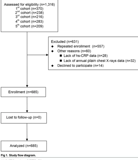
This was a prospective study performed using five pooled patient cohorts. The first three cohorts were composed of 370, 238, and 216 prevalent hemodialysis (HD) patients, respec-tively, the fourth was composed of 220 HD patients and 63 peritoneal dialysis (PD) patients
and the fifth was 209 HD patients. These patient cohorts have been described previously in more detail. [20–23] In brief, the five cohorts were collected prospectively to understand the associations between serum fetuin A level, inflammatory markers (such as, high sensitivity C-reactive protein (hs-CRP) and lipid profiles with specific outcomes in prevalent dialysis patients, at the Far Eastern Memorial Hospital from 2007 to 2014. All patients had baseline data on fetuin A at entry. The exclusion criteria of the five cohorts were as follows: (1) active infection; (2) recent hospitalization within 3 months; (3) psychotic illness or other communica-tion problems; (4) active malignancy; (5) younger than 20 years; (6) receiving HD or PD for less than 3 months; and (7) patients’refusal. In the flow diagram, we have clearly shown the reasons of exclusion in this cohort study (Fig 1). Before initiating this prospective study, we have re-evaluated all the participants in the five cohorts about the wills of being analyzed for the pre-specified outcome (fracture). For the most part, the exclusion of participants was due to repeated enrollment (88%), only 14 patients (1%) declined to participate this prospective
Fig 1. Study flow diagram.
analysis and none of the participants in the cohort were excluded because of missing fetuin A data. The final numbers of enrolled participants from the five cohorts are 354, 18, 117, 163 and 33 respectively.
All of the subjects provided written informed consent, the study complied with the World Medical Association Declaration of Helsinki—Ethical Principles for Medical Research Involv-ing Human Subjects, and the Ethics Committee of Far Eastern Memorial Hospital approved the study protocol (ClinicalTrials.gov; NCT01457625).
In total, 685 patients (mean age, 59±13 years; 348 women) who underwent prevalent HD (629) and PD (56) at the Far Eastern Memorial Hospital, Taiwan, were enrolled from February 2007 (first cohort), March 2009 (second cohort), March 2011 (third cohort), March 2013 (fourth cohort) and September 2014 (fifth cohort). The median dialysis vintage before recruit-ment was 2.5 years (range, 0.4–26.5 years).
Measurement of serum fetuin A concentrations
Serum fetuin A levels in the five cohorts were measured using three types of highly sensitive, two-site enzyme-linked immunoassays (GenWay Biotech, Inc., San Diego, CA, USA; Adipo Bioscience, Inc., Santa Clara, CA, USA and R&D Systems, Inc., Minneapolis, MN, USA). The intra-assay coefficients of variation were 4.1%, 4.0% and 4.0%, respectively, and the inter-assay coefficients of variation were 6.2%, 6% and 6.4%, respectively. The linear measurement ranges of the assays for human fetuin A levels were 0.002-2.5 g/L and 0.003-2.5 g/L and 0.002-2.5 g/L, respectively. Blood samples for the measurement of fetuin A levels were obtained once on recruitment and were immediately centrifuged and stored at−70°C until the time of the assay.
Measurements of clinical parameters
Demographic data, a concurrent medical history of CV disease and smoking status were
recorded. Venous blood was sampled in the morning after an overnight fast of more than 8 hours before the patient’s mid-week dialysis session in the HD patients, or before the first daily dwell of dialysate in the PD patients. Intact PTH levels were determined by immunoassay (Roche Modu-lar E170 analyzer). The hs-CRP levels were determined using the immunonephelometric method using a Tina-quant CRP (Latex) ultrasensitive assay (D & P Modular Analyzer, Roche Diagnos-tics GmbH, Mannheim, Germany). The geriatric nutritional risk index (GNRI) was calculated by the following formula: GNRI = [14.89 × albumin level(g/dL)] + [41.7 ×body weight/WLo], where WLo is the ideal body weight calculated from the Lorentz equation. The GNRI has previously been validated in dialysis patients, and a higher GNRI indicates better nutritional status.[24]
Outcomes
The primary outcome was the incidence of major fractures, which was defined as a new symp-tomatic fracture of the hip, pelvis, humerus, distal forearm, lower leg or vertebrae that occurred during follow-up. The occurrence of a major fracture was assessed by a clinical diagnosis (either from inpatient chart review or outpatient medical records), and concurrent roentgeno-gram, ordered for a suspicious fracture, with defined evidence of a fracture in the formal roent-genogram report by a radiologist. The outcome information was centrally assessed by trained clinicians, nephrologists and radiologists.
Assessment of VC
We defined the presence of VC as the presence of aortic arch calcification on a posterior-ante-rior plain chest X-ray at the entry of study. All of the participants received routine annual pos-terior-anterior plain chest X-rays in our hospital. Two trained physicians blinded to the patients’clinical data reviewed plain chest X-rays performed before study enrollment to assess the presence of aortic arch calcification.
Statistical analysis
Continuous data were presented as mean ± SD or median (interquartile range (IQR)), and cat-egorical data were reported as percentages. Differences in baseline characteristics and biochem-ical parameters between the HD and PD patients and subjects with/without VC were
compared using the Student’sttest and Mann-Whitney U test. Similarly, differences in base-line characteristics and biochemical parameters among the patients within the fetuin A tertiles were compared using ANOVA and the Kruskal-Wallis H test, as appropriate. The chi-square test was used for categorical variables.
Since the fetuin A level was not normally distributed in the dialysis patients (P<0.001 by
either Kolmogorov-Smirnov or Shapiro-Wilk Test), we constructed plots of the fetuin A levels and crude hazard ratios (HRs) of the incident major fractures using theLowessfunction. The results revealed a non-linear relationship, suggesting the need for stratification of the patients into tertiles according to their fetuin A level for outcome analysis, which we then performed (S1 Fig). The primary predictor variables were the fetuin A level in each tertile: patients with a fetuin A level between 0.11-0.35 g/L were in tertile 1, between 0.35-0.65 g/L in tertile 2 and between 0.66-1.89 g/L in tertile 3.
Owing to the non-linear relationship between the fetuin A levels and HRs of the incident major fractures, we performed the outcome analysis in two ways: (1) to construct an algorithm for stratifying the major fracture risks in dialysis patients with recursive partitioning analysis (RPA) [25]. We performed the RPA in order to repeatedly divide patients into subgroups whether they had major fracture or not. It ideally provided a nonparametric discriminating tree for discriminating the power of risk factors of major fracture. Once RPA selected the tree, we only selected each of the splits identified with statistical criterion ofP<0.01 for outcomes
Results
The baseline characteristics of the participants
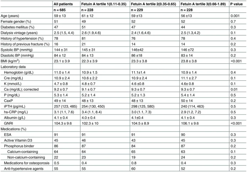
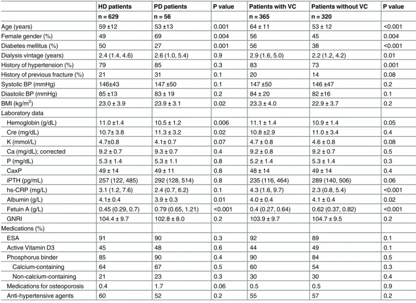
The baseline characteristics of all of the participants, those in the fetuin A tertiles are summa-rized inTable 1. The patients in the fetuin A tertiles had different age, body mass index (BMI) and nutritional status (Table 1). And the baseline characteristics of the participants receiving HD or PD and with/without VC are summarized in theTable 2. In general, more female patients received PD, fewer patients undergoing PD had diabetes, and those undergoing PD were younger, had lower hemoglobin and albumin levels, higher BMI, creatinine and fetuin A levels (Table 2). Patients with aortic arch calcification were older, had longer dialysis vintage, had higher levels of hs-CRP and lower fetuin A levels (Table 2).
Table 1. Baseline characteristics of the all patients and the patients by fetuin A tertile.
All patients Fetuin A tertile 1(0.11-0.35) Fetuin A tertile 2(0.35-0.65) Fetuin A tertile 3(0.66-1.89) P value
n = 685 n = 228 n = 229 n = 228
Age (years) 59±13 61±12 59±13 56±13 0.001
Female gender (%) 51 49 52 52 0.7
Diabetes mellitus (%) 47 51 47 44 0.3
Dialysis vintage (years) 2.5 (1.5, 4.4) 2.6 (1.9,4.6) 2.4 (1.6,4.6) 2.5 (1.3,4.2) 0.1
History of hypertension (%) 78 81 76 78 0.4
History of previous fracture (%) 16 21 14 14 0.2
Systolic BP (mmHg) 144±31 145±31 146±42 146±72 0.3
Diastolic BP (mmHg) 84±12 84±13 86±18 83±14 0.2
BMI (kg/m2) 23.1±3.9 22.3±3.9 23.3±3.8 23.8±3.8 <0.001
Laboratory data
Hemoglobin (g/dL) 11.0±1.4 10.9±1.5 11.1±1.4 10.9±1.4 0.4
Cre (mg/dL) 10.9±2.4 10.6±2.2 10.9±2.4 11.1±2.7 0.1
K (mmol/L) 4.7±0.8 4.8±0.7 4.6±0.8 4.6±0.8 0.1
Ca (mg/dL); corrected 9.2±0.7 9.1±0.7 9.3±0.7 9.3±0.7 0.01
P (mg/dL) 5.3±1.4 5.2±1.4 5.2±1.3 5.4±1.4 0.5
CaxP 49±14 48±13 48±13 50±14 0.2
iPTH (pg/mL) 257 (123, 485) 254 (130, 450) 298 (125, 580) 240 (114, 463) 0.5
hs-CRP (mg/L) 3.1 (1.1, 7.6) 3.4 (1.1, 8.4) 3.0 (1.1, 7.3) 2.9 (1.2, 7.2) 0.5
Albumin (g/L) 4.1±0.4 4.0±0.4 4.1±0.4 4.1±0.4 0.3
GNRI 104.3±9.6 102.3±10 104.5±8.9 106.1±9.6 <0.001
Medications (%)
ESA 91 91 91 90 0.3
Active Vitamin D3 45 46 43 45 0.3
Phosphorus binder 86 87 84 87 0.2
Calcium-containing 64 64 65 63 0.1
Non-calcium-containing 22 23 19 24 0.2
Medications for osteoporosis 0.5 0.4 0.8 0.4 0.3
Anti-hypertensive agents 55 55 60 52 0.2
Abbreviations: CVD, cardiovascular disease; BP, blood pressure; Cre, creatinine; BMI, body mass index; CaxP, calcium phosphate product; iPTH, intact parathyroid hormone; hs-CRP, high-sensitive C-reactive protein; GNRI, geriatric nutritional risk index; ESA, erythropoiesis-stimulating agents.
Note: Conversion factors for units: hemoglobin in g/dL to g/L, ×10; serum calcium in mg/dL to mmol/L, ×0.2495; serum phosphate in mg/dL to mmol/L, ×0.3229; serum albumin in g/dL to g/L, ×10. No conversion is necessary for serum iPTH in pg/mL and ng/L; serum potassium in mEq/L and mmol/L.
Outcomes
Overall, 177 of the participants experienced incident major fractures during a median of 3.4 years (IQR, 1.5–5.8 years) of follow-up. Thirty-seven participants had hip fractures, 10 had pel-vic fractures, 20 had humeral fractures, 28 had distal forearm fractures, 32 had lower leg frac-tures and 50 had vertebral fracfrac-tures. The incidence rate of major fracfrac-tures was 3.29 per 100 person-years. The incidence rates in the lowest to highest fetuin A tertiles were 4.83, 3.21 and 1.45 per 100 person-years, respectively.
Assessment of VC
A total of 365 participants had aortic arch calcification on the plain chest X-rays with different severity at entry, including 154, 124 and 87 participants in fetuin A tertile 1, 2 and 3,
Table 2. Baseline characteristics of the patients undergoing hemodialysis (HD) and peritoneal dialysis (PD) and patients with and without vascular calcification (VC).
HD patients PD patients P value Patients with VC Patients without VC P value
n = 629 n = 56 n = 365 n = 320
Age (years) 59±12 53±13 0.001 64±11 53±12 <0.001
Female gender (%) 49 69 0.004 56 45 0.004
Diabetes mellitus (%) 50 27 0.001 56 38 <0.001
Dialysis vintage (years) 2.4 (1.4, 4.6) 2.6 (1.0, 5.4) 0.9 2.9 (1.6, 5.0) 2.2 (1.2, 4.2) 0.01
History of hypertension (%) 79 85 0.3 83 73 0.001
History of previous fracture (%) 21 31 0.1 20 14 0.08
Systolic BP (mmHg) 146±43 147±50 0.1 147±50 146±47 0.2
Diastolic BP (mmHg) 85±13 83±19 0.2 84±20 82±16 0.1
BMI (kg/m2) 23.0±3.9 23.9±3.1 0.02 23.3±4.0 22.9±3.7 0.2
Laboratory data
Hemoglobin (g/dL) 11.0±1.4 10.5±1.2 0.006 11.1±1.4 10.9±1.4 0.05
Cre (mg/dL) 10.7±3.8 11.3±3.2 0.02 10.8±2.9 11.0±3.4 0.4
K (mmol/L) 4.7±0.8 4.1±0.7 0.07 4.7±0.8 4.6±0.8 0.08
Ca (mg/dL); corrected 9.2±0.7 9.3±0.7 0.4 9.2±0.8 9.2±0.7 0.5
P (mg/dL) 5.3±1.4 5.3±1.1 0.8 5.2±1.4 5.3±1.4 0.3
CaxP 49±14 49±11 0.8 48±14 49±14 0.4
iPTH (pg/mL) 257 (122, 485) 292 (128, 514) 0.8 235 (116, 464) 289 (140, 506) 0.06
hs-CRP (mg/L) 3.1 (1.2, 7.6) 2.4 (0.7, 6.2) 0.1 4.3 (1.6, 9.7) 2.3 (0.8, 5.4) <0.001
Albumin (g/L) 4.1±0.4 3.9±0.3 0.01 4.0±0.4 4.1±0.4 0.02
Fetuin A (g/L) 0.45 (0.29, 0.7) 0.79 (0.65, 1.21) <0.001 0.4 (0.27, 0.64) 0.62 (0.37, 0.82) <0.001
GNRI 104.4±9.7 102.8±8.0 0.2 103.9±9.7 104.7±9.5 0.2
Medications (%)
ESA 91 90 0.3 92 89 0.1
Active Vitamin D3 45 48 0.6 44 49 0.1
Phosphorus binder 85 90 0.4 90 84 0.5
Calcium-containing 64 67 0.5 60 54 0.3
Non-calcium-containing 21 23 0.3 30 30 0.4
Medications for osteoporosis 0.4 1.7 0.06 0.5 0.5 0.9
Anti-hypertensive agents 60 52 0.2 55 57 0.2
Abbreviations: CVD, cardiovascular disease; BP, blood pressure; Cre, creatinine; BMI, body mass index; CaxP, calcium phosphate product; iPTH, intact parathyroid hormone; hs-CRP, high-sensitive C-reactive protein; GNRI, geriatric nutritional risk index; ESA, erythropoiesis-stimulating agents.
Note: Conversion factors for units: hemoglobin in g/dL to g/L, ×10; serum calcium in mg/dL to mmol/L, ×0.2495; serum phosphate in mg/dL to mmol/L, ×0.3229; serum albumin in g/dL to g/L, ×10. No conversion is necessary for serum iPTH in pg/mL and ng/L; serum potassium in mEq/L and mmol/L.
respectively. The number of participants with aortic arch calcification was significantly differ-ent in the fetuin A tertiles (P<0.001 by the chi-square test). One hundred and twenty-eight
subjects with aortic arch calcification and 49 subjects without aortic arch calcification had major fractures. The incidence rates of major fractures in the participants with and without aortic arch calcification were 4.29 and 1.87 per 100 person-years, respectively.
Associations between fetuin A level and fractures
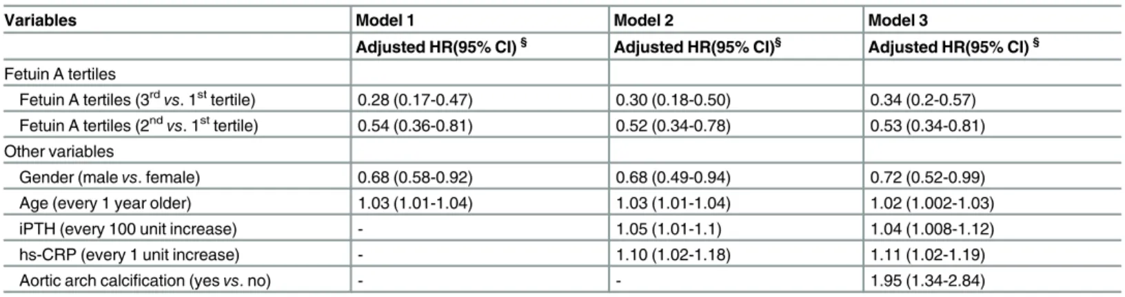
In the multivariate Cox regression model, patients with higher fetuin A levels had a lower inci-dence of major fracture (adjusted HR, 0.28; 95% CI, 0.17–0.47, tertile 3vs. tertile 1; adjusted HR, 0.54; 95% CI, 0.36–0.81, tertile 2vs. tertile 1) in Model 1. Similarly, patients with higher fetuin A levels had a lower incidence of fractures in Model 2 and 3 (Table 3). Patients with aor-tic arch calcification at study entry had a higher risk of incident fractures (adjusted HR, 1.95; 95% CI, 1.34–2.84). Fetuin A levels and the presence of VC both independently predicted the risk of major fracture in our dialysis patients.
Recursive partitioning analysis
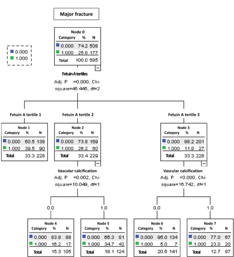
As we have shown in the analysis from the Cox proportional hazards model, fetuin A tertiles, gender, age, iPTH, hs-CRP and VC were among the significant predictive factors (Table 3) and they have selected to divide the patient population.
Two hundred and twenty-eight patients were in the tertile 3 groups. For the patients in this group, presence of VC indicated a higher incidence of fracture, and among those within fetuin A tertile 2 (N = 229), presence of VC indicated a higher incidence of fracture as well. However, the subjects in the fetuin A tertile 1, presence of VC did not further differentiate the risk of major fracture (Fig 2). The 5 groups were ultimately defined (Node 1: within fetuin A tertile 1, with or without VC; node 4: within fetuin A tertile 2, without VC; node 5: within fetuin A ter-tile 2, with VC; node 6: within fetuin A terter-tile 3, without VC and node 7: within fetuin A terter-tile 3, with VC), and the Kaplan-Meier curves for these 5 groups are shown inFig 3. Patients within the fetuin A tertile 3 and without VC had the lowest incidence of major fracture (Node 6).
Table 3. Hazard ratios (HRs) of fetuin A tertiles in predicting the occurrence of major fractures using different Cox proportional hazard regression models.
Variables Model 1 Model 2 Model 3
Adjusted HR(95% CI)§ Adjusted HR(95% CI)§ Adjusted HR(95% CI)§
Fetuin A tertiles
Fetuin A tertiles (3rdvs. 1sttertile) 0.28 (0.17-0.47) 0.30 (0.18-0.50) 0.34 (0.2-0.57) Fetuin A tertiles (2ndvs. 1sttertile) 0.54 (0.36-0.81) 0.52 (0.34-0.78) 0.53 (0.34-0.81)
Other variables
Gender (malevs. female) 0.68 (0.58-0.92) 0.68 (0.49-0.94) 0.72 (0.52-0.99)
Age (every 1 year older) 1.03 (1.01-1.04) 1.03 (1.01-1.04) 1.02 (1.002-1.03)
iPTH (every 100 unit increase) - 1.05 (1.01-1.1) 1.04 (1.008-1.12)
hs-CRP (every 1 unit increase) - 1.10 (1.02-1.18) 1.11 (1.02-1.19)
Aortic arch calcification (yesvs. no) - - 1.95 (1.34-2.84)
Abbreviations: HR, hazard ratio; CI, confidence interval; iPTH, intact parathyroid hormone; hs-CRP, high-sensitive C-reactive protein, GNRI, geriatric nutritional risk index
§Adjusted for model 1: gender, age, dialysis vintage, previous fracture history, diabetes status/hypertension status, dialysis modality, smoking/alcohol status and patient cohort; model 2: factors in model 1 and hemoglobin level, intact parathyroid hormone, GNRI, CaxP and high-sensitive C-reactive protein (hs-CRP) levels; model 3: factors in model 2 and presence of aortic arch calcification.
Associations between fetuin A level and VC
Because of the pathophysiological link between fetuin A and VC, as well as the very much interaction between fetuin A and VC (Fig 2), we further explored the interaction between fetuin A and VC in the prediction of incident fractures. We reported the interaction between
Fig 2. Classification tree based on recursive partitioning analysis.
fetuin A tertile and the presence of aortic arch calcification on the risk of incident major fracture in theTable 4. Either in patients with or without aortic arch calcification, those with higher fetuin A levels (tertile 2 and 3) had lower risk of incident fracture (P for interaction = 0.08,Table 4).
Comparison between PD and HD patients
Compared with PD patients, HD patients had lower fetuin A concentrations (0.47, 0.29- 0.7vs. 0.79, 0.65-1.21 g/L, P<0.001) (Table 2). The incidence rates of major fractures in our PD and
HD patients were 0.43 and 3.41 per 100 person-years, respectively. HD patients had a
Fig 3. Kaplan-Meier survival curves for all patients, by risk group.Node 1: within fetuin A tertile 1, with or without VC; node 4: within fetuin A tertile 2, without VC; node 5: within fetuin A tertile 2, with VC; node 6: within fetuin A tertile 3, without VC and node 7: within fetuin A tertile 3, with VC.
doi:10.1371/journal.pone.0158789.g003
Table 4. Interaction between fetuin A tertile and the presence of aortic arch calcification on the risk of incident major fracture.
Fetuin A tertile 1 (N = 228) Fetuin A tertile 2 (N = 229) Fetuin A tertile 3 (N = 228)
N with/without Major fracture
HR 95%CI N with/without Major fracture
HR 95%CI N with/without Major fracture
HR 95%CI P for trend
P for interaction
With aortic arch calcification (n = 365)
65/89 1.0 43/81 0.54
(0.33-0.87) P = 0.01
20/67 0.47 (0.26-0.99) P = 0.05
P = 0.03 P = 0.08
Without aortic arch calcification (n = 320)
25/49 0.64 (0.39-1.04) P = 0.07
17/88 0.29 (0.16-0.55) P<0.001
7/134 0.11 (0.05-0.26) P<0.001
P<0.001
Adjusted for gender, age; dialysis vintage, diabetes/hypertension status, patient cohort, intact parathyroid hormone; body mass index, albumin and high-sensitive C-reactive protein (hs-CRP) levels, fetuin A tertile and presence of aortic arch calcification
Abbreviations: N, number; HR, hazard ratio; CI, confidence interval
significant higher risk to have incident fracture compared with PD patients (adjusted HR, 4.95; 95% CI, 1.19–20.68). In addition, the incidence rates of VC in our PD and HD patients were 3.49 and 6.78 per 100 person-years, respectively. Nonetheless, no significant difference was observed in the association of fetuin A level with the risk of major fractures between the two dialysis modalities (dialysis modality interaction analysis, P = 0.8).
Sensitivity Analyses
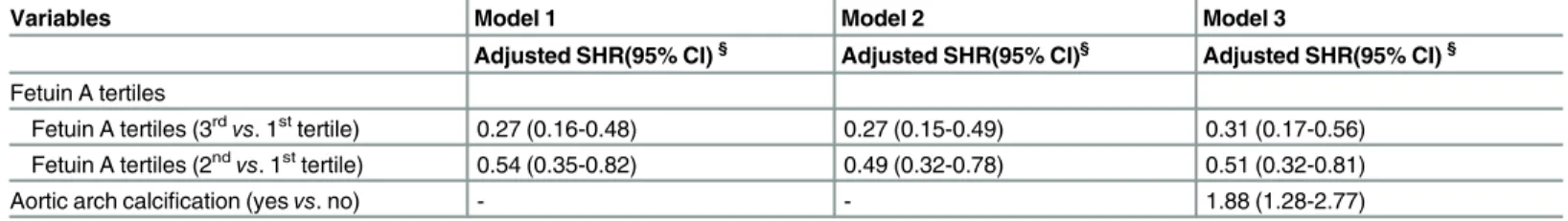
When accounting for death as a potential informative event in competing risk models, the sub-hazard ratio (SHR) for major fracture continued to be significantly less for dialysis patients with higher fetuin A level examined in a fully adjusted model as well as those without the pres-ence of aortic arch calcification (Table 5).
Discussion
The main finding of this study is that the prevalent dialysis patients with a higher baseline fetuin A level had a lower long-term risk of incident major fractures regardless of gender, dialy-sis modality (HD or PD) and nutritional or inflammatory status; besides, patients with VC at study entry also had a higher risk to have incident fracture. Interestingly, the risk of incident fractures and aortic arch calcification declined in parallel with the increase in fetuin A level; however, lower fetuin A level and the presence of VC both independently predicted the higher risk of incident fracture in dialysis patients. In addition to the inhibitory property of VC and the strong predictive power of CV death, the fetuin A level also predicted the occurrence of major fractures in the dialysis patients.
Major fractures lead to high mortality and morbidity rates in dialysis patients,[3,5] and therefore it is critical to identify high-risk patients who are susceptible to fragile fractures and to prevent potential fracture events in the long-term care of prevalent dialysis patients. Our results showed a high incidence of major fractures in the dialysis patients, around 3.29 per 100 person-years, which is nearly three times greater than that reported for elderly patients without CKD.[19] This incidence rate is similar to that presented in the Dialysis Outcomes and Practice Patterns Study [5] and the U.S. Renal Data System.[3] Of note, we found a link between fetuin A and incident fractures in our dialysis patients, that is, a higher fetuin A level predicted a lower incidence of fractures (Table 3,Fig 2). To the best of our knowledge, this is the first study to report an association between fetuin A level and incident fractures in dialysis patients. Therefore, fetuin A level in prevalent dialysis patients should be recognized as a potential marker to identify the dialysis patients who are at a higher risk of a major fracture.
Table 5. Competing risk analysis of the relative hazard of major fracture by fetuin A tertile (death is the competing event).
Variables Model 1 Model 2 Model 3
Adjusted SHR(95% CI)§ Adjusted SHR(95% CI)§ Adjusted SHR(95% CI)§
Fetuin A tertiles
Fetuin A tertiles (3rdvs. 1sttertile) 0.27 (0.16-0.48) 0.27 (0.15-0.49) 0.31 (0.17-0.56)
Fetuin A tertiles (2ndvs. 1sttertile) 0.54 (0.35-0.82) 0.49 (0.32-0.78) 0.51 (0.32-0.81)
Aortic arch calcification (yesvs. no) - - 1.88 (1.28-2.77)
Abbreviations: SHR, subhazard ratio; CI, confidence interval; iPTH, intact parathyroid hormone; hs-CRP, high-sensitive C-reactive protein, GNRI, geriatric nutritional risk index
§Adjusted for model 1: gender, age, dialysis vintage, previous fracture history, diabetes status/hypertension status, dialysis modality, smoking/alcohol status and patient cohort; model 2: factors in model 1 and hemoglobin level, intact parathyroid hormone, GNRI, CaxP and high-sensitive C-reactive protein (hs-CRP) levels; model 3: factors in model 2 and presence of aortic arch calcification.
In a landmark study using bone biopsies to estimate bone volume and multi-slice computed tomography to assess coronary artery calcification, low bone volume was found to be a signifi-cant risk factor for coronary calcification in HD patients.[10] In addition, low bone volume has been recognized as an important risk factor for the occurrence of fractures in both the general population [27–29] and dialysis patients.[30,31] Therefore, it is reasonable to assume that dial-ysis patients with evident VC are more likely to experience a fracture. Our results have clearly shown that the dialysis patients with aortic arch calcification had a higher risk of fractures (Table 3, Figs2and3). Specifically, in uremic milieu, dialysis patients tended to consume more fetuin A to prevent VC and therefore, dialysis subjects with baseline VC tended to have a lower fetuin A level concurrently. In an outstanding study which has shown that fetuin A is trafficked and exocytosed via exosome release in MV bodies. Interestingly, MV bodies containing exo-somes were easily observed in vessels, especially in calcified MVs in dialysis patients. Factors that increase exosome release will promote vascular calcification, specifically under the condi-tion of environmental calcium stress or fetuin A deficiency[16]. Therefore, the consequent gen-eralized VC can be highly anticipated.
The features of osteoporosis in CKD patients are low trabecular bone volume and disrupted micro-architecture, but no abnormalities in mineralization or bone turnover[32]. Instead, bone loss is mostly from cortical bone in subjects with CKD mineral and bone disorder
(CKD-MBD), and their iPTH, alkaline phosphatase, Klotho, sclerostin, and fetuin A levels are pronouncedly altered[33]. Due to the high prevalence of osteoporosis and CKD-MBD in CKD subjects, both conditions are commonly existent simultaneously. But, in reality, the CKD–
MBD is more complex than osteoporosis and CKD-MBD influences bone quality, contributes to high rates of fracture and most importantly, it results in VC in CKD patients. Although they both result in bone fragility, they do have different pathophysiology to destroy the bone. Osteo-porosis is induced by excessive osteoclastic bone resorption in postmenopausal woman and subjects with aging process. But CKD-MBD is related to altered mineral metabolism and the imbalance of pro- and anti-calcification factors (such as fetuin A deficiency) which induced either high or low turnover bone disease in CKD subjects [33]. Thepost hocanalyses of data from crucial osteoporosis studies suggest that in patients with mild stage 3 CKD and normal iPTH, calcium and phosphate levels, conventional treatments for osteoporosis (such as bis-phosphonates, teriparatide. . .. . .) are effective to reduce fracture rates. Nevertheless, for patients with stage 4–5 CKD, the available evidence are insufficient to determine whether these medications are effective[33,34]. Briefly speaking, low bone density and fractures induced by osteoporosis in patients with CKD differ from those with CKD-MBD.
In a recent investigation, Finket alreported that serum fetuin A level had a positive associa-tion with areal BMD, but that there was no evidence of an associaassocia-tion between fetuin A and the risk of clinical fractures in a large community cohort including 4714 elderly participants (>65
dialysis and the general population. For example, dialysis patients with a lower fetuin A level have been reported to suffer from higher CV mortality,[11,20,35] and in the general popula-tion a lower fetuin A level has also been reported to lead to higher CV calcificapopula-tion.[36] How-ever, in patients with diabetes, those with a higher fetuin A level have been reported to experience more CV complications.[37,38] It is premature to recognize this phenomenon as another“reverse epidemiology”; nevertheless, the different impact of fetuin A level on incident fractures in different populations can rationally be anticipated. Second, our participants were much younger (59 ± 13 years) than those in the Cardiovascular Health Study (74.9 ± 5.3 years). According to our results, the risk of major fractures increased by 20~30% as the dialysis patients became older (every 10-year increase) (Table 3). In addition, a classical, large cohort study reported that patients in the general population older than 75 years had a higher odds of incident osteoporotic fractures.[39] Although it was underpowered to performed the subgroup analysis in this cohort because only 216 patients were older than 65 years old, we also found a trend of declining impacts of fetuin A levels on the incidence of major fracture in patients older than 65 year old (data not shown). And as we mentioned above, elderly CKD subjects have every likelihood that having osteoporotic fracture. Therefore, the impact of fetuin A level on fractures may be altered by the aging process.
Since fetuin A inhibits the VC process,[11,40,41] fetuin A and VC may be potential factors involved in the common pathway of the pathogenesis of fragile fractures in dialysis patients. However, little is known about the physiological link between fetuin A level and the occurrence of fractures. In vitro experiments, bone re-mineralization cannot successfully be performed in fetuin A-depleted serum, although it can be achieved when the serum is reconstituted with fetuin A.[12,13] This suggests that fetuin A promotes bone mineralization in vitro. However, in fetuin A knock-out mice, the trabecular bone mass of cortical bone are unaffected by the absence of fetuin A; nevertheless, there is excess mineralization of the growth plate of long bone[17]. These conflict results from bench studies suggest that the pathophysiology between of fetuin A and bone mineralization remains unclear and it needs further works to be eluci-dated. In the view of epidemiological aspects, dialysis patients with evident VC had low bone volume[10] which led to the fragile fracture process, and concurrently, they have lower fetuin A levels as well due to a consumption process[42,43], as also seen in our results (Table 2). We hypothesize that dialysis subjects with evident VC or having low fetuin A concentration would have more incident fracture owing to either their essential low bone volume or the lack of fetuin A to promote bone mineralization. In addition, sclerostin, a Wnt signaling pathway inhibitor, has been shown to be an important messenger in the cross-talk between bone and the vasculature, and the VC and fractures as well.[44,45] In one phase II study, using romoso-zumab (a humanized monoclonal antibody to sclerostin) to treat 419 osteoporotic women, the results are promising; however the long-term (>12 months) effect is unknown.[46]
Further-more, a sclerostin inhibitor probably worsens VC in CKD rats [47] and may lead to deteriora-tion of human renal funcdeteriora-tion [33]. However, the association between fetuin A and sclerostin (or Wnt pathway) remains unknown. Therefore, further studies to investigate the potential biological pathway between fetuin A deficiency and the occurrence of incident fractures are warranted.
however, a recent study has confirmed the correlation between fetuin A level and BMD in the elderly. We could not fully confirm the correlation between fetuin A, BMD and fractures in dialysis patients from our results due to the lack of BMD measurements. Third, we only identi-fied the presence of aortic arch calcification at entry and did not further quantify the severity of calcification. Dialysis patients with different severities of VC have been reported to have differ-ent fetuin A levels,[38,42] and thereby potentially different risks of fractures. It would be more precise to quantify VC to better understand the interrelationship between fetuin A level, VC and fractures. Fourth, this was a single-center study, and all of the participants were treated by the same physicians and underwent uniform laboratory measurements during the observation period, which guaranteed the accuracy of our results. However, our conclusions cannot be gen-eralized to other ethnicities. In summary, our results suggest that lower fetuin A level and the presence of aortic arch calcification can independently predict the long-term occurrence of incident fractures in prevalent dialysis patients.
Supporting Information
S1 Fig. Distribution of fetuin A concentrations in dialysis patients in the study (n = 685).A scatter plot of log hazard ratio (HR) of major incident fracture versus fetuin A concentration with Lowess smoothed function. The plot suggested nonlinear relationships; we therefore cate-gorized patients into tertiles by fetuin A concentration for analyses.
(TIF)
Author Contributions
Conceived and designed the experiments: HYC. Performed the experiments: HYC YLC YSP. Analyzed the data: HYC JYY. Contributed reagents/materials/analysis tools: HYC YLC SPH MFP JYY. Wrote the paper: HYC.
References
1. Ball AM, Gillen DL, Sherrard D, Weiss NS, Emerson SS, Seliger SL, et al. Risk of hip fracture among dialysis and renal transplant recipients. JAMA: the journal of the American Medical Association. 2002; 288(23):3014–8. PMID:12479766.
2. Jadoul M, Albert JM, Akiba T, Akizawa T, Arab L, Bragg-Gresham JL, et al. Incidence and risk factors for hip or other bone fractures among hemodialysis patients in the Dialysis Outcomes and Practice Pat-terns Study. Kidney international. 2006; 70(7):1358–66. doi:10.1038/sj.ki.5001754PMID:16929251.
3. Beaubrun AC, Kilpatrick RD, Freburger JK, Bradbury BD, Wang L, Brookhart MA. Temporal trends in fracture rates and postdischarge outcomes among hemodialysis patients. Journal of the American Society of Nephrology: JASN. 2013; 24(9):1461–9. doi:10.1681/ASN.2012090916PMID:23744885; PubMed Central PMCID: PMC3752946.
4. Braithwaite RS, Col NF, Wong JB. Estimating hip fracture morbidity, mortality and costs. Journal of the American Geriatrics Society. 2003; 51(3):364–70. PMID:12588580.
5. Tentori F, McCullough K, Kilpatrick RD, Bradbury BD, Robinson BM, Kerr PG, et al. High rates of death and hospitalization follow bone fracture among hemodialysis patients. Kidney international. 2014; 85 (1):166–73. doi:10.1038/ki.2013.279PMID:23903367; PubMed Central PMCID: PMC3910091.
6. Danese MD, Kim J, Doan QV, Dylan M, Griffiths R, Chertow GM. PTH and the risks for hip, vertebral, and pelvic fractures among patients on dialysis. American journal of kidney diseases: the official journal of the National Kidney Foundation. 2006; 47(1):149–56. doi:10.1053/j.ajkd.2005.09.024PMID: 16377396.
8. London GM, Marty C, Marchais SJ, Guerin AP, Metivier F, de Vernejoul MC. Arterial calcifications and bone histomorphometry in end-stage renal disease. Journal of the American Society of Nephrology: JASN. 2004; 15(7):1943–51. PMID:15213285.
9. Tomiyama C, Carvalho AB, Higa A, Jorgetti V, Draibe SA, Canziani ME. Coronary calcification is asso-ciated with lower bone formation rate in CKD patients not yet in dialysis treatment. Journal of bone and mineral research: the official journal of the American Society for Bone and Mineral Research. 2010; 25 (3):499–504. doi:10.1359/jbmr.090735PMID:19594321.
10. Adragao T, Herberth J, Monier-Faugere MC, Branscum AJ, Ferreira A, Frazao JM, et al. Low bone vol-ume—a risk factor for coronary calcifications in hemodialysis patients. Clinical journal of the American Society of Nephrology: CJASN. 2009; 4(2):450–5. doi:10.2215/CJN.01870408PMID:19158372; PubMed Central PMCID: PMC2637600.
11. Ketteler M, Bongartz P, Westenfeld R, Wildberger JE, Mahnken AH, Bohm R, et al. Association of low fetuin-A (AHSG) concentrations in serum with cardiovascular mortality in patients on dialysis: a cross-sectional study. Lancet. 2003; 361(9360):827–33. doi:10.1016/S0140-6736(03)12710-9PMID: 12642050.
12. Price PA, June HH, Hamlin NJ, Williamson MK. Evidence for a serum factor that initiates the re-calcifi-cation of demineralized bone. The Journal of biological chemistry. 2004; 279(18):19169–80. doi:10. 1074/jbc.M307880200PMID:14978037.
13. Toroian D, Price PA. The essential role of fetuin in the serum-induced calcification of collagen. Calcified tissue international. 2008; 82(2):116–26. doi:10.1007/s00223-007-9085-2PMID:18097630.
14. Jahnen-Dechent W, Heiss A, Schafer C, Ketteler M. Fetuin-A regulation of calcified matrix metabolism. Circ Res. 2011; 108(12):1494–509. doi:10.1161/CIRCRESAHA.110.234260PMID:21659653.
15. Jahnen-Dechent W, Schafer C, Ketteler M, McKee MD. Mineral chaperones: a role for fetuin-A and osteopontin in the inhibition and regression of pathologic calcification. J Mol Med (Berl). 2008; 86 (4):379–89. doi:10.1007/s00109-007-0294-yPMID:18080808.
16. Kapustin AN, Chatrou ML, Drozdov I, Zheng Y, Davidson SM, Soong D, et al. Vascular smooth muscle cell calcification is mediated by regulated exosome secretion. Circ Res. 2015; 116(8):1312–23. doi:10. 1161/CIRCRESAHA.116.305012PMID:25711438.
17. Seto J, Busse B, Gupta HS, Schafer C, Krauss S, Dunlop JW, et al. Accelerated growth plate minerali-zation and foreshortened proximal limb bones in fetuin-A knockout mice. PLoS One. 2012; 7(10): e47338. doi:10.1371/journal.pone.0047338PMID:23091616; PubMed Central PMCID: PMCPMC3473050.
18. Ix JH, Wassel CL, Bauer DC, Toroian D, Tylavsky FA, Cauley JA, et al. Fetuin-A and BMD in older per-sons: the Health Aging and Body Composition (Health ABC) study. Journal of bone and mineral research: the official journal of the American Society for Bone and Mineral Research. 2009; 24(3):514– 21. doi:10.1359/jbmr.081017PMID:19016589; PubMed Central PMCID: PMC2659522.
19. Fink HA, Buzkova P, Garimella PS, Mukamal KJ, Cauley JA, Kizer JR, et al. Association of Fetuin-A with Incident Fractures in Community-Dwelling Older Adults: The Cardiovascular Health Study. Journal of bone and mineral research: the official journal of the American Society for Bone and Mineral Research. 2015. doi:10.1002/jbmr.2475PMID:25656814.
20. Chen HY, Chiu YL, Chuang YF, Hsu SP, Pai MF, Lai CF, et al. Association of low serum fetuin A levels with poor arteriovenous access patency in patients undergoing maintenance hemodialysis. Am J Kid-ney Dis. 2010; 56(4):720–7. Epub 2010/08/31. S0272-6386(10)01083-8 [pii] doi:10.1053/j.ajkd.2010. 06.015PMID:20801568.
21. Chen HY, Chiu YL, Hsu SP, Pai MF, Lai CF, Peng YS, et al. Association of serum fetuin A with truncal obesity and dyslipidemia in non-diabetic hemodialysis patients. Eur J Endocrinol. 2009; 160(5):777– 83. doi:10.1530/EJE-08-0813PMID:19228823.
22. Chen HY, Tsai WC, Chiu YL, Hsu SP, Pai MF, Yang JY, et al. Triglyceride to high-density lipoprotein cholesterol ratio predicts cardiovascular outcomes in prevalent dialysis patients. Medicine (Baltimore). 2015; 94(10):e619. doi:10.1097/MD.0000000000000619PMID:25761189; PubMed Central PMCID: PMCPMC4602469.
23. Chiu YL, Chen HY, Chuang YF, Hsu SP, Lai CF, Pai MF, et al. Association of uraemic pruritus with inflammation and hepatitis infection in haemodialysis patients. Nephrol Dial Transplant. 2008; 23 (11):3685–9. doi:10.1093/ndt/gfn303PMID:18515654.
24. Yamada K, Furuya R, Takita T, Maruyama Y, Yamaguchi Y, Ohkawa S, et al. Simplified nutritional screening tools for patients on maintenance hemodialysis. Am J Clin Nutr. 2008; 87(1):106–13. PMID: 18175743.
26. Fine JP, Gray RJ. A Proportional Hazards Model for the Subdistribution of a Competing Risk. Journal of the American Statistical Association. 1999; 94(446):496–509. doi:10.1080/01621459.1999.10474144
27. Carballido-Gamio J, Harnish R, Saeed I, Streeper T, Sigurdsson S, Amin S, et al. Structural patterns of the proximal femur in relation to age and hip fracture risk in women. Bone. 2013; 57(1):290–9. doi:10. 1016/j.bone.2013.08.017PMID:23981658; PubMed Central PMCID: PMC3809121.
28. Rabier B, Heraud A, Grand-Lenoir C, Winzenrieth R, Hans D. A multicentre, retrospective case-control study assessing the role of trabecular bone score (TBS) in menopausal Caucasian women with low areal bone mineral density (BMDa): Analysing the odds of vertebral fracture. Bone. 2010; 46(1):176– 81. doi:10.1016/j.bone.2009.06.032PMID:19747992.
29. Rudang R, Darelid A, Nilsson M, Mellstrom D, Ohlsson C, Lorentzon M. X-ray-verified fractures are associated with finite element analysis-derived bone strength and trabecular microstructure in young adult men. Journal of bone and mineral research: the official journal of the American Society for Bone and Mineral Research. 2013; 28(11):2305–16. doi:10.1002/jbmr.1974PMID:23658040.
30. Jamal SA, Gilbert J, Gordon C, Bauer DC. Cortical pQCT measures are associated with fractures in dialysis patients. Journal of bone and mineral research: the official journal of the American Society for Bone and Mineral Research. 2006; 21(4):543–8. doi:10.1359/jbmr.060105PMID:16598374.
31. Jamal SA, Hayden JA, Beyene J. Low bone mineral density and fractures in long-term hemodialysis patients: a meta-analysis. American journal of kidney diseases: the official journal of the National Kid-ney Foundation. 2007; 49(5):674–81. doi:10.1053/j.ajkd.2007.02.264PMID:17472850.
32. Toussaint ND, Elder GJ, Kerr PG. Bisphosphonates in chronic kidney disease; balancing potential ben-efits and adverse effects on bone and soft tissue. Clin J Am Soc Nephrol. 2009; 4(1):221–33. doi:10. 2215/CJN.02550508PMID:18987295.
33. Ott SM. Therapy for patients with CKD and low bone mineral density. Nat Rev Nephrol. 2013; 9 (11):681–92. doi:10.1038/nrneph.2013.182PMID:24100401.
34. Jamal SA, Bauer DC, Ensrud KE, Cauley JA, Hochberg M, Ishani A, et al. Alendronate treatment in women with normal to severely impaired renal function: an analysis of the fracture intervention trial. J Bone Miner Res. 2007; 22(4):503–8. doi:10.1359/jbmr.070112PMID:17243862.
35. Hermans MM, Brandenburg V, Ketteler M, Kooman JP, van der Sande FM, Boeschoten EW, et al. Association of serum fetuin-A levels with mortality in dialysis patients. Kidney international. 2007; 72 (2):202–7. doi:10.1038/sj.ki.5002178PMID:17342178.
36. Ix JH, Barrett-Connor E, Wassel CL, Cummins K, Bergstrom J, Daniels LB, et al. The associations of fetuin-A with subclinical cardiovascular disease in community-dwelling persons: the Rancho Bernardo Study. Journal of the American College of Cardiology. 2011; 58(23):2372–9. doi:10.1016/j.jacc.2011. 08.035PMID:22115642; PubMed Central PMCID: PMC3224791.
37. Ix JH, Shlipak MG, Brandenburg VM, Ali S, Ketteler M, Whooley MA. Association between human fetuin-A and the metabolic syndrome: data from the Heart and Soul Study. Circulation. 2006; 113 (14):1760–7. Epub 2006/03/29. CIRCULATIONAHA.105.588723 [pii] doi:10.1161/
CIRCULATIONAHA.105.588723PMID:16567568; PubMed Central PMCID: PMC2776669.
38. Laughlin GA, Cummins KM, Wassel CL, Daniels LB, Ix JH. The association of fetuin-A with cardiovas-cular disease mortality in older community-dwelling adults: the Rancho Bernardo study. Journal of the American College of Cardiology. 2012; 59(19):1688–96. doi:10.1016/j.jacc.2012.01.038PMID: 22554599; PubMed Central PMCID: PMC3345127.
39. van Meurs JB, Dhonukshe-Rutten RA, Pluijm SM, van der Klift M, de Jonge R, Lindemans J, et al. Homocysteine levels and the risk of osteoporotic fracture. The New England journal of medicine. 2004; 350(20):2033–41. doi:10.1056/NEJMoa032546PMID:15141041.
40. Schafer C, Heiss A, Schwarz A, Westenfeld R, Ketteler M, Floege J, et al. The serum protein alpha 2-Heremans-Schmid glycoprotein/fetuin-A is a systemically acting inhibitor of ectopic calcification. J Clin Invest. 2003; 112(3):357–66. doi:10.1172/JCI17202PMID:12897203; PubMed Central PMCID: PMC166290.
41. Scialla JJ, Kao WH, Crainiceanu C, Sozio SM, Oberai PC, Shafi T, et al. Biomarkers of vascular calcifi-cation and mortality in patients with ESRD. Clin J Am Soc Nephrol. 2014; 9(4):745–55. doi:10.2215/ CJN.05450513PMID:24458076; PubMed Central PMCID: PMC3974354.
42. Moe SM, Reslerova M, Ketteler M, O'Neill K, Duan D, Koczman J, et al. Role of calcification inhibitors in the pathogenesis of vascular calcification in chronic kidney disease (CKD). Kidney international. 2005; 67(6):2295–304. doi:10.1111/j.1523-1755.2005.00333.xPMID:15882271.
44. Drueke TB, Lafage-Proust MH. Sclerostin: just one more player in renal bone disease? Clinical journal of the American Society of Nephrology: CJASN. 2011; 6(4):700–3. doi:10.2215/CJN.01370211PMID: 21441122.
45. Modder UI, Hoey KA, Amin S, McCready LK, Achenbach SJ, Riggs BL, et al. Relation of age, gender, and bone mass to circulating sclerostin levels in women and men. Journal of bone and mineral research: the official journal of the American Society for Bone and Mineral Research. 2011; 26(2):373– 9. doi:10.1002/jbmr.217PMID:20721932; PubMed Central PMCID: PMC3179347.
46. McClung MR, Grauer A, Boonen S, Bolognese MA, Brown JP, Diez-Perez A, et al. Romosozumab in postmenopausal women with low bone mineral density. N Engl J Med. 2014; 370(5):412–20. doi:10. 1056/NEJMoa1305224PMID:24382002.
47. Roman-Garcia P, Carrillo-Lopez N, Fernandez-Martin JL, Naves-Diaz M, Ruiz-Torres MP, Cannata-Andia JB. High phosphorus diet induces vascular calcification, a related decrease in bone mass and changes in the aortic gene expression. Bone. 2010; 46(1):121–8. doi:10.1016/j.bone.2009.09.006 PMID:19772957.