U R O G E N I T A L I M A G I N G ( M B E R T O L O T T O , S E C T I O N E D I T O R )
Pitfalls in Imaging of Female Pelvic Masses
Mariana Horta1,2•Teresa Margarida Cunha1
Springer Science+Business Media, LLC 2017
Abstract
Purpose of Review The purpose of this review is to high-light potential magnetic resonance (MR) imaging pitfalls that may mask and simulate ovarian cancer.
Recent Findings MR imaging is the standard method for evaluating female pelvic masses of indeterminate origin, especially sonographically indeterminate adnexal masses. To define the correct origin and nature of a pelvic mass has an enormous clinical impact, namely in females of child-bearing age. This is particularly true in adnexal lesions. Ovarian cancer usually requires a cytoreductive surgery in a special-ized oncological centre. In contrast, a benign ovarian lesion may be treated by simple resection in a general hospital. To help preventing diagnostic errors and in order to guide appropriate therapeutic management, radiologists should be aware of potential MR pitfalls that may mask and simulate ovarian cancer.
Summary The first section of this article will describe the MR imaging protocol that authors perform in their onco-logical centre, highlighting how imaging techniques can be optimized in order to reduce pitfalls in the characterization of an indeterminate pelvic mass.
In the next section, authors will revise the main anatomic and organ-specific signs that may allow the radiologist to determine the ovarian origin of a pelvic mass.
Finally, benign gynaecological masses that may simulate ovarian cancer in a non-emergency setting will be dis-cussed, with emphasis on features that may provide important clues to their diagnosis.
Keywords Ovarian cancerBenign ovarian tumours
Magnetic resonance imagingFemale pelvic masses
Indeterminate adnexal massesPitfalls in gynaecological
imaging
Introduction
Radiological imaging is a key diagnostic tool of modern clinical diagnosis. It is also an important source of diag-nostic errors that can have a major impact in patients’ treatment, causing patients harm [1••].
Patient management errors have been the most cited in claims ascribed to negligence both in Radiology and in Gynaecology [2].
Major discrepancy of radiological interpretation has been reported amongst experienced radiologists when reading abdomino–pelvic computed tomography (CT) exams [3]. The retrospective inter-observer variability was reported in approximately 26% and intra-observer dis-crepancy was also estimated in 32% [3].
Although technical aspects of radiological exams may be a source of diagnostic errors, the majority of radiological mistakes are perceptual and cognitive/interpretive errors [1••,4].
Perceptual errors are responsible for the majority of diagnostic errors (up to 90%) [5–7]. They are defined as This article is part of the Topical Collection onUrogenital Imaging.
& Mariana Horta
Teresa Margarida Cunha [email protected]
1 Department of Radiology, Instituto Portugueˆs de Oncologia
de Lisboa Francisco Gentil, R. Prof. Lima Basto, 1099-023 Lisbon, Portugal
2 Instituto de Anatomia, Faculdade de Medicina da
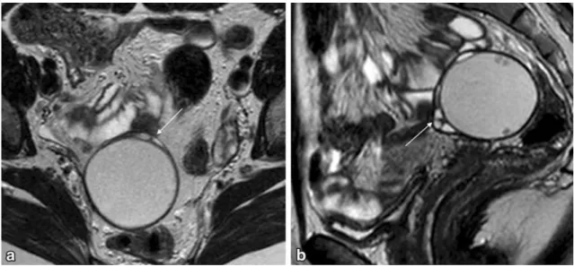
Fig. 1 Pelvic MRI for suspected endometriosis in a 32-year-old woman.aSagittal T2-weighted image;bandcAxial of the cervical canal T2-weighted images. Authors always ask clinical questions before the exam is performed, especially information that disproves
the initial diagnosis. In this case, the patient was using a contraceptive vaginal ring (arrows) that could be misinterpreted as pelvic endometriosis
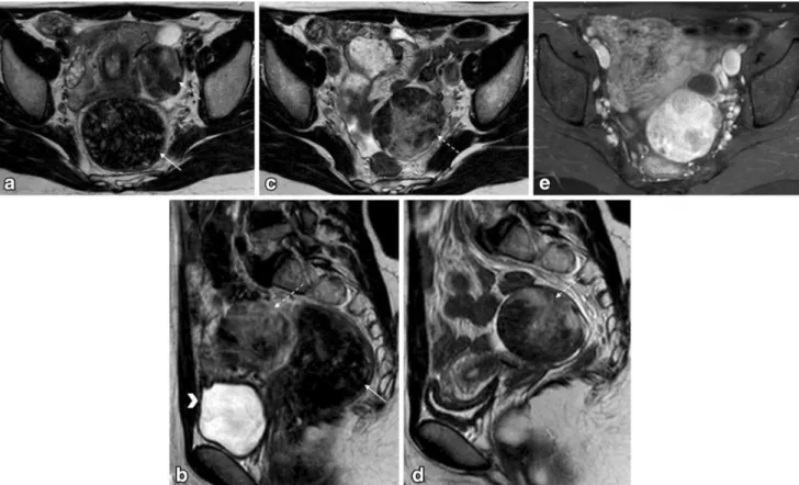
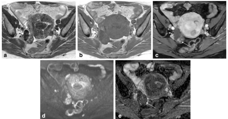
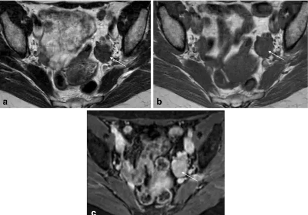
Fig. 2 Pelvic MRI for the characterization of an indeterminate adnexal mass in a 20-year-old woman.aand cAxial T2-weighted images;banddSagittal T2-weighted images;eAxial fat-suppressed contrast-enhanced T1-weighted image. Authors always encourage patients to evacuate before the exam, usually with the aid of a Microlax
. Patients also empty their bladder 1 h prior to the exam in order to achieve a moderately filled bladder. This case illustrates how rectal stools (arrow) and a filled bladder (arrowhead) impede the
correct characterization of gynaecological lesions (a and b). After evacuation and voiding, the adnexal mass (dashed-arrows) could be better characterized (c and d). It showed typical features of a sclerosing cell tumour of the ovary (benign tumour). It showed peripheral hypointense nodular areas with a spoke-wheel pattern on T2-weighted images. Dynamic contrast-enhanced study typically revealed an avid and early peripheral uptake that progresses to central areas on delayed images. The patient had a fertility-sparing surgery
those in which the radiological abnormality was not seen in the initial interpretation.
Cognitive or interpretative errors are caused by misin-terpretation due to faulty reasoning, misleading clinical information, lack of knowledge or propagation of an error that a previous radiologist had made [1••,8].
The misinterpretation of adnexal masses in women who undergo adnexal surgery is common [9].
There is a tremendous clinical impact when an inde-terminate adnexal mass is diagnosed as malignant or benign. Malignant ovarian tumours should be referred to a specialized oncological centre to assess if optimal cytore-ductive surgery is possible [10••]. On the other hand,
benign tumours can be treated in general hospitals usually by simple resection.
In general hospital and private daily-practices, pelvic gynaecological CT and magnetic resonance (MR) imaging are usually read by general abdominal radiologists with different levels of expertise. Many general radiologists are not familiarized with specific types of adnexal and pelvic tumours, which are frequent in a gynaecological oncolog-ical setting [9].
A second-opinion review is being increasingly done by radiologists in order to decrease the rate of error and misin-terpretation. One study has addressed the impact of a second-opinion review of gynaecological–oncological MR imaging Fig. 3 ‘‘Bridging vessel’’ sign in a 32-year-old woman with a
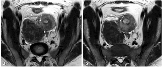
subserosal leiomyoma. a and b Axial of the uterine corpus T2-weighted images. In this case the right ovary (dashed-arrow) was compressed by the mass, making it difficult to diagnose the origin of
the pelvic mass. In order to determine if the pelvic mass is ovarian or uterine in origin, the authors perform a plane through the point of maximum contact of the mass and the uterus to look for the ‘‘bridging vessel’’ sign (arrows)
Fig. 4 Bilateral endometriomas in a 44-year-old woman.aAxial T1-weighted image;bAxial fat-suppressed T1-weighted image. Hyper-intense T1-weighted lesions are seen in both ovaries. In this case a
by sub-specialized radiologists of a gynaecological–onco-logical centre. It has shown that one-fifth of MR imaging had clinically relevant discrepancies and that second-reading was valuable in the management of these patients [11••].
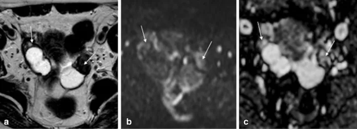
The first section of the article will describe the MR imaging protocol that authors perform in their gynaecolog-ical oncologgynaecolog-ical centre (Instituto Portugueˆs de Oncologia de Lisboa Francisco Gentil) for the characterization of a pelvic Fig. 5 Bilateral cystadenofibromas in a 56-year-old woman.aAxial
T2-weighted image;bAxial diffusion-weighted image (b=1000 s/
mm2); cADC map. Bilateral mixed ovarian masses are seen. The
solid component of these masses shows both low signal intensity on T2- weighted and on DWI images (arrows); thus the lesion is very likely to be benign and fibrotic in nature
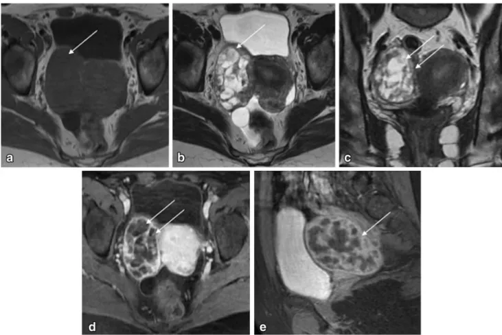
Fig. 6 Bilateral fibromas in a 55-year-old woman. a Axial T2-weighted image; b Axial diffusion-weighted image (b=1000 s/
mm2);cSemi-quantitative dynamic contrast-enhanced study;d Time-intensity curves of the myometrium (arrowhead) and of the right (arrow) and left (dashed-arrow) fibromas. Both ovarian lesions display peripheral low signal intensity on T2-weighted image and a small central cystic area. However, these tumour also display very
high signal on DWI image (b=1000 s/mm2); thus they should be
better characterized with the administration of gadolinium. Semi-quantitative dynamic study reveals a slow uptake of contrast and a low level of enhancement (type 1 curves) by both lesions, when compared to the myometrium. This curve pattern is very character-istic of benign lesions
mass of indeterminate origin. Authors will also address how imaging techniques can be optimized in order to reduce pitfalls in assessing pelvic gynaecological masses.
The next section will cover the potential pelvic anatomical pitfalls. We will revise the main radiological signs that may allow the radiologist to determine the origin of a pelvic mass.
Finally, we will demonstrate how benign gynaecology masses can mimic malignant ovarian tumours in a non-emergency setting.
MR Imaging Technique to Reduce Pitfalls
in Imaging Indeterminate Pelvic Masses
Transvaginal ultrasound complemented with transabdomi-nal approach is the first imaging modality for the assess-ment of women with a suspected gynaecological mass [12]. According to the European Society of Genitourinary Radiology (ESUR) guidelines, pelvic MR imaging should be performed to further characterize any sonographically indeterminate mass [13••]. Indeterminate adnexal masses are considered those that are complex masses with equiv-ocal malignant features, solid masses, large pelvic masses and masses adjacent to the uterus of unknown origin [13••]. The gynaecological MR imaging protocol of Instituto Portugueˆs de Oncologia de Lisboa Francisco Gentil follows
the ESUR recommendations for the characterization of sonographically indeterminate adnexal masses [13••].
Clinical questions are always asked before the exam (including age, questions regarding clinical symptoms, hormonal status, hormonal medication, recent surgeries and recent deliveries).Cognitive biases such as anchoring and framing are reduced when the radiologist seeks more clinical information from patients, especially disconfirming information that disproves the initial clinical diagnosis [4] (Fig.1).
The authors find of uttermost importance the use of strategies that limit motion artefacts. We always use anti-peristaltic agents for the improvement of image quality (20 mg butyl-scopolamine or 1 mg of glucagon intramus-cularly), unless they are contraindicated. We recommend patients to empty their bladder 1 h prior to examination in order to achieve a moderately filled bladder. Patients are also encouraged to evacuate before the exam, usually with the aid of a Microlax(Fig.2). Patients should fast (3–6 h) especially when intravenous injection of contrast is con-sidered. Scanning the patient in prone position may also help to reduce motion artefacts.
Both 3T and 1.5T magnets are valuable in the assess-ment of pelvic masses. According to ESUR guidelines, the basic MR imaging examination protocol should have a sagittal T2-weighted imaging of the pelvis and a pair of T1-weighted and T2-T1-weighted images in the same plane Fig. 7 Left extra-peritoneal solitary fibrous tumour in a 26- year-old
woman.aAxial T2-weighted image;bCoronal T2-weighted image; c Axial fat-suppressed contrast-enhanced T1-weighted image. An
through the indeterminate mass [13••]. If the origin of the mass is unknown, especially if the mass is adjacent to the uterus, a T2-weighted oblique sequence should be per-formed along the long axis of the uterus (which is the ovarian axis). Alternatively, a plane through the point of maximum contact of the mass and the uterus should be performed, in order to look for the ‘‘bridging vessel’’ sign that allows to determine the uterine origin of the lesion [13••] (Fig.3).
When a pelvic mass has high T1 signal, a fat-saturated T1-weighted sequence is mandatory to determine whether it has blood, high-protein or fat content (Fig.4).
If a pelvic mass has very dark signal on T2-weighted images, diffusion-weighted images (DWI) should be per-formed. A mass that displays both low T2 signal and DWI signal (b =800–1000 s/mm2) is very likely to be benign (ex.: fibroma; cystadenofibroma; pedunculated leiomyoma;
Brenner tumour), thus intravenous contrast is not needed [13••,14] (Fig.5).
Intermediate T2-weighted solid masses and cystic-solid masses with intermediate to high DWI signal (b =800–1000 s/mm2) can be malignant or benign (ex. cellular fibroma, degenerated pedunculated leiomyoma, tubo-ovarian abscess). In this case a T1-weighted sequence with intravenous contrast should be performed [13••] (Fig.6).
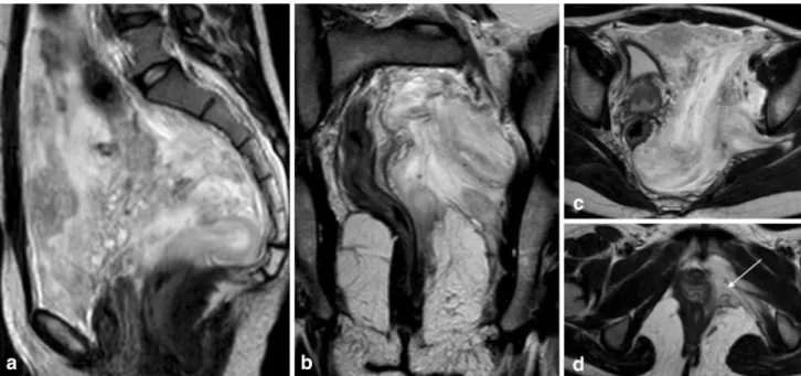
When gadolinium is administered to better characterize a T2-intermediate signal mass or a complex solid-cystic mass, authors perform a semi-quantitative study in order to obtain and compare the time-intensity curve of the solid component of the adnexal lesion with the one of the external myometrium. A rapid uptake of contrast with a rapid washout of the solid components has been strongly associated with malignant lesions (type 3 curve). On the Fig. 8 Extra-peritoneal sacral plexiform schwannoma.aSagittal
T1-weighted image;b Sagittal contrast-enhanced T1-weighted image; c and d Axial contrast-enhanced T1-weighted images. An
extra-peritoneal highly vascularized mass (arrow) is seen arising from the sacrum. It displaces the rectum anteriorly and laterally ( dashed-arrow)
other hand, a slow uptake of contrast and a low level of enhancement (type 1 curve) has a very high likelihood of benignity [13••,15–17] (Fig.6).
Pitfalls in Assessing the Anatomical Origin
of a Pelvic Mass
Large pelvic masses can originate from gynaecological organs and also from the bladder, the upper third of the rectum, the sigmoid colon, the bowel loops and from the extra-peritoneal space. It can be very challenging to define
the correct origin a pelvic mass in order to make a list of differential diagnosis. First, the radiologist should look for the displacement of anatomic landmarks that can help to diagnose the mass as intra- or extra-peritoneal in origin. Then, the radiologist should try to find specific anatomical organ-signs in order to confidently define the mass as ovarian or non-ovarian.
Is it Intra- or Extra-Peritoneal?
A detailed discussion of the pelvic anatomy is beyond the scope of this article. However, there are some anatomical Fig. 9 Vulval aggressive angiomyxoma in a 29-year-old woman
presenting as a transpatial pelvic mass.aSagittal T2-weighted image; bCoronal T2-weighted image;canddAxial T2-weighted images. A
transpatial high T2 signal lesion with a swirling appearance is seen occupying intra and extra-peritoneal spaces (arrows)
Fig. 10 ‘‘Negative embedded organ’’ sign caused by a small bowel GIST in a 37-year-old woman. aAxial T2-weighted image;bSagittal T2-weighted image;cSagittal fat-suppressed contrast-enhanced T1-weighted image. Images show an intra-peritoneal pelvic mass that
concepts that should be kept in mind when we are evalu-ating a pelvic mass of unknown origin.
The peritoneum is a serous membrane that wraps many of the abdomino-pelvic organs (visceral peritoneum) and covers the walls of the peritoneal cavity (parietal peri-toneum). The peritoneal cavity is a potential space formed by the continuous lining of these two layers. In females, the peritoneal cavity opens to the exterior through the fallopian tubes, the uterine and vaginal cavities [18].
An intra-peritoneal organ is anatomically defined as one that is almost completely covered by visceral peritoneum
(ex. the uterine corpus, the fallopian tubes, the bladder, the jejunoileum, the sigmoid colon and the upper third of the rectum). Nonetheless, the only organ that is truly intra-peritoneal is the ovary, because it is the only one that lies inside the peritoneal cavity making it a non-virtual space. The ovary is not covered by the peritoneum, so the oocyte can reach the fallopian tubes.
An extra-peritoneal structure is one that is not covered by the peritoneum or has only one surface covered by it (ex. the uterine cervix, the vagina, the lower two-thirds of the rectum, the ureter, the iliac vessels and the iliac lymph nodes).
Intra-peritoneal masses typically displace laterally or posteriorly the ureter and the iliac vessels. Conversely, extra-peritoneal masses tend to displace them anteriorly or medially (Fig.7). An extra-peritoneal mass usually also effaces the pelvic wall muscles (Fig.7).
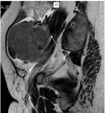
If a mass lies in the pre-sacral space it will typically cause anterior and/or lateral displacement and compression of the rectum (Fig.8).
However, one should note that there are masses that by its aggressive and invasive nature are transpatial (ex.: abscess; hematoma; angiomyxoma); therefore, these pat-terns of displacement cannot be applied (Fig.9). In our experience, both intra- and extra-peritoneal masses can cause anterior or lateral displacement of the uterus and bowel, as well as their compression, and thus we do not rely on the pattern of displacement of these organs.
Is it Ovarian or a Non-ovarian Mass?
There are four radiological signs that may help the radi-ologist to establish the origin of a pelvic mass.
Fig. 11 ‘‘Beak’’ signaAxial T2-weighted image;bSagittal T2-weighted image. The ‘‘beak’’ sign occurs when a mass deforms the edge of its organ of origin into a beak shape (arrows)
Fig. 12 ‘‘Prominent feeding artery’’ sign caused by a left Sertoli-Leydig cell ovarian tumour in a 15-year-old girl. Coronal T2-weighted image shows an engorged left ovarian artery that allowed the radiologist to diagnose this mass as a left ovarian tumour
The ‘‘embedded organ’’ sign means that at least a part of an organ seems to be embedded by the tumour [19]. On the contrary, organs that are adjacent to the mass and are not the site of origin, may display a crescent shape due to compression—‘‘negative embedded organ’’ sign (Fig.10).
A large mass may also make a small organ such as the ovary, undetectable. This has been described as the ‘‘phantom organ’’ sign [19,20].
The ‘‘beak’’ sign occurs when a mass deforms the edge of its organ of origin into a beak shape [19,20] (Fig.11). ‘‘Prominent feeding artery’’ sign is seen when an artery is enlarged because of tumour vascular supply [19, 20]. Anatomical recognition of the dilated artery may allow the radiologist to diagnose the origin of the tumour (Fig. 12). All of the described signs may be applied to both intra-or extra-peritoneal masses [19,20].
One sign that is used to determine the ovarian origin of a pelvic mass is the ‘‘ovarian vein’’ sign. The ovarian vein goes from the ovary through the ovarian suspensory liga-ment to the inferior vena cava (right ovarian vein) or to the left renal vein (left ovarian vein).
An enlarged ovarian vein is usually seen when a tumour arises from the ovary [21,22]. The depiction of a dilated ovarian vein is also very useful to establish the laterality of the tumour, especially when the mass is very large and compresses the contralateral ovary. However, sometimes a large pelvic mass of non-ovarian origin can compress the ovarian vein and make it look like it is joining it [21,22]. For this reason, the ‘‘ovarian suspensory ligament’’ sign seems to be more reliable than the ‘‘ovarian vein’’ sign (Fig.13).
The suspensory ligament of the ovary is a good ana-tomic landmark to find the ovary. It is a fold of peritoneum that connects the ovary to the pelvic wall. Both the ovarian artery and the ovarian vein cross the suspensory ligament of the ovary to reach the ovarian hilum. On axial images, it appears as a fan-shaped soft tissue structure anterior to the Fig. 13 ‘‘Ovarian suspensory ligament’’ sign caused by a Sertoli cell
tumour of the right ovary. Sagittal T2-weighted image shows a fan-shaped soft tissue structure anterior to the psoas muscles and in contiguity with the ovary (arrows). This ligament is crossed by the ovarian vessels to reach the ovarian hilum
Fig. 14 ‘‘Waist’’ sign caused by a chronic hydrosalpinx in a 74-year-old woman. a Axial T2-weighted image. b Sagittal T2-weighted image. A well-defined tubular-shaped organ dilated with fluid is seen
psoas muscle in continuity with the ovarian vessels that merges with the ovarian mass [22] (Fig.13).
When a fallopian tube is dilated it appears as a well-defined, tubular-shaped organ with folded configuration into a C or a S (Fig.14).
There are signs that may allow the radiologist to dif-ferentiate a dilated tube of an ovarian or para-ovarian cyst. The visualization of incomplete longitudinal mucosal folds or plicae is a highly specific sign of a hydrosalpinx [23] (Fig.14). The ‘‘waist’’ sign, which describes diamet-rically opposed indentations in the tube wall, can also help to confidently diagnose a dilated tube [24] (Fig.14).
The ‘‘beads-on-a-string’’ and ‘‘cogwheel’’ signs were initially described in the set of salpingitis. However, they may also be found in tumoral tube disease (particularly the ‘‘cogwheel’’ sign). The ‘‘cogwheel’’ sign describes a dila-ted tube folded on itself, which results in the visualization of thick incomplete septa. The ‘‘beads-on-a-string’’ sign describes endosalpingeal folds, which when viewed on cross-section in a dilated tube simulate small nodular mural projections.
Although an uterine mass is usually promptly diag-nosed, pedunculated leiomyomas sometimes are difficult to distinguish from broad ligament and adnexal masses (usually fibromas and thecomas). The misinterpretation especially occurs when large exophytic uterine degener-ated leiomyomas or leiomyosarcomas make the visual-ization of the adnexa difficult (Fig.15). In these cases, the differentiation from ovarian or tubal cancer might be difficult.
There are two radiological signs that may avoid this pitfall. The ‘‘bridging vessel’’ sign is present when we can clearly visualize the vessels anchoring the leiomyoma to the uterine wall (Fig.13). The ‘‘claw’’ sign describes the sharpened angles in the edge of a mass, which correspond to the myometrium that surrounds the tumour of uterine origin (Fig.15).
Benign Gynaecological Entities Mimicking
Ovarian Malignancy in a Non-emergency Setting
Fibroma and Cellular Fibroma
Fibromas are benign lesions that account for approximately 4% of all ovarian tumours [25••]. They tend to present in women in their late forties as hormonally inert tumours (Fig.6).
The diagnosis of typical fibromas is usually straight-forward. They range from small to large solid masses and characteristically present homogeneous low T2 signal and DWI signal (b =800–1000 s/mm2). According to the lat-est ESUR guidelines, these features are enough to diagnose a lesion as benign [13••]. Fibromas usually also display weak and delayed contrast uptake [26•] (Fig. 6). Occa-sionally, they can present with speckled calcifications.
In our view, there are three situations that can present as potential pitfalls when accessing a fibroma.
Fibromas may be associated with exuberant ascites and hydrothorax (Meigs’ syndrome) (Fig.16). This Fig. 15 ‘‘Claw’’ sign caused by a leiomyosarcoma in a 72-year-old woman.aSagittal T2-weighted image.bAxial T2-weighted image. The ‘‘claw’’ sign describes the sharpened angles in the edge of a mass which correspond to the myometrium that surrounds a tumour of uterine origin
combination of findings may lead the radiologist to diag-nose a fibroma as a malignant tumour, since ovarian car-cinoma typically has peritoneal spread.
Fibromas can sometimes have necrosis and haemor-rhagic degeneration (Fig.16). This is particularly true in cellular fibromas and when fibromas cause adnexal torsion.
Cellular fibromas are tumours with low malignant potential, representing approximately 10% of ovarian
fibromas [25••]. They usually present as large masses with heterogeneous signal due to haemorrhage and necrosis. A large mixed heterogeneous ovarian tumour with areas of T2 high signal or/and haemorrhagic content can be easily misdiagnosed as ovarian cancer (Fig.16).
Any large ovarian mass can cause adnexal torsion. In a subacute setting, patients may present with non-specific symptoms and with an indeterminate solid adnexal mass on ultrasound.
Fig. 16 Cellular fibroma of the right ovary in a 50-year-old female patient. a Axial fat-suppressed T1-weighted image; b Axial T2-weighted image; c Sagittal fat-suppressed contrast-enhanced T1-weighted image;d Sagittal T2-weighted image. A large, heteroge-neous and well-defined tumour is seen making the ‘‘negative embedded organ’’ sign in the uterus (arrows). This supports its
In both these scenarios, the radiologist should look for the presence of solid areas that display both low T2 signal and DWI signal (b=800–1000 s/mm2) in order to make the correct diagnosis. If adnexal torsion is suspected, contrast administration is crucial to determine the non-vi-ability of the ovary.
Fibromas are also frequently misdiagnosed as uterine leiomyomas. The discussion of this differential diagnosis will be done ahead (Fig.16).
Thecoma
Thecomas are solid benign ovarian tumours that typically occur in post-menopausal women (Fig.17). They are fre-quently associated with oestrogen-related symptoms. Therefore, women can present with uterine bleeding, endometrial hyperplasia and endometrial carcinoma.
The distinction between a thecoma and other malignant ovarian masses that may cause endometrial hyperplasia and/or cancer (endometrioid ovarian carcinoma; clear cell ovarian carcinoma; granulosa cell tumours) is usually easy; the latter are typically mixed ovarian masses. However, the differentiation from FIGO stage IIIA endometrial cancer may be challenging (invasion of the serosa and/or the adnexa through direct extension or metastases).
Endometrial cancer metastases to the ovaries may present as solid masses.
Pure thecomas do not have any particular characteristics and may mimic any malignant solid ovarian tumour.
Pure thecomas tend to display greater hyperintensity on T2-weighted images (due to oedema or cystic degenera-tion) and more avid contrast uptake when compared to more fibrous tumours, such as fibromas [27] (Fig.17).
A study by Zhang et al. showed that most thecomas displayed homogenous isointense signal on T1- and T2-weighted images and on DWI [28]. Moreover, the ADC values did not differ significantly from those of ovarian cancer [28] (Fig.17).
Cystadenofibroma and Brenner Tumour
Both cystadenofibroma and Brenner tumour are epithelial ovarian tumours with fibrous components [25••] (Figs.5,18). Cystadenofibromas are benign tumours that usually appear as complex cystic masses (Fig.15). They some-times show mild enhancing thick septa and/or nodular parietal thickening that may mimic the solid components of ovarian cancer. However, these solid components show very low signal on T2- weighted images as they are fibrous in nature (Fig.5).
Fig. 17 Thecoma of the right ovary in a 75-year-old female patient. aAxial T2-weighted image;bAxial T1-weighted image;cAxial fat-suppressed contrast-enhanced T1-weighted image;dAxial diffusion-weighted image (b=1000 s/mm2); e ADC map. A small solid
tumour is seen in the right ovary, showing both intermediate and hypo
signal on T2-weighted images and moderate contrast uptake. The lesion also displays high signal on highbvalues and low signal on ADC map. These characteristics may also be seen in ovarian cancer. No oestrogen-related symptoms were present in this case. A submucosal fundal leiomyoma is noted (dashed-arrow)
The vast majority of Brenner tumours are benign. They typically appear as asymptomatic solid masses with homogeneous low signal on T2-weighted images (Fig.18). In contrast to fibromas, they usually show moderate con-trast uptake (Fig.18).
Sclerosing Stromal Tumour
Sclerosing stromal tumours are benign ovarian neoplasms that are more likely to occur in women under 30 years of age as unilateral masses [29] (Fig.2).
Sclerosing stromal tumours appear as solid neoplasms with avid contrast uptake, mimicking ovarian malignant tumours. Cystic degeneration can also be seen and may be peripheral. When it occurs the tumour may be wrongly interpreted as an enhancing parietal solid nodule/mass of an ovarian cystic malignant neoplasm.
The distinction between these tumours and malignant ovarian lesions is particularly important, because the cor-rect diagnosis may lead to a fertility-sparing surgery in a woman of child-bearing age.
Sclerosing stromal tumours have characteristic radio-logical features. They tend to show pseudolobular
peripheral T2 hypo-/isointense areas with a spoke-wheel pattern and T2-hyperintense clefts (Fig.2). Moreover, dynamic contrast-enhanced study typically reveals an avid and early peripheral uptake that progresses to central areas on delayed images. Sometimes a peripheral capsule may be seen, reflecting their slow-growing nature [26•, 30] (Fig.2).
Struma Ovarii
Struma ovarii is a monodermal teratoma that is predomi-nantly or totally composed of thyroid tissue [25••] (Fig.19). It is benign in the majority of cases; however thyroid malignancy arising in a struma ovarii has been reported [31].
On MR imaging, struma ovarii usually presents as a multiloculated mass, with locules of variable intensity and with solid components that may avidly enhance, mimicking malignancy [31] (Fig. 19). Diagnostic clues of this entity are: its typical ‘‘stained glass’’ appearance (due the variable content of its locules) and the presence of colloid material that characteristically shows hypointensity on both T1- and T2-weighted images (Fig.19).
Fig. 18 Brenner tumour of the left ovary in a 58-year-old female patient. a Axial T2-weighted image; b Axial T1-weighted image; c Axial fat-suppressed contrast-enhanced T1-weighted image. A
Degenerated Leiomyomas
Typical leiomyomas usually do not pose diagnostic prob-lems. They present as T2- hypointense uterine tumours with delayed contrast uptake. However, pedunculated subserosal degenerated leiomyomas may be difficult to differentiate from adnexal tumours (Fig.3). This is par-ticularly true when large masses impede the visualization of the ovaries and of the vascular pedicle between the uterus and the juxta-uterine mass. Furthermore, leiomy-omas may detach from their stalk and form parasitic leiomyomas [32] (Fig.20).
When a juxta-uterine mass is of unknown origin, a T2-weighted oblique sequence should be performed along the long axis of the uterus to determine if it arises from the
ovary or from the uterus [13••,32] (Fig.20). Alternatively, a plane through the point of maximum contact of the mass and the uterus can also be done in order to look for the ‘‘bridging vessel’’ sign [13••].
About 10% of leiomyomas have histological degenera-tion [25••].This especially occurs in large tumours. Atypi-cal degenerating and cellular forms of pedunculated leiomyomas with high signal on T2-weighted sequences can be very difficult to differentiate from ovarian cancer, especially when serum levels of CA-125 are elevated [32] (Fig.20).
The types of degeneration that most resemble ovarian cancer are cystic, hyaline, myxoid and hydropic (Fig. 20). Although myxoid degenerating leiomyomas typically dis-play high signal on T2-weighted sequences, they show Fig. 19 Struma ovarii of the right ovary in a 46-year-old female
patient.aAxial T1-weighted image;bAxial fat-suppressed contrast-enhanced T1-weighted image;cAxial T2-weighted image;dSagittal T2-weighted image. A multiloculated mass with locules of variable
intensity and with solid components that avidly enhance is seen in the right ovary. Some of the locules show hypointense signal on both T1-and T2-weighted images
mild contrast uptake that is not a feature of ovarian cancer. Hyaline leiomyomas usually show a cobblestone appear-ance, with both high and hyposignal areas on T2-weighted images.
Diffuse hydropic leiomyomas are rare. Since they have abundant watery oedema, high T2-signal can be misinter-preted as myxoid matrix or as malignancy (of uterine or ovarian origin) [32]. Moreover, they may also be associ-ated with ‘‘Pseudo-Meigs’’ syndrome, another potential confounder.
Conclusion
MRI provides high resolution images and it is a powerful imaging modality for the diagnosis of female pelvic masses of indeterminate origin.
It can be very challenging to define the correct origin of a pelvic mass. The radiologist should try to look for ana-tomic signs that may help to diagnose the mass as intra- or
extra- peritoneal. Then, the radiologist should try to find specific anatomical organ-signs to define the mass as ovarian or non-ovarian in origin.
When ovarian origin is suspected, the radiologist should be aware of benign entities that mimic malignancy. In a non-emergency setting, they include: fibromas, cellular fibromas, thecomas, cystadenofibromas, Brenner tumours, sclerosing stromal tumours, struma ovarii and degenerated leiomyomas. Knowledge of the existence of these diagnostic pitfalls should help the radiologist to avoid misinterpretation of lesions in a pelvic magnetic resonance performed for the characterization of an indeterminate female pelvic mass.
Compliance with Ethical Guidelines
Conflict of interest Mariana Horta and Teresa Margarida Cunha each declare no potential conflicts of interest.
Human and Animal Rights and Informed Consent This article does not contain any studies with human or animal subjects performed by any of the authors.
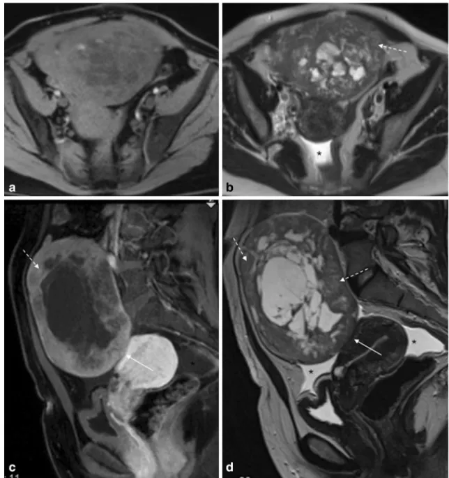
Fig. 20 Parasitic leiomyoma with cystic degeneration in a 47-year-old female patient.aAxial T1-weighted image;bAxial T2-weighted image; c Oblique T2-weighted image along the long axis of the uterus;dAxial fat-suppressed contrast-enhanced T1-weighted image; e Sagittal fat-suppressed contrast-enhanced T1-weighted image. A
References
Papers of particular interest, published recently, have been highlighted as:
• Of importance and •• Of major importance
1.•• Bruno MA, Walker EA, Abujudeh HH. Understanding and confronting our mistakes: the epidemiology of error in radiology and strategies for error reduction. RadioGraphics. 2015;35(6): 1668–76.Recent review article about different types of errors in Radiology and about strategies to reduce them.
2. Kravitz RL, Rolph JE, McGuigan K. Malpractice claims data as a quality improvement tool. I. Epidemiology of error in four spe-cialties. JAMA. 1991;266(15):2087–92.
3. Abujudeh HH, Boland GW, Kaewlai R, Rabiner P, Halpern EF, Gazelle GS, et al. Abdominal and pelvic computed tomography (CT) interpretation: discrepancy rates among experienced radi-ologists. Eur Radiol. 2010;20(8):1952–7.
4. Lee CS, Nagy PG, Weaver SJ, Newman-Toker DE. Cognitive and system factors contributing to diagnostic errors in radiology. AJR Am J Roentgenol. 2013;201(3):611–7.
5. Berlin L. Radiologic errors and malpractice: a blurry distinction. AJR Am J Roentgenol. 2007;189(3):517–22.
6. Muhm JR, Miller WE, Fontana RS, Sanderson DR, Uhlenhopp MA. Lung cancer detected during a screening program using four-month chest radiographs. Radiology. 1983;148(3):609–15. 7. Harvey JA, Fajardo LL, Innis CA. Mammograms with
impalpa-ble retrospective in breast vs blinded previous patients carci-noma: 1993 ARRS President’s award. AJR Am J Roentgenol. 1993;161(6):1167–72.
8. Berlin L, Hendix RW. Perceptual errors and negligence. AJR Am J Roentgenol. 1998;170:863–7.
9. Asch E, Levine D, Pedrosa I, Hecht JL, Kruskal J. Patterns of misinterpretation of adnexal masses on CT and MR in an aca-demic radiology department. Acad Radiol. 2009;16(8):969–80. 10.•• Woo YL, Kyrgiou M, Bryant A, Everett T, Dickinson HO.
Centralisation of services for gynaecological cancer. Cochrane Database Syst Rev. 2012; 14;(3):CD007945. Meta-analysis to assess the effectiveness of centralisation of care for patients with gynaecological cancer.
11.••Lakhman Y, D’Anastasi M, Micco` M, Scelzo C, Vargas HA, Nougaret S, et al. Second-opinion interpretations of gynecologic oncologic MRI examinations by sub-specialized radiologists influence patient care. Eur Radiol. 2016; 26(7):2089–98.Recent study that determined the impact of second-reading of gynaeco-logical MRI by sub-specialized gynaecological radiologists.
12. Harris RD, Javitt MC, Glanc P, Brown DL, Dubinsky T, Haris-inghani MG, et al. ACR appropriateness criteria? Clinically suspected adnexal mass. Ultrasound Q. 2013;29(1):79–86. 13.•• Forstner R, Thomassin-Naggara I, Cunha TM, Kinkel K,
Masselli G, Kubik-Huch R, et al. ESUR recommendations for MR imaging of the sonographically indeterminate adnexal mass: an update. Eur Radiol. 2017; 27(6):2248–57.European Society of Genitourinary Radiology updated guidelines for sonographically indeterminate adnexal masses.
14. Thomassin-Naggara I, Daraı¨ E, Cuenod CA, Fournier L, Tous-saint I, Marsault C, et al. Contribution of diffusion-weighted MR imaging for predicting benignity of complex adnexal masses. Eur Radiol. 2009;19(6):1544–52.
15. Thomassin-Naggara I, Dara E, Cuenod CA, Rouzier R, Callard P, Bazot M. Dynamic contrast-enhanced magnetic resonance imaging: a useful tool for characterizing ovarian epithelial tumors. J Magn Reson Imaging. 2008;28(1):111–20.
16. Bernardin L, Dilks P, Liyanage S, Miquel ME, Sahdev A, Rockall A. Effectiveness of semi-quantitative multiphase dynamic contrast-enhanced MRI as a predictor of malignancy in complex adnexal masses: radiological and pathological correla-tion. Eur Radiol. 2011;22(4):880–90.
17. Thomassin-Naggara I, Aubert E, Rockall A, Jalaguier-Caudray A, Rouzier R, Darai E, et al. Adnexal masses: development and preliminary validation of an MR imaging scoring system. Radi-ology. 2013;267(2):432–43.
18. Horta M, Couceiro C. Peritoneal anatomy: imaging overview. ESGAR 2013.e-Poster:EE-113.
19. Dionisio T, Gomes FV, Cunha TM. Non-ovarian pelvic masses: key concepts and useful clues to the radiologist. Eur Congr Radiol. 2014. doi:10.1594/ecr2014/C-1191.
20. Nishino M, Hayakawa K, Minami M, Yamamoto A, Ueda H, Takasu K. Primary retroperitoneal neoplasms: CT and MR imaging findings with anatomic and pathologic diagnostic clues. RadioGraphics. 2003;23(1):45–57.
21. Lee JH, Jeong YK, Park JK, Hwang JC. ‘‘Ovarian vascular pedicle’’ sign revealing organ of origin of a pelvic mass lesion on helical CT. AJR Am J Roentgenol. 2003;182(1):131–7. 22. Saksouk FA, Johnson SC. Recognition of the ovaries and ovarian
origin of pelvic masses with CT. RadioGraphics. 2004;24(suppl_1): S133–46.
23. Outwater EK, Siegelman ES, Chiowanich P, Kilger AM, Dunton CJ, Talerman A. Dilated fallopian tubes: MR imaging charac-teristics. Radiology. 1998;208(2):463–9.
24. Patel MD, Acord DL, Young SW. Likelihood ratio of sono-graphic findings in discriminating hydrosalpinx from other adnexal masses. AJR Am J Roentgenol. 2006;186(4):1033–8. 25. •• Kurman RJK, Carcangiu ML, Herrington CS, Young RH.
Classification of tumours of the ovary. In: WHO Classification of Tumours, Volume 6. 4th ed. Lyon: IARC, 2014; 11–83.Latest WHO histological classification of gynaecological tumours.
26. •Horta M, Cunha TM. Sex cord-stromal tumors of the ovary: a comprehensive review and update for radiologists. Diagn Interv Radiol. 2015;21(4):277–86.Most recent radiological review of sex cord-stromal tumours of the ovary.
27. Tanaka YO, Tsunoda H, Kitagawa Y, Ueno T, Yoshikawa H, Saida Y. Functioning ovarian tumors: direct and indirect findings at MR imaging. RadioGraphics. 2004;24(suppl_1):S147–66. 28. Zhang H, Zhang G-F, Wang T-P, Zhang H. Value of 3.0 T
dif-fusion-weighted imaging in discriminating thecoma and fibroth-ecoma from other adnexal solid masses. J Ovarian Res. 2013;6(1):1.
29. Outwater EK, Wagner BJ, Mannion C, McLarney JK, Kim B. Sex cord-stromal and steroid cell tumors of the ovary. Radiographics. 1998;18(6):1523–46.
30. Jung SE, Rha SE, Lee JM, et al. CT and MRI findings of sex cord-stromal tumor of the ovary. Am J Roentgenol. 2005;185(1): 207–15.
31. Leite IST, Cunha TM, Figueiredo JP, Fe´lix A. Papillary carci-noma arising in struma ovarii versus ovarian metastasis from primary thyroid carcinoma: a case report and review of the lit-erature. J Radiol Case Rep. 2013;7(10):1–10.
32. Horta M, Cunha TM, Oliveira R, Magro P. Hydropic leiomyoma of the uterus presenting as a giant abdominal mass. BMJ Case Rep. 2015;8:bcr2015211929–5.