On: 24 June 2008
Access Details: [subscription number 794230680] Publisher: Informa Healthcare
Informa Ltd Registered in England and Wales Registered Number: 1072954 Registered office: Mortimer House, 37-41 Mortimer Street, London W1T 3JH, UK
Acta Radiologica
Publication details, including instructions for authors and subscription information:
http://www.informaworld.com/smpp/title~content=t713394674
Magnetic Resonance Evaluation of Adnexal Masses
A. Guerraabc; T. M. Cunhaabc; A. FélixabcaDepartment of Radiology, Hospital Pulido Valente, Lisbon, Portugal
bDepartment of Radiology, Instituto Portugu s de Oncologia de Francisco Gentil,
Lisbon, Portugal
cDepartment of Pathology, Instituto Portugu s de Oncologia de Francisco Gentil,
Lisbon, Portugal First Published: 2008
To cite this Article: Guerra, A., Cunha, T. M. and Félix, A. (2008) 'Magnetic Resonance Evaluation of Adnexal Masses', Acta Radiologica, 49:6, 700 — 709 To link to this article: DOI: 10.1080/02841850802064995
URL:http://dx.doi.org/10.1080/02841850802064995
PLEASE SCROLL DOWN FOR ARTICLE
Full terms and conditions of use:http://www.informaworld.com/terms-and-conditions-of-access.pdf
This article maybe used for research, teaching and private study purposes. Any substantial or systematic reproduction, re-distribution, re-selling, loan or sub-licensing, systematic supply or distribution in any form to anyone is expressly forbidden.
Downloaded
B
y:
[
G
uerra,
A
dalgisa]
A
t:
09:
34
24
June
2008
ORIGINAL ARTICLE
Magnetic Resonance Evaluation of Adnexal Masses
A. GUERRA, T. M. CUNHA& A. FE´LIX
Department of Radiology, Hospital Pulido Valente, Lisbon, Portugal; Department of Radiology, Instituto Portugueˆs de Oncologia de Francisco Gentil, Lisbon, Portugal; Department of Pathology, Instituto Portugueˆs de Oncologia de Francisco Gentil, Lisbon, Portugal
Guerra A, Cunha TM, Fe´lix A. Magnetic resonance evaluation of adnexal masses. Acta Radiol 2008;49:700709.
Background: Accurate evaluation of adnexal masses allows correct surgical procedure, avoiding unnecessary surgery.
Purpose:To evaluate the accuracy of magnetic resonance imaging (MRI) in the diagnosis of malignancy of adnexal lesions.
Material and Methods:We retrospectively reviewed the pelvic MRI scans of 161 patients with 199 surgically confirmed adnexal masses, between November 1998 and June 2005. The criteria for adnexal malignancy were enhanced solid lesions, contrast-enhanced solid components in mixed lesions (except those with low-signal-intensity solid components on T2-weighted imaging [T2WI]), contrast-enhanced papillary projections in cystic lesions (except those with low-signal-intensity papillary projections on T2WI), or septal thickness ]3 mm. Ascites, peritoneal metastasis, and pelvic adenopathy were also regarded as criteria for malignancy.
Results: On MRI evaluation, 97 adnexal lesions were malignant and 102 were non-malignant. Thirty-two percent of patients with ascites had benign lesions. Histopatho-logic evaluation of the adnexal lesions showed that 83 were malignant (true positives), 100 were non-malignant (true negatives), and seven were uncertain malignant potential tumors; two were false negative and seven were false positive. The MRI sensitivity and specificity for malignancy were 98% and 93%, respectively. MRI reached an accuracy of 95%, with a positive predictive value of 0.92 and a negative predictive value of 0.98 for malignant adnexal lesions. The kappa coefficient was 0.906, indicating almost perfect agreement between MRI and histological results.
Conclusion:MRI is an accurate method for evaluating the malignancy of adnexal lesions.
Key words:Adnexal masses; genital; magnetic resonance imaging; ovarian tumor; pelvis
Adalgisa Guerra, Largo Maria Leonor 12 14A, 1495-144, Miraflores, Lisboa, Portugal
(fax.351 243679942, e-mail. gisaguerra@gmail.com)
Accepted for publication February 28, 2008
Ovarian cancer is the second most common gyne-cologic malignancy, and is the leading cause of death concerning gynecologic tumors (1). The majority of women with ovarian cancer present at an advanced stage of disease, largely because there are no apparent signs or symptoms at earlier stages (1). Screening examinations, including imaging modalities, are important for the early detection and evaluation of adnexal lesions. As adnexal masses are commonly found, the main goal of imaging should be the evaluation and characteriza-tion of the lesions in order to differentiate malig-nant from non-maligmalig-nant tumors. Transvaginal ultrasonography (TVUS) should be the first ima-ging approach for adnexal lesions (2), which allows
specific diagnosis in the majority of cases (3). However, magnetic resonance imaging (MRI) is superior in the characterization of adnexal masses (4) and should be used to further characterize indeterminate adnexal mass identified by US (5). Previous imaging studies have reported accuracies in the characterization of malignancy of adnexal masses ranging from 60 to 95% for US (4, 6, 7) and from 83 to 94% for MRI (3, 4, 713); the lower
figure of 83% is from a relatively early pioneer study using older equipment (3).
Reported MR imaging criteria for the diagnosis of adnexal malignant tumors include mass size larger than 4 cm, predominantly solid lesions, the pre-sence of necrosis on contrast-enhanced solid lesions,
DOI 10.1080/02841850802064995 #2008 Informa UK Ltd. (Informa Healthcare, Taylor & Francis AS) ACTA RADIOLOGICA
Downloaded B y: [ G uerra, A dalgisa] A t: 09: 34 24 June 2008
contrast-enhanced papillary projections, or septal thickness ]3 mm in cystic lesions, and bilaterality
(8, 14). The presence of ascites, peritoneal metastasis, pelvic adenopathy, and the continuous spread of the adnexal lesion to adjacent organs or the pelvic wall are also helpful criteria in the diagnosis of a malignant tumor or in cases where it is suspected (8). Adnexal lesions with signals characteristic of fat, blood, or cyst without enhancement are reported as benign lesions (15).
Our aim was to evaluate the accuracy of MRI in the diagnosis of malignancy of adnexal lesions. In order to determine the accuracy of MRI, we classified the adnexal lesions as malignant and non-malignant using MR, and compared this with the final histological diagnoses.
Material and Methods
We retrospectively reviewed data concerning the pelvic MRI exams of 161 patients with 199 adnexal lesions that underwent surgery at our hospital between November 1998 and June 2005. The mean patient age was 50.5 years (range 1583
years). In this study, lesions of ovarian and extra-ovarian origin, such as the fallopian tube and peritoneum, were included. The vast majority of the adnexal masses were ovarian in origin.
The pelvic MRI studies were performed, accord-ing to the guidelines of our hospital, for the characterization of indeterminate adnexal mass identified by US. Patient case notes included reports of previous US and, occasionally, clinical reports.
MRI examinations were performed using a 1.0-Tesla superconducting unit (Gyroscan NT10; Phi-lips Medical Systems, Eindhoven, The Netherlands) with a pelvic phased-array coil. All examinations were carried out according to the following imaging protocol: T1-weighted transverse turbo spin-echo (repetition time/echo time [TR/TE] 550/14 ms), T2-weighted transverse and sagittal turbo spin-echo (TR/TE 4000/90 ms), and T1-weighted transverse turbo spin-echo sequences were carried out imme-diately after completing the administration of 0.2 mmol/kg gadopentetate dimeglumine (Magnevist; Schering, Berlin, Germany), with a fat-suppression technique, a matrix of 256256, 5-mm slice
thick-ness, and 1-mm interslice gap. All transverse planes were acquired from the aortic bifurcation to the ischial tuberosity. If a large tumor was detected, the pelvic MRI study was extended to the rest of the abdomen to cover the entire lesion. Dynamic studies were not performed.
All patients fasted for 6 hours before the exam-ination, and 20 mg of intramuscular hyoscine butylbromide (Buscopan; Boehringer, Germany) was given to reduce motion artifacts by bowel movements.
MR imaging analysis
MRI readings were carried out by a senior radi-ologist (T.M.C.) with experience in pelvic MRI and a radiologist in training (A.G.). All images were saved on film, and were retrospectively reviewed and consensually evaluated by both. All adnexal lesions were analyzed and classified according to the following criteria: contrast-enhanced solid lesions, contrast-enhanced solid components in mixed le-sions (except those with low-signal-intensity solid components on T2-weighted images), contrast-enhanced papillary projections in cystic lesions (except those with low-signal-intensity papillary projections on T2-weighted images), or septal thickness ]3 mm. The presence of ascites,
perito-neal metastasis, and pelvic adenopathies was also an ancillary criterion for malignancy; otherwise, lesion size and bilaterality were not regarded as criteria for malignancy.
Adnexal lesions were classified into three types: cystic, solid, or mixed lesions with malignant or non-malignant features according to the following characteristics.
a. Lesions were considered cystic if they had low to slightly high signal intensity on T1-weighted images and high signal intensity on T2-weighted images compared with urine, and were classified as malignant cystic lesions if there were contrast-enhanced papillary projec-tions (Fig. 1) or septal thickness ]3 mm
Downloaded
B
y:
[
G
uerra,
A
dalgisa]
A
t:
09:
34
24
June
2008
b. Lesions were considered solid if they had variable signal intensity on T1- and T2-weighted images and showed enhancement after contrast with or without areas of necrosis. All contrast-enhanced solid lesions were con-sidered malignant, except those with low signal intensity on T2-weighted images.
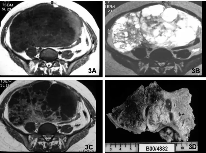
c. Lesions were considered mixed if they had solid and cystic components in harmonious portions (Fig. 3). All these lesions were considered malignant, except those with low-signal-intensity solid components on T2-weighted images (Fig. 4). If the lesion had a
predominantly cystic component with a negli-gible enhanced solid component, it was con-sidered as cystic with papillary projection. If the lesion had a predominantly enhanced solid component with a negligible cystic area, it was considered as solid with areas of necrosis/cystic areas.
Mixed, solid, or cystic lesions with very-low-signal-intensity solid components on T2-weighted images (like striate muscle) were considered as benign tumors with fibrous components (1619). Lesion
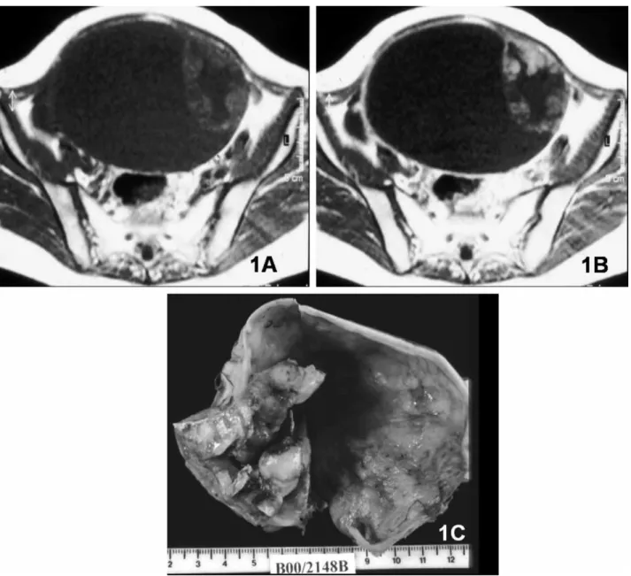
size 4 cm as a malignant criterion was not Fig. 1. Clear-cell carcinoma. A. Large cystic right adnexal lesion with intermediate-signal-intensity solid papillary projections and a thick septum on axial T1-weighted MR image. B. On contrast-enhanced axial T1-weighted image, the papillary projections and the septum show contrast enhancement. C. Gross specimen section of right oophorectomy.
Acta Radiol 2008 (6)
Downloaded
B
y:
[
G
uerra,
A
dalgisa]
A
t:
09:
34
24
June
2008
included if it did not contribute to further predic-tion of malignancy when combined with other imaging findings (9). Neither did we use bilaterality as a criterion of malignancy, because it occurs in both malignant and benign lesions, and it is mainly related to specific histological types (5, 10).
Histological analysis
Histological reports were also evaluated, and if there was any doubt concerning the histological diagnosis, the specimen was reviewed by a pathologist (A.F.) with experience in gynecological pathology. Using the World Health Organization (WHO)
classifica-tion of tumors of female genital organs (20) for the histological diagnosis, adnexal lesions were divided into three groups: malignant, non-malignant, and uncertain malignant potential tumors. All malignant neoplasms and borderline tumors were included as malignant tumors. All benign lesions, as well as functional ovarian cysts, peritoneal inclusion cysts, and adnexal inflammatory lesions, were grouped and classified as non-malignant lesions. The remaining tumors were classified as uncertain malignant po-tential tumors.
The MRI results (malignant/non-malignant) were classified as correct or not correct according to the final histological diagnoses (malignant/
Downloaded
B
y:
[
G
uerra,
A
dalgisa]
A
t:
09:
34
24
June
2008
non-malignant). All MRI and histological results were compared, except those relating to tumors classified as of uncertain malignant potential.
Statistical analysis
The kappa statistic was used to evaluate the degree of correlation between the MRI and histological results.
Results
MR results
Based on the analysis of the imaging characteristics of the 199 lesions, 97 adnexal lesions were char-acterized as malignant and 102 as non-malignant. Of these, seven lesions were excluded from the study because they were histologically classified as un-certain malignant potential tumors. The final pathological report of the remaining 192 lesions found that 83 malignant (true positive) and 100
non-malignant (true negative) lesions were correctly diagnosed. The MRI evaluation of adnexal lesions failed to detect malignancy in nine cases: two were false-negative and seven were false-positive results. On MRI evaluation of the 192 adnexal lesions, 120 lesions were found to be cystic, 33 were mixed, and 39 were solid.
Cystic lesions
Of the 120 cystic lesions, 32 were classified as malignant and 88 as non-malignant. The vast majority of non-malignant cystic lesions had vari-able signal intensity on T1- and T2-weighted images without enhanced papillary projections or thick septa. Cystic lesions with very-low-signal-intensity solid components on T2-weighted images (similar to muscle) (Fig. 5) were also included in this group. This pattern was present in six cases: five cystade-nofibromas and one borderline cystadenofibroma tumor.
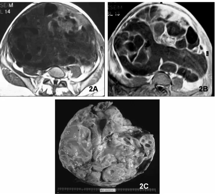
Fig. 3. Germ-cell tumor of the right ovary in a 40-year-old woman. Large mixed mass with solid and cystic components on T1-weighted (A) and T2-weighted (B) images. C. Contrast-enhanced T1-weighted image shows prominent enhancement of the solid component. D. Section of part of the resected specimen with solid hemorrhagic and necrotic tumor.
Acta Radiol 2008 (6)
Downloaded
B
y:
[
G
uerra,
A
dalgisa]
A
t:
09:
34
24
June
2008
On MRI, 99% of all non-malignant cystic lesions were histologically confirmed as benign, except the borderline cystadenofibroma (1%). The
major-ity of the non-malignant cystic lesions were simple hemorrhagic or high-protein-content cysts, endo-metriomas, and teratomas.
All 32 cases of cystic lesions defined as malig-nant by MRI (enhanced papillary projections or enhanced thick septa) were confirmed by histology as malignant, except for three (9%) lesions (one benign struma ovarii, one cystadenoma associated with benign struma ovarii, and one serous cysta-denoma).
Solid lesions
Of the 39 adnexal masses demonstrating solid pattern, 31 were malignant and eight were non-malignant in the final histological diagnoses. All five solid lesions considered as non-malignant on MRI had a non-malignant histological diagnosis. These lesions were three fibromas, one fibrothe-coma, and one thecoma. In none of the malignant lesions was this MR feature (solid lesions with low signal intensity on T2-weighted images) found. Four lesions (10%) with solid pattern and malig-nant features on MRI (two fibromas and one bilateral cystadenofibroma) were non-malignant on histologic examination. The remaining solid lesions were correctly diagnosed on MRI as malignant.
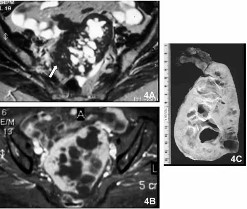
Fig. 4. Left adnexal fibroma in a 70-year-old woman. A. Mixed left adnexal lesion demonstrates heterogeneous signal intensity with high-signal-intensity cystic portions and low-high-signal-intensity solid portions (white arrow) on axial T2-weighted MR image. B. The mass shows enhancement of the solid portions on contrast-enhanced fat-suppressed axial T1-weighted image. C. Gross specimen section of left oophorectomy.
Downloaded B y: [ G uerra, A dalgisa] A t: 09: 34 24 June 2008 Mixed lesions
All 21 lesions with mixed pattern and criteria of malignancy on MRI were correctly diagnosed as malignant. Twelve lesions with mixed pattern and low-signal-intensity solid components on T2-weighted images were classified as non-malignant. Eleven (92%) were correctly diagnosed on MRI as non-malignant (seven cystadenofibromas and four fibromas), and one (8%) was a borderline Brenner tumor.
Ancillary criteria
Ascites, peritoneal nodules, and pelvic adenopathies were present in 65, 17, and eight patients, respec-tively. All patients with pelvic adenopathies and peritoneal metastasis had malignant adnexal masses. Ascites was presented in 44 patients with malignant adnexal masses and in 21 with benign adnexal masses.
Lesions with the highest accuracy rate for the diagnosis of malignancy achieved by MRI were mixed lesions; these diagnoses concurred 100% with the histological diagnoses. There were three cystic (9%) and four solid (13%) lesions that were histologically non-malignant, but had malignant features on MRI.
Histological results
Using histological evaluation, 85 masses were classified as malignant tumors (37 ovarian tumors, 16 epithelial borderline ovarian tumors [one cysta-denofibroma, 10 epithelial serous tumors, three epithelial mucinous tumors, one mixed tumor, and one Brenner tumor], 26 ovarian metastases, five primary fallopian tube carcinomas, and one carci-noma originating from the mesosalpinx). One hundred seven were classified as benign tumors (94 ovarian benign tumors, seven functional cysts, one peritoneal inclusion cyst, one cytosteatonecrosis of an epiploic appendix, three tubo-ovarian ab-scesses, and one hematosalpinx). Seven were classi-fied as uncertain malignant potential tumors (one sclerosing stromal tumor, one Sertoli-Leydig cell tumor, one granulosa cell tumor, one stromal endometrial tumor in an endometrioma, one steroid cell tumor, one Leydig cell tumor, and one ovarian Wolffian tumor).
Statistical results
A kappa coefficient of 0.906 revealed an almost perfect agreement between MRI and the histologi-cal results. The MRI sensitivity and specificity for malignancy were 98% and 93%, respectively. MRI
reached an accuracy of 95%, with a positive predictive value of 0.92 and a negative predictive value of 0.98 for malignant adnexal lesions.
Discussion
Preoperative characterization of adnexal lesions has important implications. Firstly, it is of considerable value for the gynecologist or general surgeon to know before surgery whether the lesion is benign or malignant, as this enables them to perform the most appropriate surgical procedure. Secondly, clinical and laboratory data are usually not specific enough for the characterization of the malignant nature of these lesions, especially in premenopausal women (9).
US and MRI are the most useful modalities for the assessment of adnexal lesions (4). US should be the primary imaging approach for the assessment and characterization of adnexal lesions (2). MRI, specifically contrast-enhanced MRI, provides addi-tional information, mainly in the characterization of indeterminate lesions identified by US, as contrast-enhanced MRI is significantly more accurate than US in adnexal lesion characterization, as shown by previous studies (4, 11, 21). Endovenous contrast administration is fundamental to the differentiation between solid and cystic lesions. For this reason, it is essential to apply the correct MRI protocol when evaluating adnexal lesions.
In this study, we found that MRI is a reliable method for differentiating between malignant and non-malignant adnexal lesions with high accuracy (95%), and our data concur with previously pub-lished data that report accuracies ranging from 83 to 94% (3, 4, 713). The pelvic MRI scans of 161
patients with 199 adnexal lesions who underwent surgery at our hospital were reviewed, and the MRI and histological results were compared. Of these lesions, seven were excluded (seven adnexal pa-tients) because the final histological diagnosis of these lesions was tumor with uncertain prognosis. These tumors could not be unambiguously classified as malignant or non-malignant, as the prognosis was unpredictable. It is important to point out that all these lesions had malignant features on MRI. One hundred ninety-two adnexal lesions were hence included, and well-established MRI criteria for diagnosing malignancy were applied. Some modifi-cations were introduced, such as masses with very low signal intensity (similar to muscle striate) on T2-weighted images, which were excluded from the malignant group. Although the size of the lesion (4 cm) is considered a criterion of malignancy by
Acta Radiol 2008 (6)
Downloaded
B
y:
[
G
uerra,
A
dalgisa]
A
t:
09:
34
24
June
2008
some authors (8, 4, 14), it was not included as such in our study, as we agree with others that lesion size (4 cm) when combined with other imaging
find-ings does not further contribute to the prediction of malignancy (9). The bilaterality of adnexal lesions is another accepted criterion of malignancy. This criterion was also rejected, because bilaterality can occur in both malignant and benign lesions, and is mainly related to specific histological types (5, 10). Despite bilateral ovarian involvement usually re-flecting the presence of metastases, recent studies have shown that bilaterality is not a reliable differentiating feature of primary versus secondary ovarian neoplasm (22).
All lesions studied in this report with non-malignant features on MRI were correctly diag-nosed as non-malignant, except for two borderline tumors (one cystic lesion with low-signal-intensity papillary projections on T2-weighted images and one mixed lesion with low-signal-intensity solid components on T2-weighted images). The majority of non-malignant lesions were cystic. Our study shows that non-malignant simple cystic lesions were almost all correctly diagnosed based on previously established criteria (3, 4, 15). The identification of blood and high protein content based on signal intensity characteristics on T1- and T2-weighted images contributed to the diagnosis of hemorrhagic/ high-protein-content cysts, as proven in the litera-ture (15).
All endometriomas in this study had high signal intensity on T1-weighted images with loss of signal on T2-weighted images (‘‘shading’’), as already shown by previous studies (3, 4, 15). In this series, as in the study by TOGASHI et al. (23), no case of
malignancy was misdiagnosed as an endometrial cyst. The diagnosis of mature teratoma was cor-rectly made when fatty tissue was identified (3, 4, 15, 16).
The extra-ovarian non-malignant lesions were correctly diagnosed because of their appearance*
oblong fluid-filled tubular structures, as in hemato-salpinx and tubo-ovarian abscesses (3, 4). In these cases, additional clinical history and examination were fundamental in establishing the diagnosis.
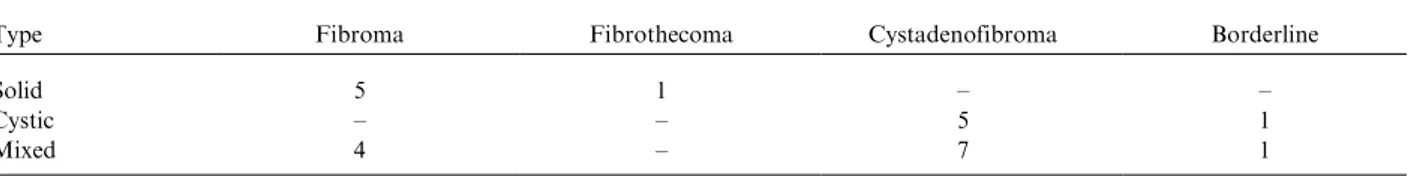
The remaining non-malignant lesions were cystic lesions with low-signal-intensity papillary projec-tions, homogeneous solid lesions with very low signal intensity on T2-weighted images, and mixed lesions with very-low-signal intensity solid compo-nents on T2-weighted images (like striate muscle). Our data proved that homogeneous solid lesions with very low signal intensity on T2-weighted images were all non-malignant. The majority of mixed and cystic lesions with very-low-signal-intensity components on T2-weighted images (like striate muscle) were non-malignant. We failed only in two borderline tumors: one borderline cystade-nofibroma and one borderline Brenner tumor (Table 1). These tumors may have extensive areas of dense fibrous tissue, and have features on MRI similar to other benign tumors with fibrous compo-nents (16). Previous studies support the thesis that fibromas, cystadenofibromas, and fibrothecomas are defined by their low-signal-intensity content on T2-weighted images. However, no previous studies have proven that all lesions with solid components with very low signal intensity on T2-weighted images (such as striate muscle) could be regarded as non-malignant (1619).
The three cystic lesions that we failed to diagnose as non-malignant on MR were one benign struma ovarii, one cystadenoma associated with benign struma ovarii, and a serous cystadenoma. The two struma ovarii were cystic lesions with septal thick-ness ]3 mm and contrast-enhanced papillary
projections without fat component tissue signal on MR. Our data are in line with previous studies that have reported that some benign ovarian tumors with enhancing solid portions such as struma ovarii could mimic malignant lesions (15). The other cystic lesion was a serous cystadenoma with a contrast-enhanced papillary projection.
The four solid lesions incorrectly diagnosed as malignant on MRI were two fibromas and one bilateral cystadenofibroma. These lesions had little fibrous tissue, which was not enough to produce as low a signal as muscle on T2-weighted imaging. In addition to the non-malignant findings referred to above, we can conclude that when an adnexal lesion
Table 1. Adnexal lesions with low-signal-intensity solid components on T2-weighted images.
Type Fibroma Fibrothecoma Cystadenofibroma Borderline
Solid 5 1
Cystic 5 1
Downloaded B y: [ G uerra, A dalgisa] A t: 09: 34 24 June 2008
has a very-low-signal-intensity solid component on T2-weighted images, it should have fibrous tissue in its composition. However, not all lesions with fibrous content have low-signal-intensity solid components on T2-weighted imaging. This feature may result from the amount of fibrous tissue in the adnexal lesion.
The ancillary inclusion criteria may be important, as they increase the diagnostic confidence of malig-nancy (15). However, caution should be used regarding the presence of ascites. Ascites is not an unusual finding associated with benign lesions, mainly fibromas. In our study, contrary to the findings of SOHAIBet al. (10), 32% of patients with
ascites had benign lesions. All patients with pelvic adenopathies and peritoneal metastasis had malig-nant adnexal tumors.
Our conclusions are limited by the following factors: the patient population was previously selected and biased by the US examination; more benign than malignant lesions were found; the study was retrospective; and the MR machine used was of only intermediate strength. However, our results are in line with previous studies performed with 1.5-Tesla unit equipment, and 1.0-1.5-Tesla machines can obtain similar results. Finally, because the number of cases with very-low-signal-intensity solid compo-nents on T2-weighted images was small, it cannot be regarded as a statistically acceptable criterion of non-malignancy. Nevertheless, it is our belief that our findings are of significant importance and should be divulged to clinicians, since they indicate that the lesion has a high probability of being non-malignant, with a great likelihood of being a cystadenofibroma, fibroma, or thecoma. We also believe that it would be important to follow up our work with a further study to confirm our findings through statistically acceptable data.
In conclusion, MRI has an excellent accuracy (95%) with a high (0.92) positive predictive value for the diagnosis of malignant adnexal lesions. Ascites is an ancillary inclusion criterion of malig-nancy, but is present in some (32%) benign adnexal lesions as well.
References
1. Moon HM. Ovarian tumors: general considerations and mode of spread. In: Kim SH, McClennan BL, Outwater EK, editors. Radiology illustrated: gynecologic imaging. 1st ed. New York: Cambridge University Press; 2005. p. 45399.
2. Kinkel K, Hricak H, Lu Y, Tsuda K, Filly RA. US characterization of ovarian masses: a meta-analysis. Radiology 2000;/217:/80311.
3. Jain KA, Friedman DL, Pettinger TW, Alagappan R, Jeffrey RB Jr, Sommer FG. Adnexal masses: compar-ison of specificity of endovaginal US and pelvic MR imaging. Radiology 1993;/186:/697704.
4. Yamashita Y, Torashima M, Hatanaka Y, Harada M, Higashida Y, Takahashi M, et al. Adnexal masses: accuracy of characterization with transvaginal US and precontrast and postcontrast MR imaging. Radiology 1995;/194:/55765.
5. Bazot M, Nassar-Slaba J, Thomassin-Naggara I, Cortez A, Uzan S, Darai E. MR imaging compared with intraoperative frozen-section examination for the diag-nosis of adnexal tumors; correlation with final histology. Eur Radiol 2006;/16:/268799.
6. Orden MR, Jurvelin JS, Kirkinen PP. Kinetics of US contrast agent in benign and malignant adnexal tumors. Radiology 2003;/226:/40510.
7. Rieber A, Nussle K, Stohr I, Grab D, Frenchel S, Kreienberg R, et al. Preoperative diagnosis of ovarian tumors with MR imaging: comparison with transvaginal sonography, positron emission tomography, and histo-logic findings. Am J Roentgenol 2001;/177:/1239. 8. Stevens SK, Hricak H, Stern JL. Ovarian lesions:
detection and characterization with gadolinium-en-hanced MR imaging at 1.5 T. Radiology 1991;/181:/ 4818.
9. Hricak H, Chen M, Coakley FV, Kinkel K, Yu K, Sica G, et al. Complex adnexal masses: detection and characterization with MR imaging multivariate ana-lysis. Radiology 2000;/214:/3946.
10. Sohaib SA, Sahdev A, Van Trappen P, Jacobs IJ, Reznek RH. Characterization of adnexal mass lesions on MR imaging. Am J Roentgenol 2003;/180:/1297304. 11. Medl M, Kulenkampff KJ, Stiskal M, Peters-Engl C, Leodolter S, Czembirek H. Magnetic resonance imaging in the preoperative evaluation of suspected ovarian masses. Anticancer Res 1995;/15:/11235.
12. Occhipinti KA. Computed tomography and magnetic resonance imaging of the ovary. In: Gynecologic ima-ging. Anderson JC, ed. London: Churchill Livingstone; 1999. p. 34559.
13. Saini A, Dina R, McIndoe GA, Soutter WP, Gishen P, deSouza NM. Characterization of adnexal masses with MRI. Am J Roentgenol 2005;/184:/10049.
14. Forstner R, Hricak H, White S. CT and MRI of ovarian cancer. Abdom Imaging 1995;/20:/28.
15. Jeong YY, Outwater EK, Kang HK. Imaging evaluation of ovarian masses. Radiographics 2000;/20:/144570. 16. Jung SE, Lee JM, Rha SE, Byun JY, Jung JI, Hahn ST.
CT and MR imaging of ovarian tumors with emphasis on differential diagnosis. Radiographics 2002;/22:/1305 25.
17. Kim KA, Park CM, Lee JH, Kim HK, Cho SM, Kim B, et al. Benign ovarian tumors with solid and cystic components that mimic malignancy. Am J Roentgenol 2004;/182:/125965.
18. Schwartz RK, Levine D, Hatabu H, Edelman RR. Ovarian fibroma: findings by contrast-enhanced MRI. Abdom Imaging 1997;/22:/5357.
Acta Radiol 2008 (6)
Downloaded
B
y:
[
G
uerra,
A
dalgisa]
A
t:
09:
34
24
June
2008
19. Troiano RN, Lazzarini KM, Scoutt LM, Lange RC, Flynn SD, McCarthy S. Fibroma and fibrothecoma of the ovary: MR imaging findings. Radiology 1997;/204:/ 7958.
20. WHO histological classification of tumours of the ovary. In Tavassoli FA, Devilee P, editors. World Health Organization classification of tumors. Pathology and genetics. Tumors of the breast and female genital organs. 1st ed. Lyon: IARC Press; 2003. p. 1145. 21. Popovich MJ, Hricak H. The role of magnetic resonance
imaging in the evaluation of gynecologic disease. In:
Callen PW, editor. Ultrasonography in obstetrics and gynecology. 3rd ed. Philadelphia: Saunders; 1994. p. 66088.
22. Brown DL, Zou KH, Tempany CM, Frates M, Silver-man S, McNeil B, et al. Primary versus secondary ovarian malignancy: imaging findings of adnexal masses in the Radiology Diagnostic Oncology Group Study. Radiology 2001;/219:/2138.