RBCCV 44205-1627 DOI 10.5935/1678-9741.20150016
Study of novel coating strategy for coronary
stents: simutaneous coating of VEGF and
anti-CD34 antibody
Estudo da nova estratégia de revestimento para stents coronários: revestimento simultâneo de VEGF e
anticorpo anti-CD34
Chun-Li Song
1, MD; Qian Li
1, MD; Yun-Peng Yu
1, PhD; Guan Wang
1, PhD; Jin-Peng Wang
1, MD;
Yang Lu
1, PhD; Ji-Chang Zhang
1, PhD; Hong-Ying Diao
1, MD; Jian-Gen Liu
1, MD; Yi-Hang Liu
1,
MD; Jia Liu
1, MD; Ying Li
1, MD; Dan Cai
1, MD; Bin Liu
1, PhD
1TheSecond Hospital of Jilin University, Changchun, Jilin, China.
This study was carried out at the Department of Cardiology, at the Second Hospital of Jilin University Changchun, Jilin, China.
Financial support: see in "Acknowledgements".
No conlict of interest.
Correspondence address: Bin Liu
The Second Hospital of Jilin University
Department of Cardiology - The Second Hospital of Jilin University, 218 Ziqiang Street - Changchun, Jilin, China - Zip Code 130041 E-mail: [email protected]
Article received on August 18th, 2014
Article accepted on February 22nd, 2015
Abstract
Introduction: Intravascular coronary stenting has been used in the treatment of coronary artery disease (CAD), with a major limitation of in-stent restenosis (ISR). The 316 stainless steel has been widely used for coronary stents. In this study, we developed a novel coating method to reduce ISR by simultane-ously coating vascular endothelial growth factor (VEGF) and anti-CD34 antibody on 316L stainless steel.
Methods: Round 316L stainless steel sheets in the D-H group were polymerized with compounds generated from condensa-tion reaccondensa-tion of dopamine and heparin using N-(3-dimethylami-nopropyl)-N’-ethylcarbodiimide (EDC) and N-hydroxysuccin-imide (NHS). Sixteen sheets from the D-H group were further immersed into 1ug/ml VEGF165 and 3mg/ml heparin sodium one after another for 10 times, and named as the D-(H-V)10 group. Eight sheets from the D-(H-V)10 group were coated with an-ti-CD34 antibody and termed as the D-(H-V)10-A group.
Immu-noluorescence assay and ELISA were used to evaluate wheth -er the 316L stainless steel disks w-ere successfully coated with VEGF and anti-CD34 antibody.
Results: The results of immunoluorescence assay and ELI
-SA showed that VEGF could be detected in the D-(H-V)10 and D-(H-V)10-A group, suggesting the steel sheets were successful-ly covered with VEGF. Anti-CD34 antibody could onsuccessful-ly be ob-served in the D-(H-V)10-A group, which was the only group coat-ed with CD34 antibody. Both results suggestcoat-ed that the 316L stainless steel sheets were successfully coated with VEGF and anti-CD34 antibody.
Conclusion: Our study developed a method to simultaneous-ly coat VEGF and anti-CD34 antibody to stainless metal steel. This research serves as a fundamental role for a novel coating strategy.
Descriptors: Coronary Artery Disease. Drug-Eluting Stents. Coronary Restenosis. Vascular Endothelial Growth Factor. An-tigens, CD34.
Resumo
INTRODUCTION
In-stent restenosis (ISR) was mainly caused by compli-cations of intracoronary stent placement, including throm-boembolic events and neointimal hyperplasia due to smooth muscle cell hyperproliferation. Drug eluting stents (DESs) have been designed mainly to reduce cellular proliferation and thus reduce ISR. Drug-eluting stents currently on the market release cytotoxic drugs such as paclitaxel and rapa-mycin to inhibit neointimal hyperplasia at the expense of delaying endothelialization[1,2].However, the incomplete
en-dothelialization of the stent surface has been suggested that may lead to the increased long-term incidence of thrombosis and ISR[3].The critical role of the vascular endothelium in
preventing thrombosis and regulating neointimal hyperplasia has resulted in restenosis prevention strategies that focus on enhancing endothelialiazation[4-6].
Vascular epithelial growth factor (VEGF), a cytokine originally described in 1983[7], is involved in processes
es-sential to the growth, maintenance and repair of vascular structures. Exogenous VEGF has been reported to show accelerated re-endothelialization of damaged arteries in the rat carotid artery and attenuated intimal hyperplasia[8]. The
delivery of VEGF using VEGF-eluting stents showed that it has been used to promote revascularization and re-endothe-lialization by stimulating endothelial progenitor cell migra-tion and maturamigra-tion [9,10]. Circulating endothelial progenitor
cells (EPCs), a subset of bone marrow-derived stem cells, possess the ability to differentiate into functional and mature endothelial cells and recently have been identiied as a key
316 tem sido amplamente utilizado para stents. Neste estudo, foi desenvolvido um novo método de revestimento para reduzir a RIS para revestir simultaneamente o fator de crescimento en-dotelial vascular (VEGF) e anti-CD34 em aço inoxidável 316L.
Métodos: Placas de aço inoxidável 316L redondas no gru-po DH foram gru-polimerizadas com comgru-postos gerados a partir da reacção de condensação de dopamina e heparina utilizando N- (3-dimetilaminopropil) -N’-etilcarbodiimida (EDC) e N-hi-droxissuccinimida (NHS). Dezesseis folhas a partir do grupo DH foram ainda imersas em 1 ug/ml de VEGF 165 e 3 mg/ml de heparina sódica, um após outro por 10 vezes, sendo
deno-minado como o grupo D-(HV)10. Oito folhas de D-(HV)10 foram revestidas com anticorpo anti-CD34 e denominado como grupo D-(HV)10-A. Testes de imunoluorescência e ELISA foram usa -dos para avaliar se os discos de aço inoxidável 316L foram re-vestidos com sucesso com VEGF e anticorpo anti-CD34.
Resultados: Os resultados dos testes de imunoluorescência
e ELISA mostraram que o VEGF pôde ser detectado nos gru-pos D-(HV)10 e D-(HV)10-A, evidenciando que as chapas de aço foram cobertas com VEGF com sucesso. O anticorpo anti-CD34 podia apenas ser observado no grupo D-(HV)10-A, o único grupo revestido com anticorpo CD34. Ambos os resultados sugerem que as chapas de aço inoxidável 316L foram revestidas com su-cesso com VEGF e anticorpo anti-CD34.
Conclusão: Nosso estudo desenvolveu um método para re-vestir simultaneamente VEGF e anti-CD34 de aço inoxidável. Esta pesquisa tem um papel fundamental para a nova estratégia de revestimento.
Descritores: Doença Arterial Coronariana. Stents Farmaco-lógicos. Reestenose Coronária. Fator de Crescimento Endotelial Vascular. Antígenos CD 34.
Abbreviations, acronyms & symbols
BSA Bovine serum albumin CAD Coronary artery disease DESs Drug eluting stents
EDC N-(3-dimethylaminopropyl)-N’-ethylcarbodiimide ISR In-stent restenosis
NHS N-hydroxysuccinimide VEGF Vascular endothelial growth factor
factor for re-endothelialization[11]. The EPC capture stents
have been developed using immobilized antibodies targeted at EPC surface antigens, such as CD34[12].
In this report, to further accelerate re-endothelialization, we aimed to develop method to simultaneously coat VEGF and anti-CD34 antibody. Our results showed that VEGF and anti-CD34 antibody were successfully coated onto the 316 stainless steel.
Experimental procedures
Preparation of the coated steel sheet
Round 316L stainless steel sheets (diameter 6 mm, thick-ness 1 mm) were used to facilitate the measurement and eval-uation of the properties of the coating, instead of bare metal stents with limited testable aspects and relatively high costs. The metal surface was polished, washed and dried at 60oC
for 24 hours, then sterilized by ultraviolet radiation. Heparin was conjugated to dopamine using N-(3-dimethylaminopro-pyl)-N’-ethylcarbodiimide (EDC) and N-hydroxysuccinimide (NHS)[13].The sterilized sheet was polymerized with
com-pounds generated from condensation reaction of dopamine (Aladdin, Shanghai, China) and heparin (Aladdin) as de-scribed[13], and termed as the D-H group.
times (5 min per time) and dried under nitrogen. These coat-ing procedure were repeated 10 times, and these sixteen steel disks were set as the D-(H-V)10 group.
The D-(H-V)10-A group was obtained by the following procedures. Eight steel disks from the D-(H-V)10 group were immersed into 0.1mg/ml protein A (Aladdin) dissolved in PBS for 30 min at RT, blocked in 10 mg/ml bovine serum albumin (BSA; BD Biosciences) for 24 hours at 4 oC and im-mersed with 2 μg/ml rabbit anti-human CD34 antibody (Ab -cam, Cambridge, UK) for 24 hours at 4 oC. After the whole
procedure, three groups of sheets were obtained (8 sheets/ each group) for further evaluations.
Immunoluorescence detection of the coated VEGF and anti-CD34 antibody
The levels of coated VEGF and anti-CD34 antibody of the stainless steel sheets were detected using immunoluorescence microscopy in the D-H group, D-(H-V)10 group and D-(H-V)10-A group. The sheets were soaked for 1 hour in blocking solution (PBS containing 3% BSA). For the detection of VEGF, all the three groups were incubated with rabbit anti-human VEGF primary antibodies (1:100; Abcam) overnight at 4 oC,
washed three times with PBS, and then incubated with Alexa Fluor® 488 Goat Anti-Rabbit IgG (Life technologies) at room
temperature for 1 hour. For the detection of rabbit anti-human CD34 antibody, secondary antibodies- Alexa Fluor® 488 Goat
Anti-Rabbit IgG were directly incubated. Wash three times to remove non-speciic binding of the secondary antibodies and observe using a Laser scanning confocal microscope (Leica TCS SP5; Leica Microsystems, Germany).
The detection of coated VEGF and anti-CD34 antibody by ELISA
Coated steel sheets from the D-H group, D-(H-V)10 group and D-(H-V)10-A group were immersed into RIPA lysis buffer for 24 hours at 4 oC to dissolve VEGF and anti-CD34 antibody.
The levels of VEGF and anti-CD34 antibody were detected using VEGF165 ELISA Kit (Life technologies) and Rabbit IgG ELISA Kit (Novus Biologicals, USA), respectively.
Statistical analysis
Statistical signiicance was evaluated by comparing mean values (±standard deviation) using the two-tailed Student’s t-test for independent groups. The probability value P<0.05 was considered to be statistically signiicant.
RESULTS
Immunoluorescence staining
To evaluate whether the 316L stainless steel sheets were successfully coated with VEGF and anti-CD34 an-tibody, immunoluorescence assay was performed. The results of immunoluorescence assay showed that VEGF
could be detected in the D-(H-V)10 and D-(H-V)10-A group, suggesting the steel sheets were successfully covered with VEGF. Anti-CD34 antibody could only be observed in the D-(H-V)10-A group, which was the only group that coated with CD34 antibody (Figure 1). Our indings suggested that the 316L stainless steel sheets were successfully coat-ed with VEGF and anti-CD34 antibody.
ELISA detection of coated VEGF and anti-CD34 antibody
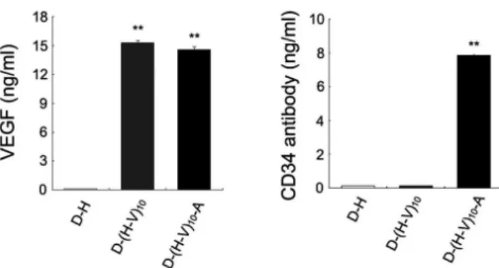
To further evaluate the coverage of VEGF and anti-CD34 antibody, ELISA was performed in the D-H group, D-(H-V)10 group and D-(H-V)10-A group. Similar to the results of immu-noluorescence assay, VEGF could be detected in the D-(H-V)10
and D-(H-V)10-A group, and anti-CD34 antibody could only be observed in the D-(H-V)10-A group (Figure 2). The results of ELISA further conirmed that we successfully coated VEGF and anti-CD34 antibody onto the 316 stainless steel.
Fig. 1 - Immunoluorescence detection of the coated VEGF and anti-CD34 antibody. The levels of coated VEGF and anti-anti-CD34 antibody of the 316L stainless steel were detected using immunoluorescence microscopy in the D-H group, D-(H-V)10 group and D-(H-V)10-A group.
DISCUSSION
Intravascular coronary stenting has been widely used for many years, and it has increased the quality of life and life ex-pectancy of patients with coronary disease. The 316 stainless steel is one of the most widely used materials for coronary stents with a board range of mechanical properties. However, the exposure of lowing blood to the bare metal stent may lead to thrombus formation and smooth muscle cell prolif-eration, and inally cause in-stent restenosis (ISR).There -fore, huge amount of recent work has attempted to develop non-thrombogenic coating for these metallic stents[14-16].
Early drug-eluting stents coating with various kind of drugs have been designed to reduce the restenosis through minimize vascular inlammation and cellular proliferation[17],
which including a polymer-based drug delivery platform and a pharmacologic agent (usually an immunosuppressant and/ or antiproliferative compound).Though early trials seem to be exciting with markedly reduction rates of ISR (5%-8%)
[18,19], long-term follow-up studies showed that DESs
implan-tation increased the long-term risk of thrombosis by 15% -35% compared with bare-metal stents implantation[20].
Based on our expanding understanding of pathophysiol-ogy of restenosis, novel stent coating strategies have been developed, such as delivery of VEGF (e.g., VEGF-eluting stents[9]) and the use of antibodies that recognize epitopes speciic to endothelial progenitor cells (e.g., anti-CD34-coat -ed stents[12]). Both coating strategies have been designed to
inhibit thrombosis mainly through promoting re-endotheli-alization of cardiovascular stents. Many clinical studies sug-gest that the Genous EPC-capture stent is a safe choice for patients with coronary disease[21-24]. However, Adrian et al.[25]
reported that a similar late luminal loss of Genous EPC-cap-ture stent to that of a bare-metal stent, despite initial opti-mism of rapid endothelialization.
In this study, our approach of surface modiication has included the combination of VEGF and anti-CD34 antibody. The goal of this combination is to further accelerate endo-thelial repair, and thus further reduce the exposure time of stents in blood, decrease the rate of long-term thrombosis and shorten the time of antiplatelet therapy for patients. We irstly used dopamine-mediated heparin coating[13] and then a layer-by-layer method was employed to build multilayer ilms composed of heparin and VEGF on metal substrates. Based on the speciic afinity of protein A and IgG antibodies, the pro -tein A allows the subsequent immobilization of the anti-CD34 antibody. Our primary results of immunoluorescence and ELISA showed that the stainless metal steel was successfully coated with VEGF and anti-CD34 antibody (Figures 1 and 2). This research serves as a fundamental role for the novel coat-ing strategy of simultaneous coatcoat-ing of VEGF and anti-CD34 antibody and further studies on the toxicity and effect of the combined coating are currently ongoing.
Authors’ roles & responsibilities
CLS Analysis and interpretation of data; statistical analysis; de-sign and study dede-sign; carried out operations and experi-ments
QL Analysis and interpretation of data; statistical analysis; car-ried out operations and experiments
YPY Analysis and interpretation of data; statistical analysis; car-ried out operations and experiments
GW Analysis and interpretation of data; statistical analysis; car-ried out operations and experiments
JPW Analysis and interpretation of data; statistical analysis; car-ried out operations and experiments
YL Analysis and interpretation of data; statistical analysis; car-ried out operations and experiments
JCZ Analysis and interpretation of data; statistical analysis; car-ried out operations and experiments
HYD Analysis and interpretation of data; carried out operations and experiments
JGL Analysis and interpretation of data; carried out operations and experiments
YHL Analysis and interpretation of data; carried out operations and experiments
JL Analysis and interpretation of data; carried out operations and experiments
YL Analysis and interpretation of data; carried out operations and experiments
DC Analysis and interpretation of data; carried out operations and experiments
BL Analysis and interpretation of data; carried out operations and experiments
REFERENCES
1. Carter AJ, Aggarwal M, Kopia GA, Tio F, Tsao PS, Kolata R, et al. Long-term effects of polymer-based, slow-release, sirolimus-eluting stents in a porcine coronary model. Cardiovasc Res. 2004;63(4):617-24.
2. Nakazawa G, Otsuka F, Nakano M, Vorpahl M, Yazdani SK, Ladich E, et al. The pathology of neoatherosclerosis in human coronary implants bare-metal and drug-eluting stents. J Am Coll Cardiol. 2011;57(11):1314-22.
ACKNOWLEDGEMENTS
3. Serruys PW, de Jaegere P, Kiemeneij F, Macaya C, Rutsch W, Heyndrickx G, et al. A comparison of balloon-expandable-stent implantation with balloon angioplasty in patients with coronary artery disease. Benestent Study Group. N Engl J Med. 1994;331(8):489-95.
4. Asai J, Takenaka H, Kusano KF, Ii M, Luedemann C, Curry C, et al. Topical sonic hedgehog gene therapy accelerates wound healing in diabetes by enhancing endothelial progenitor cell-mediated microvascular remodeling. Circulation. 2006;113(20):2413-24.
5. Ii M, Nishimura H, Iwakura A, Wecker A, Eaton E, Asahara T, et al. Endothelial progenitor cells are rapidly recruited to myocardium and mediate protective effect of ischemic preconditioning via “imported” nitric oxide synthase activity. Circulation. 2005;111(9):1114-20.
6. Finn AV, Joner M, Nakazawa G, Kolodgie F, Newell J, John MC, et al. Pathological correlates of late drug-eluting stent thrombosis: strut coverage as a marker of endothelialization. Circulation. 2007;115(18):2435-41.
7. Senger DR, Galli SJ, Dvorak AM, Perruzzi CA, Harvey VS, Dvorak HF. Tumor cells secrete a vascular permeability factor that promotes
accumulation of ascites luid. Science. 1983;219(4587):983-5.
8. Asahara T, Bauters C, Pastore C, Kearney M, Rossow S, Bunting S, et al. Local delivery of vascular endothelial growth factor accelerates reendothelialization and attenuates intimal hyperplasia in balloon-injured rat carotid artery. Circulation. 1995;91(11):2793-801.
9. Swanson N, Hogrefe K, Javed Q, Gershlick AH. In vitro evaluation of vascular endothelial growth factor (VEGF)-eluting stents. Int J Cardiol. 2003;92(2-3):247-51.
10. Lahtinen M, Blomberg P, Baliulis G, Carlsson F, Khamis H, Zemgulis V. In vivo h-VEGF165 gene transfer improves early endothelialisation and patency in synthetic vascular grafts. Eur J Cardiothorac Surg. 2007;31(3):383-90.
11. Lin HH, Chen YH, Yet SF, Chau LY. After vascular injury, heme oxygenase-1/carbon monoxide enhances re-endothelialization via promoting mobilization of circulating endothelial progenitor cells. J Thromb Haemost. 2009;7(8):1401-8.
12. Klomp M, Beijk MA, de Winter RJ. Genous endothelial progenitor cell-capturing stent system: a novel stent technology. Expert Rev Med Devices. 2009;6(4):365-75.
13. Bae IH, Park IK, Park DS, Lee H, Jeong MH. Thromboresistant and endothelialization effects of dopamine-mediated heparin coating on a stent material surface. J Mater Sci Mater Med. 2012;23(5):1259-69.
14. de Torre IG, Wolf F, Santos M, Rongen L, Alonso M, Jockenhoevel S, et al. Elastin-like recombinamer-covered stents: Towards a fully biocompatible and non-thrombogenic device for cardiovascular diseases. Acta Biomater. 2015;12:146-55.
15. Whitbourne RJ, Chamberlain AM, Hullihen DG, Rosebrough SF, Calistri-Yeh M. Medicated stent having multi-layer polymer coating. Google Patents; 2012.
16. Wright C, Llanos GH, Rakos R, King K, Falotico R. Methods and Devices for Delivering Therapeutic Agents to Target Vessels. Google Patents; 2012.
17. Gomes WJ, Buffolo E. Coronary stenting and inlammation. Rev
Bras Cir Cardiovasc. 2003;18(4):III-VII.
18. Moses JW, Leon MB, Popma JJ, Fitzgerald PJ, Holmes DR, O’Shaughnessy C, et al.; SIRIUS Investigators. Sirolimus-eluting stents versus standard stents in patients with stenosis in a native coronary artery. N Engl J Med. 2003;349(14):1315-23.
19. Stone GW, Ellis SG, Cox DA, Hermiller J, O’Shaughnessy C, Mann JT, et al.; TAXUS-IV Investigators. A polymer-based, paclitaxel-eluting stent in patients with coronary artery disease. N Engl J Med. 2004;350(3):221-31.
20. Byrne RA, Sarafoff N, Kastrati A, Schömig A. Drug-eluting stents
in percutaneous coronary intervention: a beneit-risk assessment.
Drug Saf. 2009;32(9):749-70.
21. Lee YP, Tay E, Lee CH, Low A, Teo SG, Poh KK, et al. Endothelial progenitor cell capture stent implantation in patients with ST-segment elevation acute myocardial infarction: one year follow-up. EuroIntervention. 2010;5(6):698-702.
22. Chong E, Poh KK, Liang S, Lee RC, Low A, Teo SG, et al. Two-year clinical registry follow-up of endothelial progenitor cell capture stent versus sirolimus-eluting bioabsorbable polymer-coated stent versus bare metal stents in patients undergoing primary percutaneous coronary intervention for ST elevation myocardial infarction. J Interv Cardiol. 2010;23(2):101-8.
23. Aoki J, Serruys PW, van Beusekom H, Ong AT, McFadden EP, Sianos G, et al. Endothelial progenitor cell capture by stents coated with antibody against CD34: the HEALING-FIM (Healthy Endothelial Accelerated Lining Inhibits Neointimal Growth-First In Man) Registry. J Am Coll Cardiol. 2005;45(10):1574-9.
24. Duckers HJ, Soullie T, den Heijer P, Rensing B, de Winter RJ, Rau M, et al. Accelerated vascular repair following percutaneous coronary intervention by capture of endothelial progenitor cells promotes regression of neointimal growth at
long term follow-up: inal results of the Healing II trial using
an endothelial progenitor cell capturing stent (Genous R stent). EuroIntervention. 2007;3(3):350-8.