RBCCV 44205-1629
DOI 10.5935/1678-9741.20140097
Effect of N-acetylcysteine in hearts of rats
submitted to controlled hemorrhagic shock
Efeito da N-acetilcisteína em corações de ratos submetidos ao choque hemorrágico controlado
Luiz Dantas de Oliveira Filho
1, MD; Karen Ruggeri Saad
2, MSc, PhD; Paulo Fernandes Saad
2,
MD, MSc, PhD; Marcia Kiyomi Koike
3, PhD; Sônia Maria da Silva
1, PhD; Edna Frasson de Souza
Montero
1,3, MD, PhD
1Universidade Federal de São Paulo (UNIFESP), São Paulo, SP, Brazil. 2Universidade Federal do Vale do São Francisco (UNIVASF), Petrolina, PE, Brazil.
3Faculdade de Medicina da Universidade de São Paulo (FMUSP), São Pau-lo, SP, Brazil.
This study was carried out at Laboratório de Cirurgia e Experimentação da Universidade Federal de São Paulo (UNIFESP), São Paulo, SP, Brazil.
No inancial support.
Correspondence address: Edna Frasson de Souza Montero
Laboratório de Fisiopatologia Cirúrgica - LIM-62 Faculdade de Medicina - USP
Av. Dr. Arnaldo, 455 - 2 andar
Cerqueira César, São Paulo, SP, Brazil - Zip Code: 01246-903 E-mail: [email protected]
Article received on February 17th, 2014 Article accepted on July 24th, 2014 Abstract
Introduction: Pharmacological therapy is a strategy for the prevention of complications associated with ischemia and reper-fusion injury that occurs after volume replacement in the treat-ment of hemorrhagic shock.
Objective: The aim of this study was to evaluate the effect of
N-acetylcysteine associated with luid resuscitation in cardiac
injury in a rat hemorrhagic shock model.
Methods: Mice Wister male rats were randomly and sub-jected to controlled hemorrhagic shock for 60 min. and then, subjected to resuscitation with Ringer lactate. In a group of six
animals, 150mg/kg of N-acetylcysteine were added to luid vol
-ume replacement. The animals were observed for 120 min and after this period, were euthanized and cardiac tissue was col-lected for histopathological analysis and measurement of
thio-barbituric acid reactive substances and pro-and anti-inlamma
-tory interleukin.
Results: Cardiac tissue of the group treated with N-acetyl-cysteine showed lower concentrations of thiobarbituric acid reactive substances (0.20±0.05 vs. 0.27±0.05, P=0.014) and
re-duced histopathological damage and edema when compared to the group whose volume replacement occurred only with Ringer lactate. There was no difference in the expression of cytokines interleukin 6 (2,138.29±316.89 vs. 1,870.16±303.68, P=0.091) and interleukin 10 (1.019,83±262,50 vs. 848.60±106.5, P=0.169) between the treated groups.
Conclusion: The association of N-acetylcysteine on volume replacement attenuates oxidative stress in the heart, as well myocardial damage and edema, but does not modify the
expres-sion of inlammatory cytokines.
Descriptors: Shock, Hemorrhagic. Heart. Acetylcysteine.
Oxidative Stress. Inlammation.
Resumo
INTRODUCTION
Trauma is the third death cause in the world, compro-mising mainly young and adult people.Bleeding is the ma-jor cause of the early death related to trauma.Additionally, deaths will occur due to severe injury to internal organs in the next hours, or due to multi-organ failure and sepsis, lately[1].
Hypoperfusion, following hemorrhagic shock, generates
a global hypoxia that promotes the release of inlammato -ry cytokines and neutrophils activated from the splanchnic territory, notably from the liver and intestine, which via the bloodstream or lymphatic circulation promotes injuries to distant organs. This acute phase response of the trauma is characterized by the production and release of cytokines
such as the alpha tumor necrosis factor alpha (TNF-α), in
-terleukins 1β, 6 and 8[2].Although oxygen is essential for the
survival of the tissues, during the restoration of perfusion, the cells suffer a harmful effect, characterizing the reperfu-sion injury[3].
Alterations induced by ischemia and reperfusion injury (IR) can be related to two different mechanisms. One of them, char-acterized by excessive production and subsequent release of re-active oxygen species (ROS), highly cytotoxic during the reper-fusion phase, whose oxidative state biochemical markers are the
Objetivo: O objetivo deste estudo foi avaliar a repercussão da N-acetilcisteína associada à reposição volêmica na lesão car-díaca em modelo de choque hemorrágico em ratos.
Métodos: Ratos Wistar, machos, foram randomizados e sub-metidos ao choque hemorrágico controlado por 60 minutos e, depois, submetidos à reposição volêmica com Ringer Lactato. Em um grupo de seis animais, foram adicionados 150 mg/Kg
de N-acetilcisteína ao luido de reposição volêmica. Os animais
foram observados por 120 minutos e após este período foram
submetidos à eutanásia e coleta do tecido cardíaco para análise histopatológica e dosagem de substâncias reativas ao ácido
tio-barbitúrico e interleucinas pró e anti-inlamatórias.
Resultados: Foi observada menor concentração de substâncias reativas ao ácido tiobarbitúrico (0,20±0,05 vs. 0,27±0,05, P=0,014) e menor dano histopatológico e edema no tecido cardíaco do grupo tratado com N-acetilcisteína em relação ao grupo cuja reposição vo-lêmica ocorreu somente com Ringer Lactato. Não foi observada di-ferença da expressão das citocinas interleucina 6 (2.138,29±316,89 vs. 1.870,16±303,68, P=0,091) e interleucina 10 (1.019,83±262,50 vs. 848,60±106,5, P=0,169) entre os grupos tratados.
Conclusão: A associação da N-acetilcisteína na reposição vo-lêmica atenua o estresse oxidativo no coração, assim como dano
e edema miocárdicos, porém, não modiica a expressão de cito
-cinas inlamatórias.
Descritores: Choque Hemorrágico. Coração. Acetilcisteína.
Estresse Oxidativo. Inlamação.
Abbreviations, acronyms & symbols
CONCEA Council for the Control of Animal Experimentation
IR Reperfusion injury
MAP Mean arterial pressure
NAC N-acetylcysteine
ROS Reactive oxygen species
TBARS Thiobarbituric acid reactive substances TNF-α Tumor necrosis factor alpha
end products of lipid peroxidation, among which the thiobar-bituric acid reactive substances (TBARS)[4]; the other, by the
interaction of polymorphonuclear and capillary endothelial
cells, mediated by inlammatory cytokines and cell adhesion
molecules[5].
In an attempt to minimize the damages caused by ROS, cardiac myocytes use antioxidant systems – substances that slow down or inhibit oxidative aggression. The most im-portant endogenous antioxidants are the superoxide dis-mutase, catalase, glutathione peroxidase, and vitamin E. These systems are overloaded after ischemia and reperfu-sion[6].The damage to cardiac myocytes can happen, then, by
cell-to-cell contact (neutrophils – myocyte) with the release of oxidative cytokines and proteolytic enzymes. This
accu-mulation and iniltration of neutrophils in the organ’s paren
-chyma is a fundamental step for development of the trauma’s
secondary injury[7]. The cardiac dysfunction established
con-tributes to aggravate the hypoperfusion injury in other organs during the shock and may result in death.
Associated with luid replacement therapy, the pharma -cological therapy has gained prominence in the reduction
of deleterious effects of immune-inlammatory phenomena
of bleeding and the volume replacement therapy[8]. Among
low-cost, highly available, low-adverse effects substance – must be highlighted. Widely used in a number of medical science
ields, it was initially used as a mucolytic agent. Its use was
then extended to antidote for acetaminophen poisoning and prevention of contrast-induced nephropathy[9].
The in vivo NAC is metabolized in cysteine, which is a precursor of glutathione.In its reduced and oxidized forms, the glutathione participates – together with the glutathione peroxidase – in the ROS degradation cascade, removing free radicals. Thus, NAC can help restoring depleted glutathione reserves, replenishing cellular thiols during the IR process[10].
On IR injury, the NAC mechanism of action occurs by direct reaction with nitric oxide. This effect seems to occur after ROS release, protecting endothelial cells and subse-quent activation of Kupffer cells. Its action through the sulf-hydryl groups prevents the reaction of nitric oxide with the superoxide radical, hydrogen peroxide, and hydroxyl radical, preventing the formation of peroxynitrite and its consequenc-es, such as lipid peroxidation, protein denaturation and DNA damage[11].
Despite of being widely used in medical practice and ex-perimental models of IR injury, the literature about the use of NAC in the treatment of hemorrhagic shock and its pos-sible protective effect in cardiomyocytes is scarce. As satis-factory results were observed with the use of NAC as pro-tective drug of lung and liver tissue in experimental studies with controlled hemorrhagic shock models[12,13], as well as
in other studies that used tissue IR injury models[14-16], the
aim of this study was to assess the possible cardioprotec-tive effect of adding NAC to volume replacement solution after induction and maintenance of controlled hemorrhagic shock.
METHODS
Animals
Male Wistar rats (RattusnorvegicusAlbinus), with ages between 90 and 120 days, and average weight of 319±26g, were used.
All animals were handled according to the “Guide for the Care and Use of Laboratory Animals” (Institute of Lab-oratory Animal Resources, National Academy of Sciences, Washington, D.C., 1996) and the animal experimental ethical principles of the National Council for the Control of Animal Experimentation (CONCEA).Study protocol approved by the Research Ethics Committee of Universidade Federal de São Paulo, Protocol No. 1712/11.
Anesthesia and operative procedure
The animals were weighed and anesthetized with ket-amine (50 mg/Kg) + xylazine (15 mg/kg) by intraperito-neal injection. They were considered anesthetized after be-ing in deep sleep without reaction to mechanical stimuli,
with loss of righting relexes and member withdrawal after painful stimulus caused by gripping and palpebral relex.
Additional doses of the anesthetic cocktail (half the initial dose) were provided to animals as necessary during the pro-cedure, which were also kept spontaneously ventilating in ambient air.
The right common carotid artery, right external jugular vein, and the right femoral artery were cannulated with In-tracath® 22G (Bencton-Dicknson, Sandy, EUA).Heparin and
resuscitation luids were injected with venous catheter, ac -cording to the experimental groups; arterial catheters were used to the bleeding that caused the shock and monitoring of the mean arterial pressure (MAP), whose values allowed establishing the effectiveness of the procedures employed.
Experimental groups and induced controlled hemor-rhagic shock
After the surgical procedure, the animals were divided into the following study groups:
Control group (GC, n=6): without induction of hemorrhag-ic shock, suffering euthanasia shortly after the post-operative stabilization period [15 minutes (min)];
Ringer’s lactate group (RL, n=6): induced hemorrhagic shock. 33 mL/kg of Ringer’s lactate solution (RL) plus 50%
of the blood withdrawn were used for volume replacement for 20 min.
Ringer’s lactate group combined with NAC (RLNAC,
n=6): induced hemorrhagic shock. 150 mg/kg of NAC[17]
dissolved in 33 mL/kg of RL solution plus 50% of the blood
withdrawn were used for volume replacement for 20 min. Non-fractional sodium heparin was infused before induc-tion of hemorrhagic shock (100 UI/rat). Next, blood was re-moved through the arterial catheter for an interval of 10 min, using a 10 mL previously heparinized syringe, until reaching MAP of 35 mmHg. This pressure was maintained for 60 min, removing or reinserting heparinized whole blood, in the case of ±5 mmHg change in MAP.
To control the MAP, the arterial catheter was connected to a pressure transducer, connected to a calibrated preamp and a data acquisition computerized system (Dixtal DX 2020), in which the hemodynamic data (MAP and heart rate) were stored.
After 60 min of the beginning of hemorrhagic shock, the animals were submitted to volume replacement with the
treatments speciied above. The volume resuscitation was
considered successful when the MAP remained above 80 mmHg for at least 5 min. After the shock and resuscitation stages, the animals were monitored for another 120 min; af-ter this period, euthanasia was performed by exsanguination, under anesthesia.
Euthanasia and organ removal
immediately frozen in liquid nitrogen and stored at -70° C.
Another fragment was ixed in 10% formaldehyde solution.
Next, this fragment was dehydrated in growing ethanol con-centrations according to the histological techniques for
in-clusion in parafin. The tissue fragment was cut in sections of 4 μm and stained with hematoxylin and eosin solution.
Determination of Lactate and Serum Potassium
In order to assess the metabolic changes caused by hemorrhagic shock and the effectiveness of treatments, arterial blood samples (0.3 mL/animal) were collected for evaluation of lactate and serum potassium, in pre-hepa-rinized syringes, before the shock induction, at the end of the shock period, and at the end of the stabilization after volume reanimationphase (Radiometer ABL 555, Copen-hagen, Denmark).
Determination of thiobarbituric acid reactive sub-stances in cardiac tissue
A fragment of the left ventricle was withdrawn after eu-thanasia and frozen at -70° C; subsequently, it was
homog-enized in 1 ml of KCl 1.15% with sonicator (PT3100 Poly -tron) and used to determine the TBARS.
The lipid peroxidation of cardiomyocytes’ cell mem -branes caused by the formation of free radicals was estab-lished by means of the TBARS dosage method[18], which
value was expressed as nanomoles per milligram of protein (nmol/mg of protein).For this purpose, after homogenization the aliquots were centrifuged at 10,000 rpm for 20 min at 4° C (5804® Centrifuge Eppendorf, Hamburg, Germany). For
reaction, 100 μL of supernatant, 100 μL of 8.1% sodium do
-decil sulphate, 750 µL of 20% acetic acid, and 750 μL of 0.8% thiobarbituric acid were added. The mixture was heat -ed for 50 min at 95° C. After the period establish-ed, 200 µL samples were analyzed in the 532 mn spectrophotometer (Multiscan Ex, MTX Labsystems, Virginia, USA).The
re-sults were expressed as μg/mg of protein. All analyses were
performed in duplicate.
Determination of protein Interleukin 6 and 10 (IL-6), (IL-10) in cardiac tissue
The determination of IL-6 and IL-10 in cardiac tissue previously frozen in liquid nitrogen was performed using the Duo-set ELISA method (R & D Systems, Inc., Minne-apolis, MN, EUA).Initially, the tissue samples were macer-ated and homogenized in PBS at a concentration of 1 mg/ mL. After this procedure, the samples were centrifuged at 2600 rpm (Eppendorf 5804R Hamburg, Germany) for 15 min at 6° C.The collected supernatant was used in the mea-surements.
On the 96 well plate, 100 μL/well of capture antibody
anti-IL-6 or anti-IL-10 were added. After incubation for one night at 4° C, the supernatant was discarded and the plate
was washed three times with wash buffer. Then a block
re-action was performed by adding 200 μL/well of 2% bovine
serum albumin (BSA) in PBS and incubation for one hours at room temperature (20 to 26° C).The plate was again washed three times with wash buffer. It was added in
du-plicate 100 μL/well of standard and samples and incubating
the plate for two hours at room temperature. For standard curves, recombinant IL-6 or IL-10 were used in the concen-trations of 62.50; 125; 250; 500; 1000; 2000; 4000 e 8000 pg/mL. After repeating the plate washing procedure, 100
μL/well of biotinylated detection anti-IL-6 (400 ng/mL) or
anti-IL-10 (300 ng/mL) were added, and the plate was cubated for 2 hours at room temperature. At the end of in-cubation, the plate washing process was repeated and then
100 μL/well of streptavidin peroxidase enzyme were added in the proportion of 1:200 of enzyme: PBS with 0.05% of
tween-20 and incubation for an hour at room temperature protected from light. Next, the plate wash cycle was
repeat-ed and the reaction revealrepeat-ed by adding 3.3’ tetramethylben -zidine in one well and incubation for 60 min at room tem-perature protected from light. The reaction was blocked by
adding 50 μL/well of H2SO4 (1N) and the optical density of
samples at 450 nm (Multiscan Ex, MTX Labsystems, Vir-ginia, USA) was evaluated immediately after the reaction blocking. All analyses were made in duplicate.
Histopathological Analysis
An experienced pathologist assessed the histology slides qualitatively on light microscopy (Zeiss Axio Image A2, Oberkochen, Germany), blind to the groups. At least twenty cutting areas were randomly chosen and analyzed. The severity of histological lesions was assessed through parameter-based scores: myocardial damage, assessed by the presence of
con-traction bands and eosinophils; leukocyte iniltration, assessed
by the presence of neutrophils, macrophages and lymphocytes; and interstitial edema. Each parameter was assessed by a score using the following scale: 0 – absent; 1 – slight; 2 – moderate; 3 – severe; and 4 - very severe[19].
The total score corresponding to inlammatory lesions
was performed by summing the values ascribed to each pa-rameter for each animal (total ranging from 0 to 12).
Statistical Analysis
The data are presented as mean ± standard deviation. The data were analyzed by means of the SigmaStat Statis-tical program version 3.1 (Systat Software, San Jose, USA).
The groups were compared by Variance Analysis (One-way Variance Analysis - ANOVA) or ANOVA on ranks (Kru-skal-Wallis One-way Analysis of Variance on Ranks), after normality and equality variance tests. In the event of statistical difference (P<or=0.05) the ANOVA was complemented with the appropriate post-hoc test. Differences among groups were
Linear regression analysis was also performed to assess
the correlation between the studied TBARS and interleukins’
dosages.
RESULTS
Metabolic Analysis
At the end of the shock period, the RL and RLNAC
groups showed signiicant lactate levels increases compared
to the control group (7.23±1.03 vs 6.85±1.03 vs 1.15±0,25 mmol/L respectively; P=0.002).There were no signiicant differences at the end of the stabilization after volume re-animationphase in lactate levels between the three groups (2.89±0.94 vs 2.75±0.99 vs 1.75±1.09 mmol/L, respectively; P=0.101).
Serum potassium levels also showed signiicant increase
in groups RL and RLNAC when compared with the control group after the shock period (6.68±0.44 vs 6.86±0.84 vs 4.95±0.39 mmol/L, respectively; P<0.001).However, at the end of the experiment, group RL presented the highest potas-sium level in comparison with the RLNAC group (5.95±0.75 vs 5.02±0.59 mmol/L, respectively; P=0.026).
Oxidative stress in cardiac tissue
Figure 1 shows the results concerning the quantiication of
TBARS in cardiac tissue for study groups. The TBARS dos-age in cardiac tissue at the end of the stabilization after volume
reanimation presented statistically signiicant increases in RL
groups (0.27±0.05 nmol/mg protein) and RLNAC (0.20±0.05 nmol/mg protein) in relation to the control group (0.03±0.02
nmol/mg protein); however, TBARS values decreased in RL-NAC group in relation to the RL group (P=0.014).
Protein dosage of pro- and anti-inlammatory inter -leukins in cardiac tissue
Figures 2 and 3 show the results concerning the quantii -cation of IL-6 e IL-10 in cardiac tissue for study groups.
It may be seen that the IL-6 dosages at the end of the post-treatment stabilization period were higher in RL (1.870±303.68 pg/mg protein) and RLNAC (2.138±316.89 pg/mg protein) groups, in relation to the control group (GC) (462.28±70.24 pg/mg protein), without any differences among treated groups (P=0.091). Likewise, IL-10 dosages presented increases in treated groups (848.58±106.48 and 1.019±262.51 pg/mg protein, respectively) in relation to the GC (247.31±39.82 pg/mg protein), without any differences among treated groups (P=0.169).
The linear regression analysis suggests positive asso-ciation between dosages of TBARS and IL-6 (r2=0.744, P<0.001) and TBARS and IL-10 (r2=0.638, P<0.001).
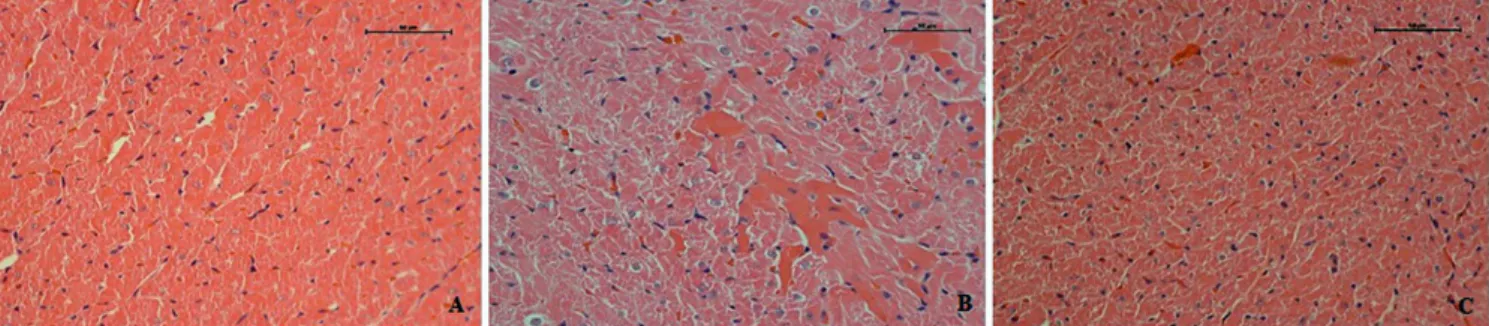
Histopathology of heart tissue
Animals in the RLNAC group presented signiicantly
lower myocardial damage when compared with the RL group (score 1 (1-2) vs. 2.5 (2-5), P=0.049), as well as for edema scores (score 0 (0-1) vs. 2 (1-2), P=0.016).There were no dif-ferences on edema scores between the RLNAC groups and the control group (P=0.935) (Figure 4 A-C).
The evaluation of myocardial inlammatory iniltrate
showed similarities between the three groups (P=0.427).
Fig. 2 - Interleukin 6 expression in cardiac tissue for the control group (CG), Ringer Lactate group (RL) and Ringer lactate with N-acetylcysteine group (RLNAC).
* Signiicant difference when compared to CG, (P<0.05) .
Fig. 4 - Photomicrographs of heart tissue stained with HE of animals subjected to hemorrhagic shock and luid resuscitation with Ringer’s lactate (RL Group, photomicrograph B) or Ringer Lactate associated with N-acetylcysteine (RLNAC Group, photomicrography C), compared to a control group (Group GC, A photomicrograph). It is observed in A, normal myocardium; B, myocardial damage by the presence of cardiomyocyte hipereosinophilic (more intense pink); C, myocardial preserved.
Fig. 3 - Interleukin 10 expression in cardiac tissue for the control group (CG), Ringer Lactate group (RL) and Ringer lactate with N-acetylcysteine group (RLNAC).
DISCUSSION
The results suggest that the NAC plays a promising role
in the pharmacological therapy combined with luid replace -ment in treating hemorrhagic shock, reducing tissue damage, edema, and oxidative stress on the cardiac tissue. To the
ex-tent of our knowledge, this is the irst study that assessed the
NAC effect on heart injury in a controlled hemorrhagic shock model in rats.
With regard to biochemistry data, the lactate – an
import-ant tissue stress predictor – presented a signiicimport-ant increase
during the shock, followed by normalization after volume
reanimation, although without NAC’s intervening. Neverthe -less, the treatment with NAC reduced the potassium levels.
After the beginning of the ischemia that follows the shock, the oxidative phosphorylation is exhausted and the anaerobic metabolism becomes the primary source of ATP
production. Such break down in the cell’s energetic condi -tion leads to an accumula-tion of extra-cell potassium. The mechanism that causes potassium accumulation is not fully explained. The Na-K pump is inhibited in ischemic muscle
cells models, contributing to reduce the K inlux parallel to
ATP-sensitive potassium channels, and it may be the main
mechanism through which potassium eflux increases during
muscle cell ischemia[20].
In an experimental study assessing secondary systemic changes to a prolonged hemorrhage hypertension condition, Torres et al.[21] noted that the potassium increase was
relat-ed to mortality and could explain sudden and early death of some animals during the experiment. While evaluating the role potassium plays as a marker of tissue hypoxia in an ex-perimental model, Rocha Filho et al.[20] noted that the increase
in potassium serum levels complied with hemodynamic
de-terioration, inding a strong correlation between potassium
and lactate levels. NAC, by acting on microcirculation and improving tissue perfusion, may take part in potassium wash-out restoring the aerobic metabolism. However, an in-depth
evaluation is necessary to clarify whether this indings may be ascribed or not to the NAC’S protection role. No data have
been found in literature to corroborate such fact.
In this study, it was noted that the myocardium damage and edema induced by hemorrhagic shock were lessened by volume replacement reanimation and NAC. Although the hemorrhagic shock was maintained for 60 minutes, no
leu-cocitary iniltrated in the cardiac tissue was noticed. Such
results agree with the studies performed by Meurs et al.[22],
who evaluated the neutrophil recruitment in several organs in hemorrhagic shock protocols. The authors pointed out that, in the heart, the early expression of adhesion molecules in the microvascular bed was not accompanied by the leucoci-tary recruitment, different from lungs, liver, and kidneys, in which the expression of adhesion molecules was accompa-nied by an expressive leucocyte migration to tissues.
However, in our study, the TBARS dosage in the cardiac tissue at the end of the stabilization after volume reanimation
presented signiicant increases, describing the lipid peroxida -tion injury, which was attenuated by NAC.
NAC effects on IR injuries were dose-dependent. While studying lung pre-conditioning with different doses of NAC
to prevent IR injury after liver injury by relow, Weinbroum
et al.[17] noted that the 100 mg/kg dose attenuated the liver
injury but not the lung one. High doses, such as 225 mg/kg, could imply a suppression of the properties that protect mac-rophages and monocytes residing in lungs, resulting in a de-crease in lungs defense. The authors have concluded that the 150 mg/kg dose was more effective to reduce accumulation of xanthine oxidase in the liver tissue, reducing the tissue damages caused by ROS.
Although this study shows the protecting effect of NAC on the oxidative stress in cardiac tissue, and that there is a positive correlation between oxidative stress and increase in
the inlammatory cytokines, it did not show tissue reduction of pro-inlammatory IL-6.
Experimental studies have shown that the expression of the ribonucleic acid messenger of IL-6 (RNAm IL-6) is in-creased based on hypoxia conditions, mainly in the lungs, liver, and intestines of rats submitted to hemorrhage, induc-ing the cardiomyocytes to produce IL-6.Kupffer cells are the most important producers of systemic IL-6 after the shock[23].
Such increase in the genic expression and IL-6 levels in the cardiomyocytes occurs mainly two hours after the hemor-rhagic shock has begun and is correlated to the cardiac dys-function[24].
The mechanism whereby IL-6 promotes cardiac dysfunc-tion has not been completely explained. Studies[24,25] suggest
that IL-6 could act in activating the κB (NF- κB) nuclear fac
-tor that, in turn, would activate the transcription of inlamma -tory cytokines, chemotaxins, and adhesion molecules, nota-bly the ICAM-1 in the heart. Such cascade of events would favor neutrophils adhesion and migration processes through the endothelial barrier to the interstitial space and parenchy-matous tissue, with consequent myocardial damage.
Despite the increased levels of IL-6 noted in the hearts of both groups submitted to hemorrhagic shock, there was
no difference in the scores for leucocitary iniltration for all
study groups, including the GC group. The experimental pro-tocol follow-up of this study is considered short to be able
to verify myocardium iniltrate, because the increase in in
-terleukins dosages takes place before inlammatory cells are
present in the tissue.
In this study, we noted that the shock protocol activated
the inlammatory cascades with signiicant increase of IL-6
and IL-10; nevertheless, there was no interference in the modulation with NAC in reducing IL-6 and increasing the expression of the IL-10.Mukherjee et al.[26] reported that the
increased plasma dosage of IL-10 in neonatal rats after two hours of induced septic shock. However, the authors state that, after 4 hours from the beginning of the experiment, the serum levels of IL-6 and IL-10 were similar in the groups, showing that the effect of the administration of NAC on interleukins expression is time-dependent. Therefore, they suggested once again that longer experimental protocols are needed to elucidate the effect of NAC on the expression of interleukins in hemorrhagic shock.
CONCLUSION
NAC showed a protective role in the cardiac tissue of rats submitted to hemorrhagic shock, mainly in lessening oxidative stress and histologic injury. Nevertheless, new studies must be performed that should consider the use of larger NAC doses associated with longer observation protocols in order to allow analyzing data regarding the late stage of the shock.
Authors’ roles & responsibilities
LDOF Analysis and/or interpretation of data, inal approval of the manuscript, design and study design, operations and/or ex-periments conduct
KRS Final approval of the manuscript, conception and design of the study, operations and/or experiments conduct, manu-script writing or critical review of its content
PFS Analysis and/or interpretation of data, inal approval of the manuscript, study design, manuscript writing or critical re-view of its content
MKK Analysis and/or interpretation of data, statistical analysis, i -nal approval of the manuscript, manuscript writing or critical review of its content
SMS Analysis and/or interpretation of data, inal approval of the manuscript, operations and /or experiments conduct EFSM Analysis and/or interpretation of data, inal approval of the
manuscript, study design, manuscript writing or critical re-view of its content
REFERENCES
1. de Knegt C, Meylaerts SA, Leenen LP. Applicability of the trimodal distribution of trauma deaths in a Level I trauma centre in the Netherlands with a population of mainly blunt trauma. Injury. 2008;39(9):993-1000.
2. Anaya-Prado R, Toledo-Pereyra LH. The molecular events underlying ischemia/reperfusion injury. Transplant Proc. 2002;34(7):2518-9.
3. Rushing GD, Britt LD. Reperfusion injury after hemorrhage: a collective review. Ann Surg. 2008;247(6):929-37.
4. Lefèvre G, Beljean-Leymarie M, Beyerle F, Bonnefont-Rousselot D, Cristol JP, Thérond P, et al. Evaluation of lipid peroxidation by measuring thiobarbituric acid reactive substances. Ann Biol Clin (Paris). 1998;56(3):305-19.
5. Zweier JL, Talukder MA. The role of oxidants and free radicals in reperfusion injury. Cardiovasc Res. 2006;70(2):181-90.
6. Garlid AO, Jaburek M, Jacobs JP, Garlid KD. Mitochondrial reactive oxygen species: which ROS signals cardioprotection? Am J Physiol Heart Circ Physiol. 2013;305(7):H960-8.
7. Vinten-Johansen J. Involvement of neutrophils in the pathogenesis of lethal myocardial reperfusion injury. Cardiovasc Res. 2004;61(3):481-97.
8. Santry HP, Alam HB. Fluid resuscitation: past, present, and the future. Shock. 2010;33(3):229-41.
9. Sochman J. N-acetylcysteine in acute cardiology: 10 years later: what do we know and what would we like to know?! J Am Coll Cardiol. 2002;39(9):1422-8.
10. Cailleret M, Amadou A, Andrieu-Abadie N, Nawrocki A, Adamy C, Ait-Mamar B, et al. N-acetylcysteine prevents the deleterious effect of tumor necrosis factor-(alpha) on calcium transients and contraction in adult rat cardiomyocytes. Circulation. 2004;109(3):406-11.
11. Glantzounis GK, Rocks SA, Sheth H, Knight I, Salacinski HJ, Davidson BR, et al. Formation and role of plasma S-nitrosothiols in liver ischemia-reperfusion injury. Free Radic Biol Med. 2007;42(6):882-92.
12. Saad KR, Saad PF, Dantas Filho L, Brito JM, Koike MK, Zanoni FL, et al. Pulmonary impact of N-acetylcysteine in controlled hemorrhagic shock model in rats. J Surg Res. 2013;182(1):108-15.
13. Saad PF, Saad KR, Oliveira Filho LD, Ferreira SG, Koike MK, Montero EF. Effect of N-acetylcysteine on pulmonary cell death in a controlled hemorrhagic shock model in rats. Acta Cir Bras. 2012;27(8):561-5.
14. Portella AO, Montero EF, Poli de Figueiredo LF, Bueno AS, Thurow AA, Rodrigues FG. Effects of N-acetylcysteine in hepatic ischemia-reperfusion injury during hemorrhagic shock. Transplant Proc. 2004;36(4):846-8.
15. Montero EF, Abrahão MS, Koike MK, Manna MC, Ramalho CE. Intestinal ischemia and reperfusion injury in growing rats: hypothermia and N-acetylcysteine modulation. Microsurgery. 2003;23(5):517-21.
17. Weinbroum AA, Kluger Y, Ben Abraham R, Shapira I, Karchevski E, Rudick V. Lung preconditioning with N-acetyl-L-cysteine
prevents reperfusion injury after liver no low-relow: a
dose-response study. Transplantation. 2001;71(2):300-6.
18. Shimizu MH, Coimbra TM, de Araujo M, Menezes LF, Seguro AC. N-acetylcysteine attenuates the progression of chronic renal failure. Kidney Int. 2005;68(5):2208-17.
19. Zingarelli B, Salzman AL, Szabó C. Genetic disruption of poly (ADP-ribose) synthetase inhibits the expression of P-selectin and intercellular adhesion molecule-1 in myocardial ischemia/ reperfusion injury. Circ Res. 1998;83(1):85-94.
20. Rocha Filho JA, Nani RS, D'Albuquerque LA, Malbouisson LM, Carmona MJ, Rocha-E-Silva M, et al. Potassium in hemorrhagic shock: a potential marker of tissue hypoxia. J Trauma. 2010;68(6):1335-41.
21. Torres LN, Torres Filho IP, Barbee RW, Tiba MH, Ward KR, Pittman RN. Systemic responses to prolonged hemorrhagic hypotension. Am J Physiol Heart Circ Physiol. 2004;286(5):H1811-20.
22. van Meurs M, Wulfert FM, Knol AJ, De Haes A, Houwertjes M,
Aarts LP, et al. Early organ-speciic endothelial activation during
hemorrhagic shock and resuscitation. Shock. 2008;29(2):291-9.
23. Yang S, Hu S, Choudhry MA, Rue LW 3rd, Bland KI, Chaudry IH. Anti-rat soluble IL-6 receptor antibody down-regulates cardiac IL-6 and improves cardiac function following trauma-hemorrhage. J Mol Cell Cardiol. 2007;42(3):620-30.
24. Yang S, Zheng R, Hu S, Ma Y, Choudhry MA, Messina JL, et al. Mechanism of cardiac depression after trauma-hemorrhage: increased cardiomyocyte IL-6 and effect of sex steroids on IL-6 regulation and cardiac function. Am J Physiol Heart Circ Physiol. 2004;287(5):H2183-91.
25. Yang S, Hu S, Hsieh YC, Choudhry MA, Rue LW 3rd, Bland KI, et al. Mechanism of IL-6-mediated cardiac dysfunction following trauma-hemorrhage. J Mol Cell Cardiol. 2006;40(4):570-9.