Instituto do Coração of the Hospital das Clínicas of the FMUSP Mailing address: José Luiz Dancini - Rua Vitória, 85/51 - Cep 09030-050 Santo André, SP, Brazil - E-mail: [email protected]
Received: 1/15/03 Accepted: 5/26/03
English version by Stela Maris C. e Gandour
Arq Bras Cardiol, volume 82 (nº 3), 243-50, 2004
José Luiz Dancini, Pablo Maria Alberto Pomerantzeff, Guilherme Sobreira Spina, Mírian Magalhães Pardi, Maria Clementina Pinto Giorgi, Roney Orismar Sampaio, Max Grinberg, Sérgio Almeida de Oliveira
São Paulo, SP - Brazil
Valve Replacement with Chordal Preservation and
Valvuloplasty for Chronic Mitral Insufficiency
After the report of favorable results with mitral valvu-loplasty for patients with chronic mitral insufficiency, mitral valve replacement with preservation of the chordae tendi-neae was revised by David et al 1 and Hetzer et al 2 in 1983.
These authors reported convincing clinical evidence favo-ring the maintenance of the annulopapillary continuity, which had already been demonstrated in the pioneering study by Lillehei et al 3 in 1964. The following years
brought clinical and experimental evidence supporting this concept and spreading the recommendation not to excise all chordae tendineae in mitral valve replacement, which raised the following question: which type of chordal preservation should be used, of both leaflets or of only the posterior leaflet? Hannein et al 4 and Straub et al 5 reported no
signifi-cant difference in ventricular performance between these 2 groups of patients. On the other hand, Hassouna and Elma-halawi 6 and Yun et al 7 reported better left ventricular
sys-tolic performance and a lower mortality rate in the group un-dergoing chordal preservation of both leaflets. As a result, many surgeons continue to express their preoccupations with the demand for higher technical complexity, longer sur-gical time, the potential interference with the mobility of a leaflet or of a prosthetic occluding disk, the eventual need for underestimating the diameter of a prosthesis for the mi-tral ring, and the possibility of creating an obstruction in the left ventricular outflow tract. The lack of consensus led us to this line of research.
Methods
This study consecutively analyzed 28 symptomatic patients who underwent surgery for isolated chronic mitral regurgitation at the Instituto do Coração of the Hospital das Clínicas of the FMUSP from April 2000 to November 2002. Their ages ranged from 17 to 78 years (mean, 54.1±15.8 years; median, 55 years), and their weight ranged from 43 to 92 kg (mean, 61±13 kg; median, 60 kg). Nineteen (67.9%) were men and 9 (32.1%) were women. All patients consented to the surgery and to take part in the study.
The inclusion criterion was the presence of significant chronic mitral insufficiency. The exclusion criteria were as
Objective - To compare, from the clinical and
labora-tory points of view, 3 groups of patients undergoing surgi-cal treatment for isolated chronic mitral insufficiency. One group underwent valvuloplasty, and the other 2 groups underwent mitral valve replacement with different techni-ques for chordal preservation.
Methods - Twenty-eight patients with a mean age of
54.1 years, no coronary or multivalvular disease, and no reoperation, underwent surgery as follows: 9 underwent valvuloplasty; 10 underwent mitral valve replacement with chordal preservation in both leaflets; and 9 under-went mitral valve replacement with chordal preservation only in the posterior leaflet. Clinical, Doppler echocardio-graphic, and radionuclide ventriculographic assessments were performed until the 6th month of follow-up.
Results - At the end, 88.8% of the patients were in
func-tional class I. One died due to intracranial hemorrhage during anticoagulant treatment. The left ventricular dias-tolic diameter (P<0.0001) and end-diasdias-tolic volume (P<0.0001) decreased in the 3 groups. Only the patients undergoing valvuloplasty had a decrease in systolic diame-ter (P=0.0003) and in end-systolic volume (P=0.0040), with no change in the ejection fraction (P=0.5586). The patients undergoing mitral valve replacement had a similar drop in ejection fraction (P=0.0001 and P=0.0296).
Conclusion - The 3 surgical techniques used provided
clinical improvement. Patients undergoing valvuloplasty had better preservation of ventricular function. No signi-ficant difference was observed in cardiac performance bet-ween the 2 groups undergoing mitral valve replacement with chordal preservation within a 6-month follow-up.
Key words: mitral valve insufficiency, surgery, valvular
follows: patients with significant coronary artery disease (any coronary artery with stenosis > 50%), associated im-portant aortic or tricuspid valve disease, reoperations, and significant mitral stenosis. The surgical indication was deci-ded by the clinical team based on clinical, Doppler echocar-diographic, hemodynamic, and angiographic criteria. Signi-ficant mitral insufficiency was shown in all patients on Dop-pler echocardiography and on left ventriculography, when applicable.
As personal antecedents, we found 5 (17.9%) patients with a history of rheumatic fever, 3 (10.7%) with arterial hy-pertension, 2 (7.1%) with diabetes mellitus, 1 (3.6%) with a history of stroke with no motor sequelae, 1 (3.6%) with a his-tory of infective endocarditis, and 1 (3.6%) with epilepsy.
Physical activity was classified according to the 4 functional classes (FC) of theNew York Heart Association. Only 1 (3.6%) female patient was in FC IV (case 26); the re-maining 27 (96.4%) were in FC III. The postoperative recor-dings were obtained at 3 and 6 months of follow-up. The car-diac rhythm was analyzed on ordinary 12-lead electrocardio-graphy. Five (17.9%) patients had atrial fibrillation and 23 (82.1%) had sinus rhythm. Follow-up recordings were obtai-ned at 3 and 6 months.
Cardiac catheterization was performed only in the preo-perative phase in all patients aged ≥ 40 years (23 patients, 82.1%). Three (10.7%) patients aged < 40 years had already undergone the procedure by the time of inclusion in the study, adding up to 26 patients studied on cardiac catheteri-zation. Right and left chamber pressure recordings were used to compare the groups operated on only in this phase.
Doppler echocardiography was performed with the Philips – ATL device, HDI 3000 model, and the Philips – HP 1500 device (Bothell, WA, USA) with 2.0- and 3.0-megahertz transducers. The images were obtained in parasternal, apical, and subcostal views in several planes. The following measurements were taken: left ventricular systolic diameter (LVSD); left ventricular diastolic diameter (LVDD); left ven-tricular ejection fraction (EF); left venven-tricular end-diastolic volume (EDV); and left ventricular end-systolic volume (ESV). Detection and quantification of mitral reflux were per-formed with color flow mapping obtained on the left paras-ternal and 2- and 4-chamber apical views, and with pulsed-wave Doppler.
All patients underwent radionuclide ventriculography (gated) at rest up to 1 month before surgery. The images were obtained in an ADAC-TranScan gamma-chamber cou-pled to a Unix (SUN) computer model Pegasys. The follo-wing parameters were analyzed: end-diastolic volume (EDV); end-systolic volume (ESV); right ventricular ejection fraction (RVEF); and left ventricular ejection fraction (LVEF) calculated according to the equation: EF = (CD – CS) / CD, where CD and CS refer to the radioactive countings corres-ponding to maximum diastole and systole.
After induction of anesthesia, a thermodilution bal-loon catheter was introduced through puncture of the right internal jugular vein and was advanced as far as its distal impaction in the pulmonary tree. The system was connec-ted to a computer for recording and calculating the follo-wing hemodynamic variables: cardiac index (CI); left
ven-tricular systolic work index (LVSWI); pulmonary vascular resistance index (PVRI); and systemic vascular resistance index (SVRI). The measurements were taken 4 times as fol-lows: IO1 – before median sternotomy; IO2 – prior to hepa-rin administration; IO3 – after protamine sulfate administra-tion; and IO4 – after suture of the sternum.
A transesophageal transducer was positioned for in-traoperative qualitative Doppler echocardiographic assess-ment twice, as follows: during chest opening and after dis-continuation of extracorporeal circulation.
All surgeries were performed through a median sterno-tomy. Extracorporeal circulation was established through cannulation of the ascending aorta and both venae cava se-parately, and disposable membrane oxygenators were used. The left atrium was longitudinally opened. The site of greatest reflux was checked with injection of saline solution inside the left ventricle. The intraoperative decision regarding the choice of valvuloplasty or valvular replacement was made up based on the anatomical status of the mitral valve, which, whenever possible, was repaired. However, when the anatomic conditions indicated the need for prosthetic implan-tation, a list with previous randomization was consulted, and the patient underwent one or the other techniques of chordal preservation (preservation of both leaflets or of only the posterior leaflet). Based on this, the 3 following groups of patients were formed: 1) the valvuloplasty group, comprising the patients undergoing valvuloplasty; 2) the group of both leaflets, comprising the patients undergoing mitral valve replacement according to the technique of Miki et al 8, which
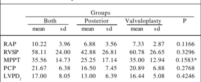
preserves the chordae tendineae of both leaflets; and 3) the posterior leaflet group, comprising the patients undergoing mitral valve replacement with chordal preservation of only the posterior leaflet. The 3 groups were compared in regard to data obtained on cardiac catheterization and Doppler echo-cardiography in the preoperative period (tab. I and II). Frag-ments of the leaflets could be removed for anatomicopatho-logical examination in 27 patients.
Functional class recordings and analysis of the cardiac rhythm were obtained at 3 and 6 months of follow-up, as were transthoracic Doppler echocardiographies, also performed prior to hospital discharge. A second radionuclide ventriculo-graphy at rest was performed 6 months after surgery.
All continuous variables were reported as minimum, maximum, median, mean, and standard deviation values,
Table I - Preoperative cardiac catheterization data
Groups
Both Posterior Valvuloplasty P mean sd mean sd mean sd
RAP 10.22 3.96 6.88 3.56 7.33 2.87 0.1166 RVSP 58.11 24.00 42.88 26.81 60.78 26.65 0.3296 MPPT 35.56 14.73 25.25 17.14 35.00 12.94 0.1583* PCP 21.67 6.38 16.50 7.45 20.89 6.88 0.2768 LVPD2 17.00 8.05 13.00 6.39 16.44 5.08 0.4246
and the categorical variables were reported as absolute and relative frequencies. The hypotheses of equality of the co-variance matrices between the groups and the normal distri-bution of the data were assessed, and the values obtained were compared among the 3 groups of patients by using analysis of variance for data with normal distribution and the Kruskal-Wallis test 9 for data that did not satisfy the
supposition of normality. The Doppler echocardiographic data relating to radionuclide ventriculography and the he-modynamic measurements were compared at several mo-ments in each group, and among the groups, through analy-sis of variance for repeated measures 10. Within this context,
the 3 following hypotheses were tested: interaction, group effect, and effect of time. Qualitative variables were analy-zed using the Fisher exact test 11. A descriptive level (P)
lo-wer than 0.05 was considered statistically significant.
Results
Of the 28 patients operated on, 9 (32.1%) were in the valvuloplasty group as follows: 7 (25%) underwent qua-drangular resection according to the double-Teflon techni-que® 12, and 2 (7.1%) underwent posterior annuloplasty with
a bovine pericardial patch. Ten (35.7%) patients comprised the group of both leaflets, and 9 (32.1%) comprised the pos-terior leaflet group. Three (10.7%) patients underwent the following complementary procedures: 1 (3.6%) patient derwent De Vega tricuspid valve annuloplasty; 1 (3.6%) un-derwent commissurotomy and decalcification of the aortic valve; and another (3.6%) underwent cerclage of the aortic ring related to the noncoronary leaflet.
The intraoperative transesophageal Doppler echocar-diographic evaluation prior to extracorporeal circulation re-vealed the following findings: rupture of the chordae of the posterior leaflet in 11 (39.3%) patients; lack of coaptation of the leaflets in 8 (28.5%) patients; rupture of the chordae of the anterior leaflet in 5 (17.9%) patients; prolapse of the posterior leaflet in 2 (7.1%) patients; prolapse of the anterior leaflet in 1 (3.6%) patient; and prolapse of both leaflets in 1 (3.6%) patient. No thrombus was detected inside the left atrium or left auricle. The result of valvuloplasty was consi-dered good on transesophageal Doppler echocardiography after extracorporeal circulation in all cases. Neither regurgi-tation through the prosthesis nor obstruction in the left
ventricular outflow tract was observed in any patient in the groups of both leaflets and of the posterior leaflet.
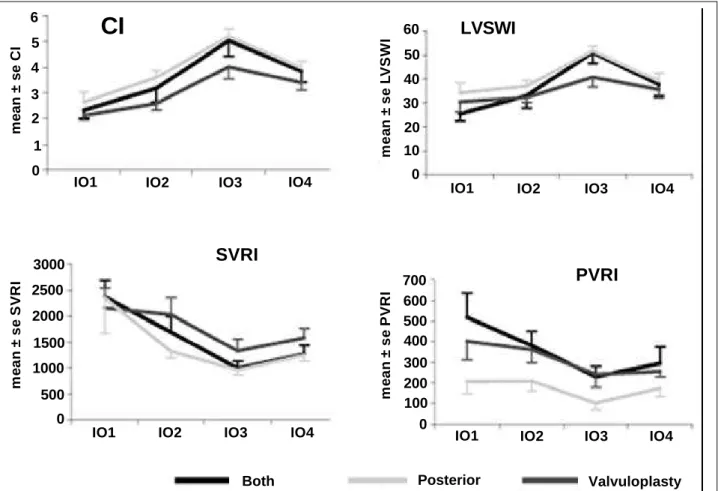
In regard to the hemodynamic variables, an increase in the CI (P < 0.0001) was observed, mainly after completion of the procedure, accompanied by an increase in the LVSWI (P < 0.0001) in the 3 groups. No significant difference was observed among the groups regarding the behavior of SVRI (P = 0.0599) and PVRI (P =0.0644), which decreased after surgery (chart I).
No in-hospital mortality was observed. Seven (25%) patients had the following in-hospital complications: acute atrial fibrillation in 2 (7.1%) patients; pericardial effusion in 2 (7.1%); phrenic paresis in 1 (3.6%); transient total atrioven-tricular block in 1 (3.6%); and infective endocarditis in 1 (3.6%) (chart II). The 2 patients with acute atrial fibrillation were pharmacologically reverted, and none of them had had it before. The 2 patients with pericardial effusion underwent surgical drainage with no recurrence. The patient with total atrioventricular block was maintained with an external pace-maker until the fourth postoperative day, when sinus rhythm was spontaneously resumed. The patient with clini-cal findings of endocarditis in the prosthesis belonged to the group of the posterior leaflet and underwent replace-ment of the mitral bioprosthesis for another of the same type on the 30th postoperative day. After the second surgery, the evolution was satisfactory, and the patient was dischar-ged on the 14th postoperative day. Two (7.1%) patients had complications 2 months after surgery. The complications were of the hemorrhagic type in both, enterorrhagia in one and fatal stroke in another (case 17). The latter patient be-longed to the group of both leaflets and was discharged from the hospital with a prescription of oral anticoagulant (dicoumarin) in a total dose because he had chronic atrial fibrillation.
Table III shows comparative data of the 3 groups re-garding the duration of extracorporeal circulation, duration of aortic cross-clamping, intensive care unit length of stay, mediastinal drainage volume, duration of hospitalization, and number of patients using vasoactive drugs for more than 24 hours. The patients undergoing valvuloplasty had a shorter time of extracorporeal circulation (P = 0.0016) and of aortic cross-clamping (P < 0.0001). No statistically signi-ficant difference was observed between the 2 groups under-going mitral valve replacement with chordal preservation.
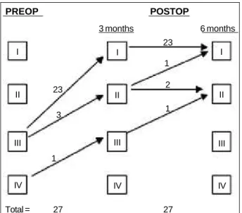
Three months after surgery, 23 (85.2%) of the 27 patients studied were in FC I; 3 (11.1%) in FC II; and only 1 (3.7%) in FC III. Six months after surgery, 24 (88.9%) patients were in FC I, and 3 (11.1%) were in FC II. The evolu-tion of all patients according to funcevolu-tional class is shown in chart III. No significant difference in functional class distri-bution was observed in the groups studied 3 months (P = 1.0000) and 6 months (P = 0.7508) after surgery. When assessing cardiac rhythm, 4 of the 5 patients with atrial fibril-lation in the preoperative period remained with arrhythmia 3 and 6 months after surgery, because they had chronic atrial fibrillation. No thromboembolism was observed up to the 6th month of follow-up.
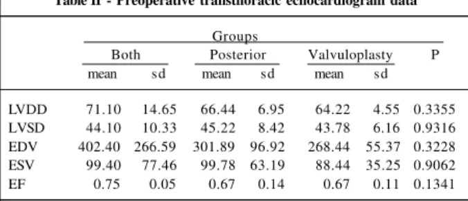
In regard to the evolution of Doppler echocardiogra-phic variables, the LVDD and EDV curves were similar in the Table II - Preoperative transthoracic echocardiogram data
Groups
Both Posterior Valvuloplasty P mean sd mean sd mean sd
LVDD 71.10 14.65 66.44 6.95 64.22 4.55 0.3355 LVSD 44.10 10.33 45.22 8.42 43.78 6.16 0.9316 EDV 402.40 266.59 301.89 96.92 268.44 55.37 0.3228 ESV 99.40 77.46 99.78 63.19 88.44 35.25 0.9062 EF 0.75 0.05 0.67 0.14 0.67 0.11 0.1341
P = descriptive level of the analysis of variance; LVDD = left ventricular diastolic diameter (mm); LVSD = left ventricular systolic diameter (mm); EDV = end-diastolic volume of the LV (cm3); ESV = end-systolic volume of the LV (cm3);
3 groups, with a significant decrease already in the in-hospi-tal phase, and a progressive and lower intensity reduction up to the 6th month of follow-up. However, only the pa-tients in the valvuloplasty group had a significant drop in LVSD and ESV (P = 0.0017 and P = 0.0101, respectively). The ejection fraction curves were different in the 3 groups stu-died: the patients in the group of both leaflets had a signifi-cant drop already in the in-hospital phase (P < 0.0001), with a tendency towards recovery in the following phases; the
patients in the posterior leaflet group had the greatest drop at 3 months after surgery; and the patients in the valvulo-plasty group had no significant difference at all phases analyzed (P = 0.5586). Chart IV shows the evolutionary cur-ves of EDV, ESV, and EF in each group.
The radionuclide ventriculography also showed a de-crease in the EDV in the 3 groups. However, only the patients
Complications N (%) Cases
In-hospital
Acute AF 2 7.1 13 e 27
Pericardial effusion 2 7.1 5 e 21
Phrenic paresis 1 3.6 9
Transient TAVB 1 3.6 12
Infective endocarditis 1 3.6 10
Total 7 25
Late
Enterorrhagia 1 3.6 9
Hemorrhagic stroke 1 3.6 17
Total 2 7.1
Chart II - In-hospital and late complications.
Table III - Comparative data of the in-hospital period in each group
Groups
Both Posterior Valvuloplasty P
mean sd mean sd mean sd
TECC 99.50 20.34 90.00 18.37 73.56 8.71 0.0016 TCCAo 79.00 16.01 72.11 16.34 47.33 7.52 <0.0001 TICU 2.40 1.07 2.11 0.33 2.44 1.01 0.8091* TINT 13.30 6.99 13.56 4.75 11.89 2.20 0.5711 Drain 564.00 188.57 702.22 220.72 608.89 316.80 0.3586 VAD 3 patients 2 patients 1 patients 0.8452*
P = descriptive level of the analysis of variance; P* = descriptive level of the Kruskal-Wallis test; TECC = time of extracorporeal circulation (minutes); TCCAo = time of cross-clamping of the aorta (minutes); TICU = time of ICU (days); THOSP = time of hospitalization (days); drain = mediastinal drainage volume (milliliters); VAD = need for vasoactive drugs for more than 24 hours.
m
e
a
n
±
s
e
C
I
6
5
4
3
2
1
0
IO1 IO2 IO3 IO4
CI
m
e
a
n
±
s
e
L
V
S
W
I 60 50
40
30
20
10
0
IO1 IO2 IO3 IO4
LVSWI
m
e
a
n
±
s
e
S
V
R
I
3000
2500
2000
1500
1000
500
0
IO1 IO2 IO3 IO4
SVRI
m
e
a
n
±
s
e
P
V
R
I 700 600 500 400
300 200 100 0
IO1 IO2 IO3 IO4
PVRI
Both Posterior Valvuloplasty
CI = cardiac index (L/min/m2); LVSWI = left ventricular systolic work index (g/L/m2); SVRI = systemic vascular resistance index (dyne/cm5/m2); PVRI = pulmonary vascular resistance index (dyne/cm5/m2)
.
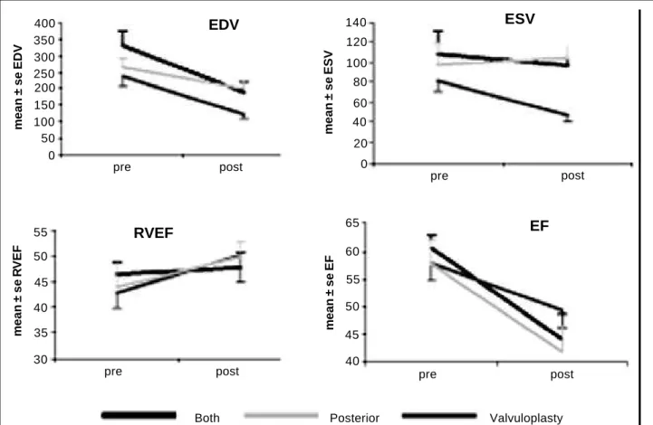
in the valvuloplasty group had a significant drop in ESV, reaching a 42% reduction at 6 months after surgery as com-pared with the preoperative value (P = 0.0012). Although a mild increase in absolute values occurred, no statistically significant changes were observed in RVEF (P = 0.0675). In regard to ejection fraction, that examination showed a drop in all groups (P < 0.0001), with no statistically significant difference among them (chart V).
Table IV shows the distribution of the anatomicopa-thological diagnosis in the 3 groups studied. No statistical-ly significant difference was observed in the incidence of myxomatous degeneration, rheumatic disease, and endo-carditis (P = 0.9416). In 1 patient, whose material was not collected for anatomicopathological study, the gross ap-pearance was that of myxomatous degeneration.
Discussion
Mitral insufficiency is a valvular heart disease that causes complex hemodynamic changes. In chronic mitral in-sufficiency, the left ventricle initially dilates, and, subse-quently, undergoes hypertrophy, recovering the relation between ventricular mass and end-diastolic volume. The ve-locity of the increase in the end-diastolic volume depends on the importance and nature of the regurgitation. The end-systolic volume is also increased, but has a considerable in-dividual variation. The effect of regurgitation on hemodyna-mic balance depends on the intensity of regurgitation and left atrial compliance. Significant insufficiency, most of the time, is oligosymptomatic for a long period and may have a high incidence of ventricular dysfunction at the time of sur-gery, which may affect postoperative survival 13.
After correction of mitral insufficiency, both through valvuloplasty and prosthesis implantation, the first conse-quence was a reduction in the left ventricular diameter and end-diastolic volume, due to the elimination of the blood vo-lume that circulated only between the left atrium and the left ventricle. A maximal reduction occurred already in the first
postoperative week, detected on transthoracic Doppler echocardiography. Although the drop remained significant for up to 6 months, in this period its magnitude was small. The radionuclide ventriculography also detected a reduc-tion in the end-diastolic volume at 6 months, with significan-tly lower values in the valvuloplasty group. These data are in accordance with the improvement in clinical findings and in functional class observed in the postoperative period.
Although removal of the regurgitating volume has a direct repercussion in the left ventricular end-diastolic me, the same does not occur in regard to end-systolic volu-me, which is more dependent on the degree of preoperative myocardial contractility and afterload. It was not observed to change in patients undergoing mitral valve replacement with chordal preservation, which led to a reduction in ejec-tion fracejec-tion. Only the patients in the valvuloplasty group had a significant drop in end-systolic volume, with no re-duction in the ejection fraction. In accordance with these results, Corin et al 14 reported reductions in the EDV, both in
patients undergoing valvuloplasty and in those undergoing mitral valve replacement with chordal preservation. Rozich et al 15 reported a drop in end-systolic volume with
mainte-nance of the annulopapillary continuity. In this group, we solely observed a tendency towards such behavior. Our data do not agree with those by Yun et al 16, who observed
an increase in end-systolic volume in the group undergoing chordal preservation of the posterior leaflet, and a reduction in that with chordal preservation in both leaflets. Usually, this effect is observed only in the postoperative period of mitral valvuloplasty, as in our case series.
The predictive value of the preoperative ejection frac-tion in late survival is consistent with some studies and has proved to be a strong indicator of left ventricular function in the postoperative period. The drop in ejection fraction below 0.55 in significant mitral insufficiency may be a clear indication of heart failure 17. According to Depace et al 18,
only the end-systolic diameter has an independent predicti-ve value for the postoperatipredicti-ve evolution in mitral valpredicti-ve re-placement with chordal preservation. Six patients had left ventricular end-systolic diameter > 55 mm in the preoperati-ve period. Of them, 3 had ejection fraction < 0.55 and only 2 improved their indices in the postoperative period. Al-though not using indices independent of the load, our re-sults suggest that ventricular performance drops immedia-tely after mitral valve replacement with chordal preservation (in both groups), which does not occur with the patients undergoing valvuloplasty.
Data about right ventricular ejection fraction observed on radionuclide ventriculography agree with those repor-ted by Le Tourneau et al 19 about a drop in the ejection
frac-tion and unaltered right ventricular ejecfrac-tion fracfrac-tion in 23 patients undergoing mitral valve replacement with chordal preservation of the posterior leaflet.
The myxomatous degeneration severely affects the mechanical properties of the mitral chordae. Barber et al 20
mechanically tested segments of normal and myxomatous chordae, and reported that the myxomatous chordae were defective with half the load of the normal chordae, explai-ning the frequent rupture of myxomatous valves. The fin-dings suggest that the procedures of chordal preservation
PREOP POSTOP
3 months 6 months
I
II
III
IV
23
3
1
2
Total = 27
I
II
III
IV
23
1
1
27
I
II
III
IV
m
e
a
n
±
s
e
E
D
V
600
500 400
300
200
100
0
EDV
pre PO1 PO2 PO3
m
e
a
n
±
s
e
E
S
V 140
120 100 80 60 40 20 0
pre PO1 PO2 PO3
ESV
m
e
a
n
±
s
e
E
F
0.80 0.75 0.70 0.65 0.60 0.55 0.50 0.45 0.40
pre PO1 PO2 PO3
EF
Both Posterior Valvuloplasty
EDV = end-diastolic volume (mm3); ESV = end-systolic volume (mm3); EF = LV ejection fraction
Chart IV - Evolutionary curves of EDV, ESV, and EF on echocardiography.
should be carefully performed, because they are abnormal and have impaired mechanical strength. In this study, only 6 of the 21 valves with the anatomicopathological diagnosis of myxomatous degeneration could be preserved, due to their intraoperative anatomical characteristics; the remai-ning underwent prosthesis implantation.
Of the 9 patients undergoing valvuloplasty, 7 under-went quadrangular resection and 2 underunder-went posterior an-nuloplasty with bovine pericardial patch. These procedures are justified by the fact that they correspond to 61.3% of all valvuloplasty procedures at the institution in the 17-year study period, according to Pomerantzeff et al 21.
Reports on dynamicor permanent obstruction of the left ventricular outflow tract emphasize that it is more fre-quently caused by overestimation of the size of the prosthe-sis or unnecessary retention of a large portion of the anteri-or leaflet that cannot be plicated. Prostheses of the bascula-te disk are susceptible to obstruction of the free movement of the disk by chordal remnants, and it is important to avoid locking of the disk caused by an eventual elongated or re-dundant chord, when techniques for chordal preservation are used 22. In addition, locking of the disks after rupture of
preserved chordae relating to the posterior leaflet has been
reported 23. The experience of the surgeons with
double-valve (aortic and mitral) replacement procedures enabled the visualization of the left ventricular cavity through aortic opening and the observation of the relation of the retained structures of the anterior leaflet to the left ventricle outflow tract. The obstruction becomes a problem only when an ex-cessive amount of tissue of the anterior leaflet remains in the ventricular side of the mitral ring. The posterior leaflet, almost always flexible, may be completely preserved, as may the chordae tendineae.
fi-m
e
a
n
±
s
e
E
D
V
400 350 300 250 200 150 100 50 0
pre post
EDV
m
e
a
n
±
s
e
E
S
V
140 120 100 80 60 40 20 0
pre post
ESV
m
e
a
n
±
s
e
R
V
E
F
55 50 45 40 35 30
pre post
RVEF
m
e
a
n
±
s
e
E
F
65
60
55
50
45 40
pre post
EF
Both Posterior Valvuloplasty
EDV = end-diastolic volume (mm3); ESV = end-systolic volume (mm3); RVEF = right ventricular ejection fraction; EF = LV ejection fraction.
Chart V - Evolutionary curves of EDV, ESV, RVEF, and EF on gated radionuclide ventriculography.
xation of the groups of chordae relating to the anterior leaflet in each respective commissure and through the re-section of an eventual excess of tissue in the posterior leaflet; 4) to reinforce possible areas of dehiscences created by resections and plications, the use of additional “U” or “8” stitches anchored in Teflon® is recommended in the
suspicious areas; 5) we emphasize the value of intraoperati-ve transesophageal Doppler echocardiography, which faci-litates the identification and quantification of an occasional obstruction still in a phase in which stitch placement could be reviewed and modified. Taking this into consideration and that the left ventricular outflow tract gradient was not recorded, the qualitative analysis required no reinterven-tion in any patient in the series.
A surgical mortality ranging from zero to 9.5% for the
groups undergoing mitral valve replacement with chordal preservation has been reported 24 ,25. In our study, the only
patient who died belonged to the group of both leaflets and the cause of death was not directly related to the heart; our mortality rate, however, was 5.3% (1/19 patients with chor-dal preservation), similar to the overall mean. Six patients re-quired vasoactive drugs for more than 24 hours after surge-ry; none, however, had prolonged low output syndrome.
In conclusion, considering physical activity, the 3 te-chniques of surgical correction provided an improvement in patients, and better preservation of ventricular function was observed in the patients undergoing valvuloplasty. No significant difference was observed in the laboratory results in the 2 groups of patients undergoing valve repla-cement with chordal preservation up to the sixth month of follow-up.
Acknowledgments
We thank Drs. Eduardo G. Rossi, Filomena R.B.G. Ga-las, and Paula H. Tamanaka for their support; Mariana Curi, Alessandra D.J.C. Almeida, Élida C. Rezende, Monica U.J. Kondo, Adriana M. Quadros, and Sergio Spezzia for their te-chnical participation; and Marília, Renato, and Elvira Maria P.A. Dancini for their contribution.
Table IV - Distribution of the anatomicopathological diagnosis
Groups
Diagnosis Both Posterior Valvuloplasty Total (%)
Myxomatous degeneration 8 7 6 21 (77.8)
Rheumatic disease 1 2 2 5 (18.5)
Infective endocarditis 1 - - 1 (3.7)
Total 10 9 8 27 (100)
1. David TE, Uden DE, Strauss HD. The importance of the mitral apparatus in left ven-tricular function after correction of mitral regurgitation. Circulation 1983; 68: 76-82. 2. Hetzer R, Bougiokas G, Franz M, Borst HG. Mitral valve replacement with pre-servation of papillary muscles and chordae tendinae – revival of a seemingly for-gotten concept. Thorac Cardiovasc Surg 1983; 31: 291-6.
3. Lillehei CW, Levy MJ, Bonnabeau RC Jr. Mitral valve replacement with preserva-tion of papillary muscles and chordae tendinae. J Thorac Cardiovasc Surg 1964; 47: 532-43.
4. Hennein HA, Swain JA, Mcintosh CL, Bonow RO, Stone CD, Clark RE. Compa-rative assessment of chordal preservation versus chordal ressection during mi-tral valve replacement. J Thorac Cardiovasc Surg 1990; 99: 828-37. 5. Straub U, Huwer H, Kalweit G, Volkmer I, Gams E. Improved regional left
ventri-cular performance in mitral valve replacement with orthotopic refixation of the an-terior mitral leaflet. J Heart Valve Dis 1997; 6: 395-403.
6. Hassouna A, Elmahalawi N. Valve replacement in rheumatic mitral incompetence: to-tal versus posterior chordal preservation. Thorac Cardiovasc Surg 1998; 6: 133-8. 7. Yun KL, Sintek CF, Miller DC, et al. Randomized trial comparing partial versus complete chordal-sparing mitral valve replacement: effects on left ventricular vo-lume and function. J Thorac Cardiovasc Surg 2002; 123: 707-14.
8. Miki S, Kusuhara K, Ueda Y, Komeda M, Okita Y, Tahata T. Mitral valve replace-ment with preservation of chordae tendineae and papillary muscles. Ann Thorac Surg 1988; 45: 28-34.
9. Rosner B. Fundamentals of Biostatistics, PWS Publishers, Massachusetts, 2nd Edition, 1986: 467-72.
10. Timm NH. Multivariate Analysis with Applications in Educations and Psycho-logy. Belmont: Wadsworth Publishing Company, 1975: 444-71.
11. Rosner B. Fundamentals of Biostatistics, PWS Publishers, Massachusetts, 2nd Edition, 1986: 326-32.
12. Pomerantzeff PM, Brandao CM, Souza LR et al. Posterior mitral leaflet repair with a simple segmental annulus support: the ‘double-Teflon technique’. J Heart Valve Dis 2002; 11(2): 160-4.
13. Braunwald E. Valvular heart disease. In: Heart Disease: A Textbook of Cardiovas-cular Medicine, vol 2, 5th ed, Braunwald E, ed, WB Saunders and Co, Philadel-phia, 1997: 1017-29.
References
14. Corin WJ, Monrad ES, Murakami T, Nonogi H, Hess OM, Krayenbuehl H. The re-lationship of afterload to ejection performance in chronic mitral regurgitation. Circulation 1987; 76: 59-67.
15. Rozich JD, Carabello BA, Usher BW, Kratz JM, Bell AE, Zile MR. Mitral valve re-placement with and without chordal preservation in patients with chronic mi-tral regurgitation. Mechanisms for differences in postoperative ejection perfor-mance. Circulation 1992; 86: 1718-26.
16. Yun KL, Sintek CF, Miller DC, et al. Randomized trial of partial versus complete chordal preservation methods of mitral valve replacement. A preliminary report. Circulation 1999; 100 (Suppl. II): II 90-4.
17. Michel PL. Mitral insufficiency. In: Textbook of Acquired Heart Valve Disease, 1st ed, Acar J, Bodnar E eds, ICR Pub, London, 1995: 403-32.
18. Depace NL, Ren JG, Iskandrian AS, Kolter MN, Hakki AH, Segal BL. Correla-tion of echocardiographic wall stress and left ventricular pressure and funcCorrela-tion in aortic stenosis. Circulation 1983; 67: 854-9.
19. Le Tourneau T, Grandmougin D, Foucher C et al. Anterior chordal transection im-pairs not only regional left ventricular function but also regional right ventricu-lar function in mitral regurgitation. Circulation 2001; 104 (12 Suppl 1): I 41-6. 20. Barber JE, Ratliff NB, Cosgrove DMIii, Griffin BP, Vesely I. Myxomatous mitral
valve chordae: mechanical properties. J Heart Valve Dis 2001; 10: 320-4. 21. Pomerantzeff PM, Brandao CM, Faber CN et al. Plástica da valva mitral:
resulta-dos aos 17 anos de experiência. Rev Bras Cir Cardiovasc 1999; 14: 185-90. 22. Dottori V, Barberis L, Lijoi A et al. Initial experience with mitral valve replacement
with total preservation of both valve leaflets. Texas Heart Inst. J 1994; 21: 215-9. 23. Mok CK, Cheung DLC, Chiu CSW et al. An unusual lethal complication of
pre-servation of chordae tendineae in mitral valve replacement. J Thorac Cardiovasc Surg 1988; 95: 534.
24. Popovic Z, Barac I, Jovic M, Panic G, Miric M, Bojic M. Ventricular performance following valve replacement for chronic mitral regurgitation: importance of chor-dal preservation. J Cardiovasc Surg 1999; 40: 183-90.