Hospital Universitário Clementino Fraga Filho, Department of Internal Medicine –Neurology, Faculty of Medicine, Universidade Federal do Rio de Janeiro: *Associate Professor, Medical Doctor; **Medical Doctor; ***Bioestatistician; ****Professor, Medical Doctor. Aceite: 29-junho-1998.
Dra. Marleide da Mota Gomes - Caixa Postal 68008 - CCS Bloco K Cidade Universitária - 21944-970 Rio de Janeiro - Brasil.
EMERGENCY PHYSICIANS DIAGNOSIS OF STROKE SUBTYPE
AN ACCURACY STUDY
MARLEIDE DA MOTA GOMES*, MONICA FONSECA COSTA**, CHARLES ANDRÉ*, RAQUEL GOMES***, SÉRGIO A. P. NOVIS****
ABSTRACT - Objective: To evaluate the accuracy of clinical unstructured and structured diagnosis of acute stroke subtypes – cerebral haemorrhage (CH), cerebral infarction (CI), subarachnoid haemorrhage (SAH).
Methods: Sixty consecutive patients with acute stroke admitted to the Emergency Ward of a Brazilian University
Hospital were examined by emergency physicians and computerised tomography (CT). We also compared it (physician’s unstructured diagnosis) to two published clinical scoring systems (structured diagnosis - Guy’s Hospital and Siriraj Hospital) applied to three other populations – regarding the operational characteristics of the tests. Results: In our personal data, among 9 variables that could discriminate CH and CI, three have statistically significant difference (p <0.05): headache (p=0.0002) and vomiting (p=0.02) occurred more frequently in CH patients, but previous stroke in those with CI (p=0.04). Unstructured diagnosis proved valid for SAH, with a +LHR= 39.7; and to a smaller degree for CI (-LHR= 0.1). However, it exhibited low sensitivity for the diagnosis of CH. Structured tests (Guy’s Hospital and Siriraj Hospital) also failed to confidently diagnose stroke subtypes, especially CH. Conclusions: Both clinical diagnosis (made by emergency physicians) and the available diagnostic tests fail to confidently discriminate CH and CI.
KEY WORDS: cerebrovascular disease, diagnosis, medical education.
Diagnóstico dos subtipos de acidente vascular cerebral: um estudo de acurácia com médicos de emergência
RESUMO - Objetivo: Avaliar a acurácia do diagnóstico clínico não estruturado e do estruturado dos subtipos do acidente vascular cerebral (AVC): hemorragia cerebral (HC), infarto cerebral (IC), hemorragia subaracnóide (HSA). Método: Sessenta pacientes com AVC consecutivamente admitidos em emergência de hospital universitário brasileiro foram examinados por médicos da emergência e por tomografia computadorizada (TC). Nós também comparamos isso (diagnóstico clínico não estruturado) com os resultados de dois testes diagnósticos estruturados sobre AVC da literatura (Guy’s Hospital e Siriraj Hospital) aplicados a três outras populações – com atenção às características operacionais dos testes. Resultados: Em nossos dados pessoais, entre nove variáveis que poderiam discriminar HC e IC, três apresentaram diferenças significantes estatisticamente (p<0.05): cefaléia (p=0.0002) e vômito (p=0.02) ocorreram mais frequentemente naqueles com HC, mas AVC prévio naqueles com IC (p=0.04). Diagnóstico não estruturado mostrou-se válido para HSA, com +LHR= 39.7; e em menor grau para IC (-LHR= 0,1). No entanto, ele exibiu baixa sensibilidade para o diagnóstico de HC. Testes estruturados (Guy’s Hospital and Siriraj Hospital) também falharam para o diagnóstico seguro dos subtipos de AVC, especialmente da HC.
Conclusão: O diagnóstico clínico (feito por médicos de emergência) e os instrumentos diagnósticos estudados
não diferenciam de modo confiável HC e IC.
PALAVRAS-CHAVE: doença cerebrovascular, diagnóstico, educação médica.
1-5%2, diagnostic errors being most likely in patients with malignant tumours and subdural bleeding3.
However, diagnosis of stroke subtype is considered less accurate. The distinction between cerebral infarction (CI) and intracerebral haemorrhage (ICH) is now seen as crucial in determining prognosis and acute care and to promote secondary stroke prevention. Certain drugs or procedures may benefit patients with CI but they are potentially dangerous in those with haemorrhage. The classic clinical features of ICH (sudden onset with severe headache, rapid deterioration of consciousness, lack of previous transient events) are frequent and reasonably specific only in massive haemorrhage. However, distinction between CI and small or superficial haemorrhages using these simple criteria seems to be considerably more difficult. To deal with this problem, clinical scoring systems including several variables have been constructed and are considered to be more accurate than unstructured clinical diagnosis as usually made in clinical practice. Instruments commonly used for this purpose include those from the Guys Hospital2, from Siriraj Hospital4 and from the Grenoble University Center5.
In many hospitals, emergency ward physicians are the first to examine acute stroke patients. We conducted a study comparing the accuracy of the diagnosis of the acute stroke syndrome and its subtypes as made by general physicians working in an emergency ward of a University Hospital or diagnostic score systems. Diagnosis based on clinical and CT data analysed by two neurologists experienced in the management of stroke was used as a reference standard.
SUBJECTS AND METHODS
The present cross-sectional study is based on prospectively collected data and was carried out at the emergency ward of a 500-bed University Hospital from May 1992 until May 1993. Patients were consecutively seen, no more than two days after symptom onset, and were included if they had clinical evaluation, an inhospital CT made shortly after admission, and enough data in relation to the variables studied.
Stroke was defined as focal cerebral dysfunction of acute onset and lasting more than 24 h or leading to death, with no apparent cause other than of vascular origin6. Diagnosis of stroke subtype was compared with final diagnosis based on clinical and CT findings analysed by two neurologists (MFC and CA). Sixty patients were studied for accuracy of stroke subtype diagnosis (two cases of subdural haematoma incorrectly diagnosed as CI were excluded). CT was routinely performed in a Somaton DR-2 equipment (Siemens, 256x256 pixels), with 4 mm (posterior fossa) and 8 mm slices. The likelihood ratios (LHR) of the clinical diagnosis based on this first evaluation and on the Guy’s Hospital and Siriraj’s Hospital scoring systems3,4 in the original settings and in Glasgow7 were compared. Predictive characteristics of the clinical scoring systems were based on given or calculated sensibility and sensitivity: (+)LHR= sensibility/ (1-especificity); (-)LHR= (1-sensibility)/especificity). Jaeschke and col.8 considered that the LH values > than 10 or less than 0.1 generate conclusive changes from pretest to post-test probability. These criteria were considered in the present study. The Grenoble test5 did not provide enough data for the calculations of the likelihood ratios, which are of primordial importance for the proposed evaluation9.
RESULTS
Sixty-two patients were studied. Final diagnosis was CI in 34, ICH in 17, subarachnoid haemorrhage (SAH) in 9, and subdural haematoma in 2. Table 1 shows the characteristics of the sample.
A univariate analysis of nine variables that might differentiate infarction from hemorrhage was done (Table 2). Headache, vomiting and a history of previous stroke helped in the differentiation between ischaemic and haemorrhagic stroke. Multivariate analysis was not performed because of the small sample size.
Table 1. Distribution of demographic characteristics in different stroke subtypes.
Stroke subclassification Age Gender Total
mean (sd) p value* male n% female n% n% Cerebral infarction 65.8 (15.2) 0.03 17 (59.4) 17 (53.6) 34 (56.7) Primary intracerebral Haemorrhage 56.3 (13.9) 9 (28.1) 8 (28.6) 17 (28.3)
Subarachnoid Haemorrhage 48.1 (10.8) 4 (12.5) 5 (17.8) 9 (15)
* ANOVA (cerebral infarction differs from subarachnoid haemorrhage).
Table 2. Univariate analysis of 9 variables that might distinguish cerebral infarction and cerebral haemorrhage.
Infarction Haemorrhage p value *
(n=34) (n=17)
Headache 5 11 0.0002
Vomiting 3 6 0.02
Previous stroke 16 3 0.04
Loss of consciousness 6 7 0.07
Babinski sign 17 13 0.07
Hypertension 9 8 0.14
Motor deficit
1 2 3
2 + 3 20 8
4 12 6 0.38
Diabetes or
coronary artery disease 14 5 0.41
*Qui-square or Fisher’s exact test.
Table 3. Validity of the unstructered clinical diagnosis compared with CT (n=60, including subarachnoide hemorrhage).
Clinical Diagnosis CT Diagnosis
Cerebral infarction Other
Cerebral infarction 32 11
other 2 15
Results: sensitivity=94,1 specificity=57,7 +LHR=2.2 -LH=0.1 (CI= 80.32-99.28) (CI 36.92-76.65)
Clinical Diagnosis CT Diagnosis
Intracebral hemor. Other
Intracebral hemor 7 2
other 8 41
Results: sensitivity=41.2 specificity=95.3 +LHR=8.9 -LH=0.6 (CI= 80.32-99.28) (CI 36.92-76.65)
Clinical Diagnosis CT Diagnosis
Subarachn. Hemor. Other
Subarachn. Hemor. 7 1
other 2 50
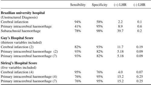
Table 4. Operational characteristics of clinical diagnosis of stroke subtypes made by emergency medicine physicians and stroke classification systems*.
Sensibility Specificity (+) LHR (-) LHR
Brazilian university hospital (Unstructured Diagnosis)
Cerebral infarction 94% 58% 2.2 0.1
Primary intracerebral haemorrhage 41% 95% 8.9 0.6
Subarachnoid haemorrhage 78% 98% 39.7 0.2
Guy’s Hospital Score (thirteen variables included)
Cerebral infarction (2) 82% 93% 11.7 0.19
Primary intracerebral haemorrhage (2) 93% 82% 5.18 0.09
Primary intracerebral haemorrhage (7) 93% 82% 5.18 0.09
Siriraj’s Hospital Score (five variables included)
Cerebral infarction (4) 95% 76% 4.0 0.07
Primary intracerebral haemorrhage (4) 76% 95% 15.2 0.25
Primary intracerebral haemorrhage (7) 76% 95% 15.2 0.25
*Cut-off point defined in each study by means of a ROC curve.
(2) in Guy Hospital – UK; (4) in Siriraj Hospital – Thailand; (7) Glasgow - UK.
DISCUSSION
Diagnosis of the acute stroke syndrome is relatively easy, but diagnosis of stroke subtype in the acute setting is certainly difficult. The sample in this study was restricted to cases of definite stroke, as in other studies concerning structured diagnosis3,8-10. The prevalence of haemorrhagic
stroke in our sample (33%) was higher than in the United Kingdom (18%) and smaller than in Thailand (69%). These differences may explain the different operational characteristics of the tests according to the population to which the clinical scoring systems were applied. Differences in stroke severity also must be considered: strokes were more likely to be severe in patients from Thailand and milder in British patients than in Brazilian patients.
The unstructured clinical diagnosis (emergency ward physician’s diagnosis) proved accurate mainly for the diagnosis of SAH, but less so for CI and especially for ICH. The diagnostic scores did not prove more accurate than unstructured clinical diagnosis (mainly in relation to the CH).
The objective of an educational policy is to improve accuracy in diagnosing the acute stroke syndrome based on clinical variables at the bedside: simple, reliable, and safe diagnostic model as compared with others, highly elaborate instruments in biomedical sciences. This probabilistic diagnostic approach has to be considered and it is very important, but the clinical reasoning has other components, such as the causal reasoning, and the deterministic reasoning, as mentioned by Kassirer, 198910.
In conclusion, the use of systematic diagnostic approaches such as those here evaluated is worthwhile as a screening procedure if there is not a proved specific therapy available. Our study shows, however, that these clinical scoring systems do not exhibit enough accuracy to be applied safely if the use anticoagulant or thrombolytic therapy should be considered, and their strict use may in fact prove dangerous if neuroimaging techniques are not routinely required.
REFERENCES
1. Kothari RU, Brott T, Broderick JP, Hamilton CA. Emergency physicians: accuracy in the diagnosis of stroke. Stroke 1995;26:2238-2241.
2. Sandercock, Allen CMC, Harrison MJG, Warlow CP. Clinical diagnosis of intracranial haemorrhage using Guy’s hospital score. BMJ 1985;291:1675-1677.
3. Bamford J. Clinical examination in diagnosis and subclassification of stroke. Lancet 1992;339:400-402.
4. Poungvarin N, Viriyavejakul A, Komontri C. Siriraj stroke score and validation study to distinguish supratentorial intracerebral haemorrhage from infarction. BMJ 1991;302:1565-1567.
5. Besson G, Robert C, Hommel M, Perret J. Is it clinically possible to distinguish nonhemorrhagic infarct from hemorrhagic stroke? Stroke 1995;26:1205-1209.
6. Aho K, Harmsen P, Hatano S, Marquardsen J, Smirnov VE, Strasser T. Cerebrovascular disease in the community: results of a WHO collaborative study. Bul WHO 1980;58:113-130.
7. Weir CJ, Murray CJ, Adams FG, Grosset DG, Lees KR. Poor accuracy of stroke scoring systems for differential clinical diagnosis of intracranial haemorrhage and infarction. Lancet 1994;344:999-1002.
8. Jaeschke R, Guyatt GH, Sackett DL. User’s guides to the medical literature: III. How to use an article about diagnosic test. Are the results of the study valid? JAMA 1993;271:389-391.
9. Jaeschke R, Guyatt GH, Sackett DL. User’s guides to the medical literature. III. How to use an article about diagnostic test. What are the results and will they help me in caring for my patients? JAMA 1993;271:703-707.