Free-hand placement of high
thoracic pedicle screws with
the aid of luoroscopy
Evaluation of positioning by CT scans
in a four-year consecutive series
Bruno Perocco Braga1,2, Josaphat Vilela de Morais2, Marcelo Duarte Vilela3
ABSTRACT
Objective: To evaluate the feasibility, safety and accuracy of pedicle screw placement
in the upper thoracic spine using the free-hand technique with the aid of fluoroscopy; to analyze the methods used to verify correct screw positioning intra and postoperatively.
Method: All patients with instability of the cervicothoracic or upper thoracic spine and at least one screw placed in the segment T1-T6 as part of a posterior construct entered the study. Only C-arm intraoperative fluoroscopy was used to guide screw placement.
Results: We obtained excellent positioning in 98.07% of the screws. CT scans precisely
demonstrated pedicle wall and anterolateral body violations. There was no hardware failure, no neurological or vascular injury and no loss of alignment during the follow-up period.
Conclusion: Pedicle screws can be safely placed in the upper thoracic spine when strict technical principles are followed. Only a CT scan can precisely demonstrate vertebral body and medial pedicle cortical violations.
Key words: pedicle screws, transpedicular fixation, thoracic spine, cervicothoracic junction, spinal instability.
Colocação de parafusos pediculares na coluna torácica alta utilizando fluoroscopia: avaliação do posicionamento dos parafusos por tomografia computadorizada em uma série de casos durante quatro anos
RESUMO
Objetivo: Avaliar a factibilidade, segurança e eficácia da colocação de parafusos
pediculares na coluna torácia alta utilizando apenas a fluoroscopia; analisar os métodos intra e pós-operatórios de verficação do posicionamento de parafusos. Método: Todos os pacientes com instabilidade da coluna cervico-torácica ou torácica alta e pelo menos um parafuso colocado no segmento T1-T6 foram incluídos no estudo. Apenas fluoroscopia intra-operatória foi utilizada para guiar a colocação dos parafusos. Resultados: Obtivemos excelente posicionamento em 98,07% dos parafusos. TC axial mostrou precisamente violações pediculares e da parede anterolateral do corpo vertebral. Não houve falência do instrumental, lesões neurológicas ou vasculares, ou perda do alinhamento sagital no período de seguimento. Conclusão: Os parafusos pediculares podem ser colocados com segurança na coluna torácica alta desde que técnicas operatórias precisas sejam executadas. Somente a TC pode demonstrar precisamente violações do corpo vertebral e da parede pedicular.
Palavras-chave: parafusos pediculares, fixação transpedicular, coluna torácica alta, junção cervico-torácica, instabilidade espinhal.
Correspondence
Marcelo Duarte Vilela Rua Aimores 2480 / 1°andar 30140-072 Belo Horizonte MG - Brasil E-mail: [email protected]
Received 27 October 2008 Received in final form 27 August 2009 Accepted 29 August 2009
1Neurosurgeon, Hospital Mater Dei, Belo Horizonte MG, Brazil; 2Neurosurgeon, Hospital da Baleia, Belo Horizonte MG,
Brazil; 3Assistant Professor, Department Of Neurological Surgery, Harborview Medical Center, University of Washington,
Techniques for stabilization of the thoracic spine have included the use of wires, hooks, rectangles, rods, plates, screws and combinations thereof. heoretical advantag-es of pedicle screw ixation include three-column sup-port1-4 greater rotational stability5,6, possibility of instru-mentation in the absence of posterior column integri-ty1-4, avoidance of neural canal dissection, decreased op-erative time3,5,7 and decreased blood loss7. More recent-ly, superior eicacy of thoracic pedicle screws over other systems has been demonstrated5-9. hey ofer higher pull-out strength9, sustain greater loads to failure8 and facili-tate a better correction of deformities5. heir use in the thoracic spine has already been described in the treat-ment of trauma2,4 deformities2, spinal tumours2 and in-fection-related instability2. Nevertheless, their placement, especially in the upper thoracic segments, is not without hazards. here is risk of injury to the spinal cord, nerve roots, lung and vascular beds. Recently, some surgeons have advocated computer-assisted or three-dimensional systems to aid proper screw placement10,11. In contrast, others have warranted safe positioning by use of only lu-oroscopy and anatomic landmarks12.he accuracy of tho-racic screw placement, deined as screws placed total-ly within the pedicle, varies from 27.6% to 91.5%2,10,13,14, even in the hands of experienced surgeons14. Interestingly enough, only a small number of complications from mal-positioned screws have been reported2,4,5, 13,15. Most litera-ture reports have not described the methods used to de-termine intraoperatively whether the screw position was considered accurate.
herefore, this is the irst study to evaluate screw po-sitioning using CT scans in all patients and to correlate the CT indings with intraoperative luoroscopy so as to establish criteria for the precise determination of screw positioning intraoperatively.
METHOD
Patient population and evaluation
During the four-year period from November 2003 through November 2007, all consecutive patients who had at least one thoracic pedicle screw placed in the up-per thoracic spine (deined here as the segment from T1 through T6) entered the study (Table 1). All were rated according to the ASIA classiication (Table 2). Imaging studies consisted of at least AP and lateral radiographs and computed tomographic scanning in all patients. MRI was obtained for neoplasms, infectious diseases, cervico-thoracic spine injuries or when the neurological exami-nation did not accurately correspond to the level of inju-ry. Surgical indications included patients with neurolog-ic deneurolog-icits with the exception of a nerve root lesion, sig-niicant anterior spinal cord compression, vertebral body collapse and kyphotic deformities of more than 35 de-grees, and lexion-distraction or lexion-dislocation inju-ries (types B or type C).
All patients were available for follow-up, which ranged from ive to 34 months. All patients had a CT scan per-formed no more than three days postoperatively to con-irm adequate placement of the screws. All but 2 patients with neoplastic lesions wore either a TLSO or a CTLSO postoperatively for at least 12 weeks. Stability was doc-umented on an upright x-ray without the orthosis, per-formed during follow-up to verify maintenance of align-ment. he study was analyzed and approved by the hos-pitals ethics committees.
Operative technique
A three-point head holder was used in all cases. After adequate exposure, the entry point for the pedicle screws was deined as the intersection of a horizontal line pass-ing through the superior margin of the transverse process
Table 1. Number of screws per level instrumented.
Level instrumented T1 T2 T3 T4 T5 T6
Number of screws 58 50 66 84 82 75
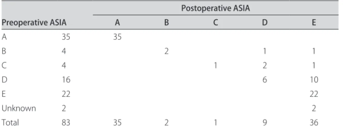
Table 2. ASIA status pre and postoperatively.
Preoperative ASIA
Postoperative ASIA
A B C D E
A 35 35
B 4 2 1 1
C 4 1 2 1
D 16 6 10
E 22 22
Unknown 2 2
and a vertical line passing 3 mm medial to the lateral bor-der of the superior facet. A pedicle probe was carefully advanced under luoroscopic guidance in the straight-for-ward or anatomical trajectory16. At least 80% of the verte-bral body was cannulated using the pedicle probe so as to place the longest screw possible, taking care not to perfo-rate the anterior cortex. In all cases we only used lateral
luoroscopic imaging and anatomical landmarks as guides to cannulate the pedicle and vertebral body.
For patients with very narrow pedicles, we used the technique of placing the screws with the entry point in-side the costovertebral joint. For the latter, the entry point was usually 2 mm lateral to the lateral edge of the supe-rior facet, converging more medially than the usual tech-nique for that level.
A small ball-tip probe was used to confirm pedi-cle and vertebral body walls integrity and measure the length of the screw. Self-tapping titanium screws
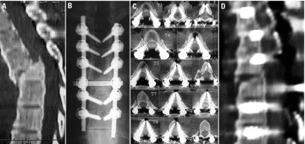
mea-Fig 2. Images of patient #27: this 32-year-old male sustained a T6-T7 fracture-dislocation. [A] Preoperative CT scan showing a T6-T6-T7 fracture-dislocation with marked anterolisthesis and kyphotic de-formity. [B] Postoperative X ray showing good coronal alignment and good screw positioning. [C] Postoperative X ray showing good sagittal alignment and good screw positioning. [D] Postoperative CT scans conirming optimal placement of pedicle screws at all levels instrumented, from T4-T6 and T8-T10, with the exception of the left T5 screw, which perforated the anterolateral cortex and put the aorta at risk. [E] CT scan after repositioning of left T5 screw showing adequate placement.
Fig 3. Images of patient # 12: a 16-year-old girl was involved in a motor-vehicle accident and was admitted complaining of back pain (ASIA E). [A] Preoperative CT scan showing a T5-T6 lexion-distraction injury with kyphotic deformity. [B] Postoperative X rays showing good coronal and sagital alignment and good screw posi-tioning. [C] Postoperative CT scans conirming optimal placement of pedicle screws at all levels instrumented, from T4-T5 and T7-T8.
suring from 3.5 to 6.25 mm were inserted. We only used 3.5 mm screws when the construct crossed the cervico-thoracic junction. When the construct was sited just on the thoracic spine, the smallest screw used was 5.5 mm. he screws were inserted using the same angulation used to cannulate the vertebral body.
In those patients in whom a costotransversectomy was done, the rod was placed on the left side irst and then a vertebrectomy with its substitution for a cage with either bone graft (in infection or trauma) or cement (in tumours) was done, prior to placement of the right rod. Anteroposterior luoroscopy was then used to conirm correct screw positioning and coronal alignment.
RESULTS
A total of 415 pedicle screws were placed in 83 pa-tients (Table 1). Neurological status improved in 10 out of 16 patients with incomplete injuries and no worsening of function was observed (Table 2). All patients had satis-factory correction of the deformity conirmed both intra and postoperatively, with no loss of correction or hard-ware failure on subsequent follow-up.
Adequate placement was accomplished in 407 screws, giving a correctness rate of 98.07%. Screw placement was veriied with postoperative CT scan in all cases. It was considered adequate if the screw did not perforate the an-terolateral cortex of the vertebral body more than 4 mm and did not violate the medial pedicle wall more than 2 mm or did not result in neurological deicits or vascular lesion. Five screws violated the spinal canal consequent to medial wall penetration of 3-4 mm, but did not re-sult in cerebrospinal luid leak or neurological deicit; al-though not considered adequate, they were still consid-ered acceptable and we did not reposition them. We did not consider pure lateral pedicle wall violations to be of signiicance. Two patients (one screw each) had screws with their tips lateral to the vertebral body but were not considered to be posing a high risk (less than 3 mm per-foration) and therefore not repositioned. Both patients remain well twenty six months after surgery. One screw in patient 28 was repositioned because it had perforated the anterolateral vertebral cortex more than 4mm and was abutting the aorta. here were 7 cases of supericial wound infection, including one of them who had men-ingitis; all were cured with antibiotics and debridement. One patient (#31) died fourth months after surgery fol-lowing deep vein thrombosis and pulmonary embolism. No loss of correction, hardware failure or instability was noted during follow-up.
Veriication of screw positioning
We analyzed the postoperative CT scans and retro-spectively correlated those with the intraoperative AP
and immediate postoperative AP and lateral x-rays. On the intraoperative AP images the screw tips should be aligned with the lateral cortex of the spinous process for the screw position to be considered excellent. his al-most always ensures that the screw tip has not perforat-ed the anterolateral cortex of the vertebral body and is not too medial, provided it is not too long on the lateral x-ray. When the screw tip crosses to the other side of the midline, the screw may be too medial, violating the me-dial pedicle wall, and probing of the track should be per-formed. It is important to note that even if the screw tip is not crossing the midline on the AP images, if the entry point is too medial, the screw may be traversing the spi-nal caspi-nal. herefore, strict attention to the entry point is of utmost importance. Sometimes the midline orienta-tion is lost, especially when a laminectomy has been per-formed or when the spinous processes are fractured. Ad-ditionally, rotational deformities in fracture dislocations may also disorient the midline. In those cases, it is dii-cult to evaluate the position of the screws on the AP im-age. It is always a good idea to draw an imaginary line from the spinous process above the instrumentation to the spinous process below the instrumentation and cor-relate that with the screw position.
We have observed that it is very diicult to clearly state whether the screws have perforated the anterolateral cor-tex of the vertebral body using only AP images, especially when the screw is not aligned with the lateral cortex of the spinous process. In those cases, one must review the pre-operative axial CT scans and determine the relationship between the anterolateral vertebral body cortex and the pedicle walls. If, on the preoperative CT scan, the anter-olateral cortex is medial to the medial wall of the pedicle (triangular-shaped vertebral body), then the entire screw tip must be placed medial to the medial pedicle wall line on a perfect AP image. If, on the preoperative CT, the ante-rolateral vertebral body cortex is lateral to the lateral pedi-cle wall (round shaped vertebral body), the entire screw tip should be at least medial to the lateral pedicle wall. One should account for the screw length and diameter.
Case examples
Patient 06 – A 50-year-old female was involved in
a motorcycle accident and sustained a complete spinal cord injury (ASIA A). CT scan showed marked T5-T6 fracture-dislocation, with striking anterolisthesis and se-vere kyphotic deformity. She underwent a posterior seg-mental instrumentation using 5.5 mm pedicle screws at T3 through T8 with excellent deformity correction and screw positioning (Fig 1).
Patient 27 –his 32-year-old male sustained a
spinous processes, a feature seen as well in the postoper-ative x-rays. Postoperpostoper-ative CT, however, showed the left screw at T5 had penetrated the anterolateral cortex and was in close contact with the aorta. he screw was repo-sitioned (Fig 2).
Patient 12 – A 16-year-old girl was involved in a
mo-tor-vehicle accident and was admitted complaining of back pain (ASIA E). A T5-T6 lexion-distraction was vi-sualized on imaging studies. A T4 through T8 posteri-or segmental instrumentation was perfposteri-ormed, with good correction of the deformity and excellent screw position-ing (Fig 3).
DISCUSSION
Technical advantages of pedicle screws in the tho-racic spine include avoidance of neural canal dissection, decreased operative time3,5,7 and less blood loss7. Biome-chanical advantages include three-column support1-4, possibility of instrumentation in the absence of posteri-or column integrity1-4 and greater rotational stability in the transverse axis5,6. An additional beneit from a screw-rod construct is the use of cross-links, forming a trian-gle in the transverse plane, which signiicantly improves screw pullout strength and rotational and lateral bending stifness17,18. Despite the narrowness of thoracic pedicles, placing screws with diameters greater than the pedicle itself has already been proven safe and eicacious2; and it is known that the greater the minor screw diameter the greater the bending strength and the larger the major screw diameter the greater the pullout strength19.
Placing pedicle screws in the upper thoracic spine is hazardous. Penetration of the medial pedicle wall may in-jure the spinal cord or dura-mater; inferior penetration may harm the nerve roots, lateral violation may damage the lung, vessels and/or sympathetic chain and perfora-tion of the anterolateral vertebral body may also cause le-sion to the great vessels and esophagus14.
he short and triangular vertebral bodies and thin and medially oriented pedicles from T1 to T6 are the major factors responsible for the diiculties in the technique of placing upper thoracic pedicle screws. Anatomic studies determined the thinnest pedicles to be between T3 and T6 (from 4.5 to 5.1 mm), compared with mean widths of 5.9 to 6.5 mm for T1-T23,6,20, and the pedicle transverse angle to be greatest at T1 and T2, measuring 28.2o and 16.6o, respectively2,20. In order to overcome these diicul-ties, it is important to know the safe margins of cortical violation. In the upper thoracic spine, the closest distance between the aorta and the vertebral body is 6 mm, at T4 through T621. Between the pedicle and the dural sac, Uğur reported no distance from T3 to T6 and only 0.5 and 0.2 mm of distance at T1 and T2, respectively3.
Attempting to bypass the challenges of narrow
pedi-cle screw ixation in the thoracic spine, an extrapedicu-lar technique has been described by Husted1. In fact, we used this technique in two children, allowing the use of 5.5 mm screws in the pedicles of T3 through T5.
Probing the pedicle tract is the only way to assess proper screw placement prior to its insertion; it is the only method that can actually prevent misplacement22. Nonetheless, even in the hands of an experienced tho-racic spine surgeon, it has an accuracy of 82%, sensitivi-ty of 81% and speciicisensitivi-ty of 93%, with medial wall viola-tions being the most diicult to assess22. We found that when probing one pedicle if the surgeon feels the tip of the contralateral screw and there is no anterior wall vio-lation, for sure both screws are in the vertebral body; one screw might be too medial, though, especially if it mea-sures longer than the contralateral one.
The most difficult but also most important step in cannulating the vertebral body is to correctly aim the probe medially. It is more diicult to cannulate the pedi-cles when using the pointing down technique, since the smallest diameter of the vertebral body is at the mid-por-tion of the body. he widest diameter is close to the disc space and therefore our preference is to cannulate using the straight technique, with the probe being parallel to the endplate, which is also better biomechanically16.
After surgery, x-rays and computed tomography scans can be used to confirm correct positioning of screws. Plain ilm accuracy depends on the experience of the in-terpreter, varying from 73% to 83%23. Routine anteropos-terior and lateral views are inadequate to evaluate screw position23,24. CT is the most accurate study; however, its sensitivity and speciicity varied from 76% to 86% and 75% to 88%, respectively25-27. Inferior wall violations are the most diicult to detect on CT scans25.
sig-niicant by others as well13,28,29 and a 4 mm safe zone has been suggested by some as the upper limit of intraspinal violation29. Inferior pedicle wall violations should be rare since the entry point is on the upper half of the pedicle but should always be checked, especially at the T1 level.
We believe the free hand technique by use of thorough knowledge of the spinal anatomy, judicious exposure and standard luoroscopy suice for the correct positioning of transpedicular screws in the upper thoracic spine. Image-guided systems can surely improve the accuracy of pedi-cle screw placement10, but the clinical outcome as evalu-ated by vascular and neurological complications, correc-tion of deformities and hardware failure seems to be un-afected, as shown in our own study and in several series in which only luoroscopic imaging or radiographs were used2,4,5,13,30. he extra surgical time and cost demanded by these new technological devices may thus not justify their use in substitution of an old established technique that produces the same clinical results.
Although other studies have shown that thoracic pedicle screws are safe and feasible, this is the irst that attempts to correlate intraoperative images with postop-erative CT scans so as to accurately identify whether a screw is accurately positioned or not.
In conclusion, pedicle screws ease better correction of spinal deformities and provide greater stability than other ixation systems, mainly through their ability to provide three-column support and rotational rigidity. Attention to anatomical landmarks and entry points, proper medi-al angulation and careful pmedi-alpation of the pedicle tract are essential for adequate placement. Satisfactory placement by use of the free hand technique with the aid of stan-dard luoroscopy can be achieved in practically all cases with a minimal incidence of screw malpositioning. Only CT scans can accurately identify anterolateral vertebral body and medial pedicle wall violations.Correlation of the intraoperative luoroscopic images with the preop-erative axial CT scan images helps conirming adequate screw positioning intraoperatively. Despite the techni-cal diiculties and risks of injury to crititechni-cal surrounding structures, the use of pedicle screws in the upper thoracic spine can be done with great eicacy and safety.
REFERENCES
1. Husted DS, Yue JJ, Fairchild TA, Haims AH. An extrapedicular approach to the placement of screws in the thoracic spine: an anatomic and radiographic as-sessment. Spine 2003;28:2324-2330.
2. Kuntz IV C, Maher PC, Levine NB, Kurokawa R. Prospective evaluation of tho-racic pedicle screw placement using luoroscopic imaging. J Spinal Disord Tech 2004;17:206-214.
3. Uğur HÇ, Attar A, Uz A, Tekdemir I, Egemen N, Genç Y. Thoracic pedicle:
sur-gical anatomic evaluation and relations. J Spinal Disord 2001;14:39-45. 4. Yue JJ, Sossan A, Selgrath C, et al. The treatment of unstable thoracic spine
fractures with transpedicular screw instrumentation: a 3-year consecutive series. Spine 2002;27:2782-2787.
5. Suk SI, Lee CK, Min HJ, Cho KH, Oh JH. Comparison of Cotrel-Dubousset
pedi-cle screws and hooks in the treatment of idiopathic scoliosis. Int Orthop 1994; 18:341-346.
6. Vaccaro AR, Rizzolo SJ, Allardyce TJ, et al. Placement of pedicle screws in the thoracic spine - part 1: morphometric analysis of the thoracic vertebra. J Bone Joint Surg (Am) 1995;77:1193-1199.
7. Polly DW, Potter BK, Kuklo T, Young S, Johnson C, Klemme WR. Volumetric spinal canal intrusion: a comparison between thoracic pedicle screws and thoracic hooks. Spine 2003;29:63-69.
8. Heller JG, Shuster JK, Hutton WC. Pedicle and transverse process screws of the upper thoracic spine: biomechanical comparison of loads to failure. Spine 1999;24:654-658.
9. Liljenqvist UR, Hackenberg L, Link TM, Halm H. Pullout strength of pedicle screws versus pedicle and laminar hooks in the thoracic spine. Acta Orthop Belg 2001;67:157-163.
10. Youkilis AS, Quint DJ, McGillicuddy JE, Papadopoulos SM. Stereotactic navi-gation for placement of pedicle screws in the thoracic spine. Neurosurgery 2001;48:771-779.
11. Schwarzenbach O, Berlemann U, Jost B, et al. Accuracy of computer-assisted pedicle screw placement: an in vivo computed tomography analysis. Spine 1997;22:452-458.
12. Carbone JJ, Tortolani J, Quartararo LG. Fluoroscopically assisted pedicle screw ixation for thoracic and thoracolumbar injuries. Spine 2003;28:91-97. 13. Belmont Jr PJ, Klemme WR, Dhawan A, Polly Jr DW. In vivo accuracy of
tho-racic pedicle screws. Spine 2001;26:2340-2346.
14. Vaccaro AR, Rizzolo SJ, Balderston RA, et al. Placement of pedicle screws in the thoracic spine. Part 2: An anatomical and radiographic assessment. J Bone Joint Surg (Am) 1995;77:1200-1206.
15. Heini P, Scholl E, Wyler D, Stefan E. Fatal cardiac tamponade associated with posterior spinal instrumentation: a case report. Spine 1998;23:2226-2230. 16. Lehman RA, Polly DW, Kuklo TR, Cunningham B, Kirk KL, Belmont Jr PJ.
Straight forward versus anatomic trajectory technique of thoracic pedicle screw ixation: a biomechanical analysis. Spine 2003;28:2058-2065. 17. Carson WL, Duield RC, Arendt M, Gaines Jr RW. Internal forces and moments
in transpedicular spine instrumentation: the efect of pedicle screw angle and transixation: the 4R-4Bar linkage concept. Spine 1990;15:893-901. 18. Ruland CM, McAfee PC, Warden KE, Cunningham BW. Triangulation of
pedic-ular instrumentation: a biomechanical analysis. Spine 1991;16:270-276. 19. Krag M. Biomechanics of thoracolumbar spinal ixation: a review. Spine 1991;
16:84-99.
20. Zindrick MR, Wiltse LL, Doornik A, et al. Analysis of morphometric character-istics of the thoracic and lumbar pedicles. Spine 1987;12:160-166. 21. Liljenqvist UR, Allkemper T, Hackenberg L, Link TM, Steinbeck J, Halm HFH.
Analysis of vertebral morphology in idiopathic scoliosis with use of magnetic resonance imaging and multiplanar reconstruction. J Bone Joint Surg 2002; 84:359-368.
22. Lehman RA, Potter BK, Kuklo TR, et al. Probing for thoracic pedicle screw tract violation(s): is it valid? J Spinal Disord Tech 2004;17:277-283.
23. Ferrick MR, Kowalski JM, Simmons Jr ED. Reliability of roentgenogram eval-uation of pedicle screw position. Spine 1997;22:1249-1253.
24. Weinstein JN, Spratt KF, Spengler D, Brick C, Reid S. Spinal pedicle ixation: re-liability and validity of roentgenogram-based assessment and surgical fac-tors on successful screw placement. Spine 1988;13:1012-1018.
25. Fayyazi AH, Hugate RR, Pennypacker J, Gelb DE, Ludwig SC. Accuracy of com-puted tomography in assessing thoracic pedicle screw malposition. J Spinal Disord Tech 2004;17:367-371.
26. Rao G, Brodke DS, Rondina M, Dailey AT. Comparison of computadorized to-mography and direct visualization in thoracic pedicle screw placement. J Neurosurg 2002;97:223-226.
27. Yoo JU, Ghanayen A, Petersilge C, Lewin J. Accuracy of using computed to-mography to identify pedicle screw placement in cadaveric human lumbar Spine 1997;22:2268-22671.
28. Belmont Jr PJ, Klemme WR, Robinson M, Polly Jr DW. Accuracy of thoracic pedicle screws in patients with and without coronal-plane deformities. Spine 2002;27:1558-1566.
29. Gertzbein SD, Robbins SE. Accuracy of pedicular screw placement in vivo. Spine 1990;15:11-14.