PURE NEURAL LEPROSY
Steroids prevent neuropathy progression
Márcia R. Jardim
1, Ximena Illarramendi
1, Osvaldo J.M. Nascimento
2,
Jose Augusto C. Nery
1,Anna M. Sales
1, Elizabeth P. Sampaio
1, Euzenir N. Sarno
1ABSTRACT - Multidrug therapy (MDT), with rifampicin, dapsone, and clofazimine, treatsleprosy infection but is insufficient in arresting or preventing the nerve damage that causes impairments and disabilities. This case-series study evaluatesthe benefits of the combined use of steroids and MDT in preventing nerve dam-age in patients with pure neural leprosy (PNL). In addition to MDT, 24 patients (88% male dam-aged 20-79 years, median=41)received a daily morning dose of 60 mg prednisone (PDN) that was gradually reduced by 10 mg during each of the following 5 months. PNL was clinically diagnosed and confirmed by nerve histopatholo-gy or PCR. A low prevalence (8.3%) of reaction was observed after release from treatment. However, most of the clinical parameters showed significant improvement; and a reduction of nerve conduction block was observed in 42% of the patients. The administration of full-dose PDN improved the clinical and electrophys-iological condition of the PNL patients, contributing to the prevention of further neurological damage.
KEY WORDS: peripheral neuropathy, pure neural leprosy, steroids.
Corticosteróides previnem a neuropatia na hanseníase
RESUMO - A poliquimioterapia (PQT), com rifampicina, dapsona, e clofazimina, trata a infecção na hanse-níase, mas é insuficiente para interromper ou prevenir o comprometimento neurológico que causa as inca-pacidades e desabilidades, nesta enfermidade. Este estudo de série de casos avalia o benefício do uso com-binado de prednisona e PQT na prevenção do dano neurológico em pacientes com a forma neural pura da hanseníase (FNP). Além do PQT, 24 pacientes (88% homens, com idade variando entre 20-79, mediana=41) receberam uma dose diária de 60 mg prednisona que foi reduzida gradualmente na dose de 10 mg duran-te cada um dos 5 meses subseqüenduran-tes. FNP foi diagnosticada clinicamenduran-te e confirmada através do estu-do histopatológico ou PCR. Baixa prevalência de reação (8,3%) foi observada apenas após o final estu-do trata-mento. A maioria dos parâmetros clínicos mostrou melhora significativa e redução do bloqueio de condu-ção foi observada em 42% dos pacientes. A administracondu-ção de doses altas de prednisona melhora a evolu-ção clínica e eletrofisiológica de pacientes com a FNP de hanseníase, contribuindo na prevenevolu-ção de novos comprometimentos neurológicos.
PALAVRAS-CHAVE: neuropatia periférica, forma neural pura da hanseníase, corticosteróides.
1Leprosy Laboratory, Oswaldo Cruz Institute, FIOCRUZ, Rio de Janeiro RJ, Brazil; 2Department of Neurology, Hospital Universitário Antônio Pedro, Fluminense Federal University, Niterói RJ, Brazil.
Received 29 January 2007, received in fi nal form 28 June 2007. Accepted 29 August 2007.
Dra. Márcia R. Jardim - Laboratório de Hanseníase / Instituto Oswaldo Cruz, FIOCRUZ - Avenida Brasil 4365 - 21040-360 Rio de Janeiro RJ - Brasil. E-mail: mmjardim@ioc.fi ocruz.br
Pure neural leprosy (PNL) presents a diagnos-tic challenge. PNL patients have nerve defi cit or en-largement of peripheral nerves with or without ten-derness in the absence of any sign of skin manifesta-tion or history of skin patches. In India, it has been re-ported that from 5.5%-17.7% of all leprosy cases are PNL1. Leprosy neuropathy classically presents as acute
neuritis characterized by nerve enlargement and pain that may be followed by neurological dysfunction2.
Neurological alteration without nerve pain, known as “silent neuritis”, has also been documented3,4.
Ac-cording to some authors, in PNL, M. leprae causes
pe-ripheral nerve damage leading to neuropathy, which may remain undiagnosed for an extended period of time, even years5. Furthermore, in all clinical forms
of leprosy, the irreversible motor and sensory alter-ations may lead to increasingly severe secondary im-pairments long after the disease has been arrested as peripheral neuropathy may be present before the patient notices any symptoms of nerve function im-pairment6. Interventions that prevent, reverse, or
of nerve damage. Multidrug therapy (MDT) alone is aimed at treating the infection but is insuffi cient in arresting or preventing the nerve damage responsible for impairment and disabilities4. The deformities seen
in patients who were diagnosed reasonably early and, so, received timely MDT5, clearly indicate the need for
using more intensive measures to recognize and treat recent nerve damage as expeditiously as possible.
Prednisone (PDN) remains the drug of choice for neuritis due to its ability to reducenerve oedema, ex-ert an immunosuppressive effect, and decrease post-infl ammatory scar formation – all important for im-proving nerve function3. Moreover, when detected
and treated in time with corticosteroids, peripheral neuropathy may not progress into deformity and may even reverse initial impairments1.
This study evaluates the benefi ts of the combined use of steroids and MDT in preventing and arresting nerve damage in PNL patients.
METHOD
A prospective longitudinal study was performed in a group of 24 PNL patients, of whom 88% weremale rang-ing from 20-79 years of age(median=41), diagnosed at the Leprosy Outpatient Clinic, Oswaldo Cruz Institute, Rio de Ja-neiro RJ, Brazil, between 1998 and 2000. PNL was clinically diagnosed and confi rmed by nerve histopathology or PCR, as described by Jardim et al7. All patients received MDT (ri-fampicin, dapsone, clofazimine) for paucibacillary (PB) lep-rosy for 6 consecutive months in accordance with WHO rec-ommendations8 plus a daily morning dose of 1 mg/kg ofPDN for one month followed by a progressive10 mg/monthly re-duction over the remainingfi ve months. Clinical and elec-trophysiological examinations were performed at diagnosis and 12 monthsafter beginning MDT. The research was car-ried out in strict compliance with the International Norms on Ethics in Human Research; and all patients were duly in-formed prior to providing their written consent.
Neurological examination – Pain and paraesthesia were evaluated by way ofvisual analogue scales (VAS). Senso-ry impairment, motor defi cit, and disability status were as-sessed by standard methods. Values were given to ther-mal and pain sensations (0=anaesthesia, 1=hypoaesthesia, 2=normal) including the monofi lament force as subjective-ly felt by the patients (0=no sensation, 1=300g, 2=4g, 3=2g, 4=0.2g, and 5=0.05g). The bilateral grades of 13 nerves were added to form the sensory score (normal=234). Individual muscle strength was graded according to the Medical Re-search Council of London9 recommendations and added to the motor score (normal score=80, since 8 nerves were eval-uated bilaterally).
Electrophysiological examination – All nerve conduction assessments were performed by the same specialist on a Ni-hon-Koden–Neuropack 2. Standard nerve conduction tech-niques were utilized10 to evaluate the median, radial, ulnar,
and sural sensory nerves as well as the median, ulnar, and peroneal motor nerves (total of 14 nerves). Partial conduc-tion block (CB) (with or without temporal dispersion) was defi ned as a 50% or more reduction of the proximal as com-pared to the distal amplitudes. Abnormal temporal disper-sion (TD) was defi ned as a proximal distal compound mus-cle action potential (CMAP) duration increase ofmore than 30%. A prolonged latency and/or 85% reduction in senso-ry conduction velocity (SCV) or motor conduction veloci-ty (MCV) was considered as a demyelinating lesion; and an axonal lesion was defi ned as >30% reduction in amplitude with/without a <30% reduction in conduction velocity11. All other patterns of amplitude, latency, and velocity not corre-spondingto any of these defi nitions were considered non-classifi able, as suggested by Tankisi et al.12
Statistical analysis – Data were analyzed using SPSS for Windows v. 11.5. Unless stated otherwise, all results were expressed as median because of the non-gaussian distri-bution of variables. Maximum and minimum values are in parentheses. McNemar and Wilcoxon tests were used to compare variables and p values of less than 0.05 were con-sidered statistically signifi cant.
RESULTS
Findings at diagnosis – The referredpatients had
been symptomatic for a period of 2-120 (median=14) months before diagnosis clinical and laboratory data are shown in Table 1. The frequency of the signs and symptoms are shown in Tables2 and 3. Disability grade 2, i.e., eye, hand or foot deformities such as ulcers, claw fi ngers/toes, foot or hand drop, lagophthalmos, or amyotrophy, was conferred on 18(75%) patients. In the sensory evaluation, the median nerve was the most frequently impaired (42%) while motor dysfunc-tion occurred predominantly in the ulnar nerve (38%).
CB always accompanied by TD was observed in 10% of the patients, most often in the ulnar nerve. All patients demonstrated demyelinating nerve lesions in a varying number of nervesranging from 1 to a maxi-mum of 10 (median=4.5).Axonal lesions were present in 46% of the patients at a maximum of 2 affected nerves out of the 14 assessed in each patient. A com-bined pattern(simultaneous axonal and demyelinat-ing fi ndings in the same nerve) was found in 38% of the patients, with a maximum of 2 affectednerves. Furthermore, the nerves of83% of the patients was found to havenon-classifi able lesions.
Table 1. Clinical and laboratory data.
ID Age
(years)
Gender SS DG Lepromin Histopathological
fi ndings
Reaction Presentation symptom (months)
3193 46 F 0.00 3 8 UII/F N Paresthesia (60)
3197 55 M 0.00 2 5 EG/Fibrosis N Paresthesia (6)
3214 25 M 0.00 2 9 EG N Paresthesia (2)
3233 55 M 0.00 2 0 UII/Fibrosis N Sensory impairment (8)
3244 28 M 0.00 0 11 UII/Fibrosis N Paresthesia (12)
3262 55 M 0.00 2 0 Fibrosis N Paresthesia (30)
3285 45 F 0.00 2 0 Normal* N Paresthesia (6)
3291 61 M 0.00 2 4 UII N Paresthesia (7)
3316 66 M 0.00 2 5 UII/Fibrosis N Paresthesia (8)
3369 39 M 0.00 2 0 UII N Paresthesia (23)
3374 23 M 0.00 2 12 UII/Fibrosis N Paresthesia (6)
3386 25 M 0.00 2 0 AFB N Paresthesia (58)
3401 79 M 0.00 0 9 Fibrosis N Paresthesia (84)
3418 20 M 0.00 2 9 UII/Fibrosis N Amiotrophy (72)
3432 29 M 0.00 2 9 UII/Fibrosis N Paresthesia (8)
3435 61 M 0.00 0 0 EG N Paresia?? (12)
3436 22 M 0.00 0 5 Normal N Sensory impairment (24)
3441 56 M 0.00 2 7 Normal N Motor impairment (12)
3450 42 M 0.00 0 6 Fibrosis N Paresthesia (24)
3467 25 M 0.00 2 9 EG N Paresthesia (14)
3469 37 M 0.00 2 6 Fibrosis N Pain (120)
3476 28 M 0.00 0 3 Fibrosis N Pain (20)
3275 66 F 0.00 2 6 Normal Y(N) 21m Paresthesia (24)
3382 24 M 0.00 2 5 UII/Fibrosis Y(N) 24m Paresthesia (20)
ID, identifi cation; F, female; M, male; SS, slit skin smears; DG, disability grade; UII, unspecifi c infl ammatory infi ltration; EG, epitelioid granu-loma; AFB, acid fast bacilli.
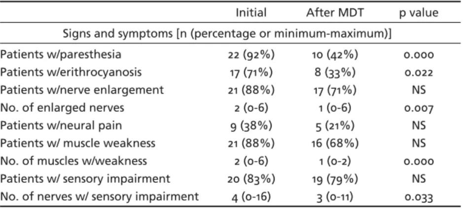
Table 2. Variations in clinical parameters.
Initial After MDT p value Signs and symptoms [n (percentage or minimum-maximum)] Patients w/paresthesia
Patients w/erithrocyanosis Patients w/nerve enlargement No. of enlarged nerves Patients w/neural pain Patients w/ muscle weakness No. of muscles w/weakness Patients w/ sensory impairment No. of nerves w/ sensory impairment
22 (92%) 17 (71%) 21 (88%) 2 (0-6) 9 (38%) 21 (88%) 2 (0-6) 20 (83%)
4 (0-16)
10 (42%) 8 (33%) 17 (71%) 1 (0-6) 5 (21%) 16 (68%)
1 (0-2) 19 (79%)
3 (0-11)
0.000 0.022 NS 0.007
NS NS 0.000
NS 0.033
Table 3. Nerve conduction parameters: percentage of patients with altered amplitude, velocity, and latency.
Initial After MDT p value
CMAP AMP
MCV ML
79% 92% 92%
79% 79% 79%
0.673 0.306 0.306
SNAP AMP
SCV
92% 71%
90% 90%
0.602 0.132
CB 38% 11% 0.046
CMAP, compound muscle action potential; AMP, amplitude; MCV, motor conduction velocity; ML, motor latency; SNAP, sensory nerve action po-tential; SCV, sensory conduction velocity; CB, conduction block.
The nerve conduction evaluation did not show signifi cant differences in the combined nerve ampli-tudes, latencies, or velocities of the 19 patients as-sessed (Tables 2 and 3). However, CB signifi cantly de-creased in 42% (8/19). Although the demyelinating le-sions of all patients remained, the number of affect-ed nerves (maffect-edian=6, maximum of 12 nerves) actual-ly decreased. Moreover, 94.7% of the patients ended treatment with at least one normal nerve and 1 pa-tient tested normal for all 14.
DISCUSSION
PLNis a form of the disease that presents as an infl ammatory neuropathy (neuritis) with secondary nerve dysfunction in the absence of skin lesions. Very few PNL patients show any nerve damage improve-ment at the end of treatimprove-ment. Consequently, it would appear that antibacterial therapy alone does not pre-vent new nerve damage either during or after chemo-therapy. Clearly, in leprosy, the permanent disabilities that often accompany nerve damage are the major concern13. Thus, new treatments and prophylactics are
urgently needed.
The use of corticosteroids was initiated at least fi f-ty years ago14 but continues to be the drug of choice
in treating reaction. In one report, six-month steroid therapy had a satisfactory effect in reversing motor paralysis caused by neuritis in about 75% of the af-fected nerves3. In a PNL case study, treatment with
the previous scheme in conjunction withMDT pre-vented the establishment of nerve trunk paralysis5.
In the present study, full-dose prednisone and MDT were administered to a group of PNL patients with positive results.
Steroids appear to act as treatment and prophylac-tics at the same time. Van Brakel et al.15, who
admin-istered 40 mg/kg prednisone to patients with various clinical forms of leprosy for four consecutive months, detected a reduced risk of reaction and nerve func-tion impairment solely within this period. The present results, however, indicated that higher doses of pred-nisone were necessary to recover nerve function.
During neuritis, either isolated or associated with reversal reaction or erythema nodosum leprosum, there is an induction or worsening of disabilities3. In
this study, the patients receiving steroids developed fewer reactions, even long after MDT had ended.
After treatment, all the parameters of patients re-ceiving corticosteroids showed improvement. Steroids prevented progressive nerve damage by interrupting the infl ammatory process that is produced in PNL. In
addition, steroids protectedother nerves from being damaged by new episodes of neuritis. Corticosteroids may be acting as both prophylactics and treatment at the same time. Van Brakel et al.15 administered
pred-nisolone (40 mg/daily) for four months but found no improvement of tactile sensation or reduction in risk of leprosy reaction or nerve function impairment beyond the initial four-month treatment phase. The present results indicated that higher doses of predni-sone were necessary to recover nerve function.
Electrophysiological examination provides invalu-able information for diagnosing and recommending the most appropriate therapeutic treatment for neu-ropathies. Naafs has reported that, during reversal re-action with neuritis, immunosuppressive therapy with corticosteroids led to a biphasic response3. Initially
(within days), the oedema regressed and, after sev-eral months, remyelination and nerve regeneration took place. Since recovery time usually takes more than six months (especially recovery from axonal le-sions), nerve conduction testing should be performed not less than one year after follow-up has begun.
In this study, the most outstanding electrophysio-logical fi nding was the reduction in conduction block / temporal dispersion. Although demyelinating lesions predominated when analyzing each nerve separately, the observed conduction block reduction is indicative of the regeneration of demyelinating lesions. This event has the same physiopathological signifi cance as that reported aboutMVC improvement when us-ing corticosteroids16.
Although demyelinating lesions remained in most patients, the significant reduction of CB seen was probably related to a reduction of infl ammatory ede-ma. The worsening of nerve conduction velocity was unreal because the nerve conduction study on the fi ve lost patients demonstrated minimal changes.
The improvement observed in this group of pa-tients indicated that the administration of full-dose PDN together with MDT was both safe and useful for PN patients. It is clear, however, that to defi ne the most appropriate use of steroids as a prophylactic drug for leprosy neuropathy, further evaluations need to be performed on a larger number of patients in a double-blind placebo study for longer follow-up time periods
Acknowledgements – We would especially like to thank Dr. Cairns Smith for his timely suggestions and Judy Grevan for editing the text.
REFERENCES
2. Kiran KU, Stanley JN, Pearson JM. The out patient treatment of nerve damage in patients with borderline leprosy using a semi-standardized steroid regimen. Lepr Rev 1985;56:127-134.
3. Naafs B. Treatment of reactions and nerve damage. Int J Lepr Other My-cobact Dis 1996;64(Suppl):S521-S528.
4. Srinvasan H, Rao KS, Shanmigan N. Steroid therapy in recent “quiet nerve paralyses” in leprosy: report of a study of twenty-fi ve patients. Indian J Lepr 1982;54:412-419.
5. Jaiswal AK, Shet y MK. Steroid therapy in recent “Quiet nerve paraly-sis” in pure neuritic leprosy. Indian J Lepr 1991;63:223-225.
6. Becx-Bleumink M, Berhe D. Occurrence of reactions, their diagnosis and management in leprosy patients treated with multidrug therapy; expe-rience in the leprosy control program of the All-Africa Leprosy and re-habilitation Training Center (ALERT) in Ethiopia. Int J Lepr Other My-cobact Dis 1992;60:173-184.
7. Jardim MR, Antunes SLG, Santos AR, et al. Criteria for diagnosis of pure neural leprosy. J Neurol 2003;250:806-809.
8. World Health Organization. Chemotherapy of leprosy: report of a WHO study group. Geneva: WHO technical report series 847, 1994. 9. Anonymous. Aids to investigation of peripheral nerve injuries. Medical
Research Council War Memorandum no.7, 2.Ed. London: HMSO, 1962. 10. Delisa JA, Lee HJ, Baran EM, Lai KS. Manual of nerve conduction ve-locity and clinical neurophysiology. Philadelphia: Lippincot Williams & Wilkins, 3.Ed, 1994.
11. Falk B, Stalberg E. Motor nerve conduction studies: measurement princi-ples and interpretation of fi ndings. J Clin Neurophysiol 1995;12:254-279. 12. Tankisi H, Johnsen B, Fuglsang-Frederiksen A, et al. Variation in the
clas-sifi cation of polyneuropathies among European physicians. Clin Neu-ropysiol 2003;114:496-503.
13. Solomon S, Kurian N, Ramadas P, Rao PSS. Incidence of nerve damage in leprosy patients treated with MDT. Int J Lepr 1998;66:451-455. 14. Lowe J. ACTH and cortisone in treatment of complications of leprosy.
Br Med J 1952;4:746-749.
15. Van Brakel W, Anderson AM, Withington SG, et al. The prognostic im-portance of detecting mild sensory impairment in leprosy: a random-ized controlled trial (TRIPOD 2). Lepr Rev 2003;74:300-310.