Arq Neuropsiquiatr 2008;66(1):99-100
99
Intrasellar Internal carotId aneurysm
coexIstIng wIth gh-secretIng pItuItary
adenoma In an acromegalIc patIent
Lauro Seda Jr
1, Arthur Cukiert
1, Kátia C. Nogueira
2, Martha K.P. Huayllas
2, Bernardo Liberman
2aneurIsma Intraselar da carótIda Interna coexIstIndo com adenoma secretor de gh em pacIente acromegálIco
Departments of Neurosurgery1 and Endocrinology2, Hospital Brigadeiro and Clínica Neuroendócrina de São Paulo, São Paulo SP, Brazil.
Received 14 May 2007, received in inal form 18 October 2007. Accepted 1 December 2007.
Dr. Arthur Cukiert – Rua Dr. Alceu de Campos Rodrigues 247 / 121 - 04544-000 São Paulo SP - Brasil. E-mail: [email protected]
The coexistence of pituitary adenoma and cerebral aneurysm is rare, although its prevalence is higher then would be expected in general population1-5. We report on
an extremely rare condition where growth hormone (GH) -secreting adenoma coexisted with an intrasellar internal carotid artery (ICA) aneurysm.
case
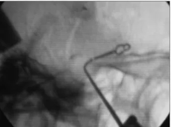
A 58 years old female presented with headache and acrome-galic phenotype. She had hypertension for 10 years and galac-torrhea for 3 years. Mean plasma GH was 8.1 ηg/mL and mean IGF-1 level was 703 ηg/mL (Reference: 78–258 ηg/mL by immu-noradiometric assay). Results obtained during pre- and postop-erative glucose tolerance tests (OGTT) and octreotide response test (100 μg administered subcutaneously every six hours) can be seen in Table 1 and 2, respectively. Prolactin baseline level was 28.6 ηg/mL and pituitary function was otherwise intact. Mag-netic resonance imaging (MRI) of the sellar region disclosed a 1.2 cm ICA aneurysm occupying the left hemisella and a 0.7 cm pituitary adenoma just bellow it (Fig 1A). Angiography disclosed left ICA aneurysm posterior to the emergency of the ophthal-mic artery (Fig 1B). The patient tolerated well a balloon occlusion test and was submitted to aneurysm exclusion through ICA oc-clusion in the neck and transcranial clipping of the supraclinoid ICA immediately below the ophthalmic artery’s exit. One month afterwards, she was submitted to complete adenoma removal
through a transsphenoidal approach (Fig 2). The excluded aneu-rysm was easily seen occupying the entire left hemisella. Immu-nohistochemical examination conirmed the presence of GH-secreting adenoma. Immediate postoperative GH and prolactin levels were 3.7 and 5.6 ηg/mL, respectively. Four days later, IGF-1 was 543 ηg/mL (78–258 ηg/mL). One month after adenoma removal, GH and IGF1 levels were 5.0 ηg/mL and 527 ηg/mL, re-spectively. After 3 months of follow-up, patient’s GH and IGF-1 levels were 4.IGF-1 ηg/mL and 413 ηg/mL, respectively. It was not possible to perform a follow-up MRI since the clip used in sur-gery was not MRI-compatible.
She was then started on Sandostatin® LAR 30 mg / month. Four months later, IGF-1 and GH levels were 107 ηg/mL
(Refer-Table 1. Pre-/ postoperative oral glucose tolerance test.
Time (minutes) 0 30 60 90 120
Glucose level (mg/dL) 93 / 92 193 / 169 181 / 204 150 / 174 108 / 111
GH (ηg/mL) 11.4 / 2.7 6.6 / 1.1 6.8 / 2.7 6.8 / 2.5 7.1 / 2.7
Table 2. Octreotide acute response test (100 μg sc 6/6h).
Time (hours) 0 2 4 6 8 24 26
GH (ηg/mL) 5.5 0.44 0.52 0.63 0.71 0.37 0.22
Arq Neuropsiquiatr 2008;66(1)
100
Intrasellar aneurysm and pituitary disorder Seda et al.
ence: 81–225 ηg/mL) and 0.9 ηg/mL, respectively. By this time, patient underwent radiosurgery. After two years of follow-up, the patient remained in remussion and receiving Sandostatin LAR.
This case report was agreed by the local ethic committee.
dIscussIon
The prevalence of sellar-region’s aneurysm among oth-ers is 1-2%2. The prevalence of the coexistence of pituitary
adenoma and cerebral aneurysm is higher then that with other benign brain tumors in the general population5.
Ap-proximately 50% of these patients have acromegaly1,3,5-8,
suggesting that high GH and IGF-1 levels or their biologi-cal effects might be implicated in the aneurysm’s genesis. High IGF-1 induces artery dilation8, atherosclerotic and
degenerative changes of the artery wall8,9, tumor invasion
and tumor-directed neovessels9. Hypertension and
diabe-tes are very likely to be involved in the process5. Patients
with intrasellar (not intracranial) aneurysms and pituitary adenomas are extremely rare.
Mostly, the diagnosis of such aneurysms is incidental, and occurs when performing the preoperative investiga-tion for adenomas7. However, different clinical
presen-tations may occur, such as fatal epistaxis10 or pituitary
apoplexy11 , as a result of aneurismal bleeding into the
adenoma. Misdiagnosis of this condition may have hazard-ous hemorrhagic consequences.
These two conditions must be treated. Although si-multaneous microsurgical treatment of the aneurysm and the adenoma through a pterional or a supraorbital keyhole approach has been advocated in different re-ports6,12,13, approaching the vascular lesion first is
usu-ally the best choice. In our case, we performed an ICA entrapment by endovascular occlusion in the neck and transcranial clipping of ICA just bellow the ophthalmic
artery’s emergence. This treatment was possible because the patient tolerated well a previous balloon occlusion test. One month later, the adenoma was completely re-moved through a transsphenoidal route. Nevertheless, the patient did not achieved endocrinological remission and needed adjuvant therapy with Octreotide and radio-surgery. Clinical and endocrinological control were then obtained.
Although the results obtained while treating this pa-tient were good, a minimally invasive therapeutic option would also be available for such patients: the vascular le-sion could be treated by endovascular trapping of the in-ternal carotid artery and the GH-secreting tumor could have been treated primarily with somatostatin analogs or transsphenoidal surgery. Primary clinical treatment with somatostatin should be considered especially in patients with known cavernous sinus invasion by the tumor and no mass effect directed to the optic apparatus14-15.
We believe that the treatment of this dual-pathology should be carried out in two steps: vascular pathology should be treated irst to avoid potential future catastroph-ic hemorrhage and the pituitary pathology afterwards.
references
1. Jakubowski J, Kendall B. Coincidental aneurysms with tumors of pitu-itary origin. J Neurol Neurosurg Psychiatry 1978;41:972-979. 2. Acqui M, Ferrante L, Fraioli B. Association between intracranial
an-eurysms and pituitary adenomas: etiopathogenic hypotheses. Neuro-chirurgia 1987;30:177-181.
3. Pant B, Arita K, Kurisu K. Incidence of intracranial aneurysms associ-ated with pituitary adenoma. Neurosurg Rev 1997;20:13-17. 4. Heshmati HM, Fatourechi V, Dagam SA, Piepgras DG. Hypopituitarism
caused by intrasellar aneurysms. Mayo Clin Proc 2001;76:789-793. 5. Wakai S, Fukushima T, Furihata T. Association of cerebral aneurysm
with pituitary adenoma. Surg Neurol 1979;12:503-507.
6. Hori T, Muraoka K Hokama Y. A growth-hormone producing pituitary adenoma and an internal carotid artery aneurysm. Surg Neurol 1982;18: 108-111.
7. Sade B, Mohr G, Tampieri D, Rizzo A. Intrasellar aneurysm and a growth hormone-secreting pituitary macroadenoma. J Neurosurg 2004; 100:557-559.
8. Weir B. Pituitary tumors and aneurysms case report and review of the literature. Neurosurgery 1992;30:585-591.
9. Mangiardi JR, Aleksic SN, Lifshitz M. Coincidental pituitary adenoma
and cerebral aneurysm with pathological indings. Surg Neurol 1983;19:
38-41.
10. Imamura J, Okuzono T, Okuzono Y. Fatal epistaxis caused by rupture of an intratumoral aneurysm enclosed by a large prolactinoma: case re-port. Neurol Med Chir 1998;38:654-656.
11. Suzuki H, Muramatsu M, Murao K. Pituitary apoplexy caused by rup-tured internal carotid aneurysm. Stroke 2001;32:567-569.
12. Fujiwara S, Fujii K, Nishio S. Diagnosis and treatment of pituitary ade-noma with adjacent carotid artery aneurysm. J Neurosurg Sci 1991;35: 41-46.
13. Revuelta R, Arraiada-Mendicoa N, Ramirez-Alba J. Simultaneous treat-ment of a pituitary adenoma and an internal carotid artery aneurysm through a supraorbital keyhole approach. Minim Invasive Neurosurg 2002;45:109-111.
14. Vieira JO, Cukiert A, Liberman B. Evaluation of MRI criteria for cav-ernous sinus invasion in patients with pituitary adenoma: logistic
re-gression analysis and correlation with surgical indings. Surg Neurol
2006;65:130-135.
15. Vieira JO, Cukiert A, Liberman B. MRI of cavernous sinus invasion by
pituitary adenoma: diagnostic criteria and surgical indings. Arq Neu
-ropsiquiatr 2004;62:437-443.