Surgical treatment of primary intramedullary
Spinal cord tumorS in adult patientS
Mario Augusto Taricco
1, Vinicius Monteiro de Paula Guirado
2,
Ricardo Bragança de Vasconcellos Fontes
2, José Pindaro Pereira Plese
1Abstract – Background: Primary spinal cord intramedullary tumors are rare and present with insidious symptoms. Previous treatment protocols emphasized biopsy and radiation/chemotherapy but more aggressive protocols have emerged. Objective: To report our experience. Method: Forty-eight patients were diagnosed with primary intramedullary tumors. The cervical cord was involved in 27% and thoracic in 42% of patients. Complete microsurgical removal was attempted whenever possible without added neurological morbidity. Results:
Complete resection was obtained in 33 (71%) patients. Neurological function remained stable or improved in 32 patients (66.7%). Ependymoma was the most frequent tumor (66.7%). Conclusion: Neurological outcome is superior in patients with subtle findings; aggressive microsurgical resection should be pursued with acceptable neurological outcomes.
KEy words: spinal cord, spastic paraparesis, spinal cord neoplasms, ependymoma; microsurgery.
tratamento cirúrgico de tumores intramedulares primários em adultos
Resumo – Introdução: Tumores intramedulares primários são raros e apresentam-se com sintomas insidiosos. Protocolos de tratamento anteriores enfatizavam biópsia e radio/quimioterapia, mas protocolos mais agres-sivos têm surgido. Objetivo: relatar nossa experiência. Método: Tumores intramedulares foram diagnosticados em 48 pacientes. o segmento cervical estava envolvido em 27% e torácico em 42% dos pacientes. remoção completa foi tentada quando possível sem aumento da morbidade neurológica. Resultados: ressecção total foi obtida em 33 (71%) pacientes. Função neurológica: permaneceu inalterada/melhorou em 32 (66,7%) pacientes. o tumor mais freqüente foi ependimoma (66,7%). Conclusão: o prognóstico é melhor em pacientes oligossintomáticos; ressecção microcirúrgica agressiva deve ser tentada sempre, com resultados clínicos aceitáveis.
PALAVrAs-CHAVE: medula espinhal, paraparesia espástica, neoplasias da medula espinhal, microcirurgia.
department of Neurology and Neurosurgery, Faculdade de Medicina da Universidade de são Paulo, são Paulo sP, Brazil: 1Md, Phd; 2Md. received 24 August 2007, received in inal form 19 october 2007. Accepted 13 November 2007.
Dr. Ricardo B.V. Fontes – Rua Jandiro J. Pereira 389 - 05658-000 São Paulo SP - Brasil. E-mail: [email protected]
spinal cord primary intramedullary tumors are rela-tively rare and account for approximately 2% of all cen-tral nervous system tumors and one third of primary spi-nal tumors. The diverse cell types which may be typical-ly found in the spinal cord are responsible for the similar variety of histological subtypes of intramedullary tumors. Astrocytes, oligodendrocytes, neurons, ependymal lining and blood vessels may all give rise to intramedullary tu-mors. The most frequent of these tumors are of glial ori-gin, astrocytomas and ependymomas comprising the ma-jority of them. These are slow-growing lesions that may involve several cord levels without exuberant symptoms arising. This behavior often leads to considering these tu-mors as “benign” when compared to intramedullary me-tastases which are usually aggressive and quickly induce severe neurological signs1-4.
These growth characteristics are the main determi-nants of the usual clinical presentation of primary intra-medullary tumors. These patients most frequently present with insidious, non-speciic symptoms that often elude primary care physicians, neurologists and patients them-selves into ignoring these complaints or attributing them to other factors. By the time evident neurological signs are present, neurological compromise is irreversible and these tumors have often grown to an extent that makes surgical resection morbid or impossible5. since the irst
re-port of successful resection of a primary intramedullary tumor in 1907 by Van Eiselberg, only a few surgeons initial-ly reported good outcomes6. several treatment protocols
ad-vances signiicantly decreased surgical morbidity. The irst few successful series pointed toward increased aggres-siveness when dealing with these tumors3,4. Greenwood
was one of these pioneering neurosurgeons and since his 1954 paper was an advocate of total resection of these tumors7. He was later joined by other accomplished
sur-geons such as yasargil, Malis, stein and de sousa8-10. Even
though they had shown improved results with the help of the surgical microscope and bipolar coagulation, diagno-sis was frequently established late in the clinical course of this condition, a situation that would only improve in the mid-1980s with the advent of magnetic resonance imag-ing (MrI). The number of patient series in the 1990s sub-sequently increased but treatment protocols still varied signiicantly between neurosurgical centers. Even though the concept of total aggressive resection was increasingly becoming popular as shown by the good results of Brot-chi, Nadkarni and rekate and Jallo et al. among others11-13,
some centers still emphasized the role of adjuvant ther-apies and limited surgical intervention, arguing that neu-rological function should be preserved at all costs con-sidering a clinical course that is frequently lenient1,14-18.
Al-though considering the general trend toward a more ag-gressive surgical stance, a ‘consensus’ surgical orientation is far from being elaborated. The question of whether pa-tients with only subtle clinical indings should be subject-ed to aggressive surgical procsubject-edures in particular is still open to debate.
The objective of this study is to present the clinical aspects and surgical results of a series of forty-eight pa-tients treated in the city of são Paulo, Brazil according to the principles of aggressive total resection, especially try-ing to correlate preoperative neurological condition, the surgeons´ intraoperative impression concerning resection extent and functional recovery.
method
Forty-eight adult patients were operated on at the Hospi-tal das Clinicas da Faculdade de Medicina de são Paulo (39 pa-tients) and the Hospital Alemão oswaldo Cruz (9 papa-tients), both located in são Paulo, Brazil, in the period between July 1992 and November 2005. Patient data are listed in Table 1; all subjects underwent full clinical investigation and MrI of the spinal cord segment in question before every surgical procedure. The most common presenting symptom was pain usually related to the affected cord segment (Table 1). All patients were grouped in-to the McCormick clinical classiication for patients with spi-nal cord tumors19. seven patients had already been submitted to some form of surgical procedure at another institution, usu-ally biopsies and thus already had their histological diagnosis established but due to worsening clinical conditions were re-ferred to our group.
All patients were operated on in the prone position follow-ing general anesthesia and antibiotic (irst-generation
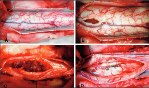
cephalo-sporin) and 10 mg dexamethasone IV bolus. Patients with upper thoracic or cervical cord tumors had their heads immobilized in a three-point Mayield-Kees device. surgical technique empha-sized a longitudinal skin incision, large enough to allow the ex-posure of normal cord above and below the level of the neo-plastic lesion. Laminectomies were performed when one or two levels were operated; when three or more laminae had to be re-moved, a laminotomy was performed instead, in order to pre-vent future deformities. The dura was opened longitudinally and a median longitudinal myelotomy was usually employed; only in those cases with a distinctive lateral cord topography was a paramedian myelotomy performed. our initial surgical goal was always aggressive, complete tumor removal. The tumor was resected in the caudal direction, en bloc whenever possi-ble without added morbidity (Fig 1). when severe morbidity was anticipated based on intraoperative indings, a subtotal or par-tial tumor resection was conducted. Patients were usually dis-charged on the fourth postoperative day and followed indei-nitely, including a new evaluation utilizing the McCormick scale at 6 months postoperatively.
statistical analysis was performed with sPss software (sPss Inc., Chicago, IL, UsA) with a signiicance level (p) of 0.05.
reSultS
The most common presenting symptom was pain lo-cated to the dorsum in half (24) of our patients (Table 1). This complaint varied immensely in nature and intensity but was correctly related to the affected cord segment in most patients. other subjective neurological complains such as paresthesia were the initial symptom in 35% (17 patients) while objective neurological signs including mo-tor weakness were the irst symptom in only a minority of our patients (7 patients – 15%). The 48 cases in our se-ries included 20 (42%) thoracic, 13 (27%) cervical and 15 (31%) cervico-thoracic cord tumors. Ependymoma was the most frequent histological diagnosis obtained (32 patients – 66.7%), followed by cavernous angioma (7 patients – 14.6%), three astrocytomas (6.3%), two lipomas and one ganglioglioma and hemangioblastoma each (Table 2).
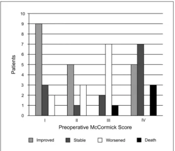
surviv-ing patients have been followed for at least six months, in-cluding four patients for more than 13 years. Late postop-erative evaluation (>6 months) of the surviving patients has shown that 19 of them had improved McCormick scores compared to their preoperative status (39.6%) and another 13 (27.1%) had maintained the same score while the remaining 12 patients (25%) were clinically worse than before the surgical procedure. when stratiied according to preoperative neurological function, a statistically sig-niicant trend of clinical improvement after surgery was evident in patients with McCormick I and II scores (Fig 2). Eighteen out of 23 patients with McCormick scores I and II were neurologically stable or better postoperative-ly; even patients with only discrete symptoms (Group I) clearly beneited from the surgical procedure. on the oth-er hand, clinical worsening could be expected from grade III patients; none of them improved while only 2 out of 10 maintained preoperative function. Those patients with severe lifestyle restrictions preoperatively (grade IV) re-mained largely stable but a small subset of them (ive pa-tients) exhibited some degree of improvement. deaths were present only in class III and IV patients.
It is also noteworthy that only 5 of the 13 patients which underwent partial resection eventually worsened and exhibited a decreasing McCormick score during the irst two years of follow-up. These ive patients were thus submitted to a second-stage procedure and in three of these patients total aggressive resection was obtained this time. Another three patients died but the surviving ive patients maintained stable neurological conditions and thus chose not to undergo another resection. All surviv-ing 44 patients underwent postoperative imagsurviv-ing control with Mr in the irst six months. out of the 33 surviving patients that underwent complete resection, MrI dem-onstrated remaining neoplastic tissue in only three (9.1%).
Table 1. Patient’s data.
Mean age 35.0
Age distribution <25 y: 11 (23%) 25–40 y: 25 (52%)
>40 y: 12 (25%)
Gender Male: 29 (60%)
Female: 19 (40%) Presenting symptom Back pain: 24 (50%)
Paresthesias: 17 (35%) Motor weakness: 7 (15%) Tumor location Cervical: 13 (27%)
Cervico-thoracic: 15 (31%) Thoracic: 20 (42%) Preoperative McCormick grade I: 14 (24%)
II: 9 (19%) III: 10 (21%) IV: 15 (31%)
Table 2. Postoperative results and complications.
Histological subtype Ependymoma 32 (66.7%) Cavernous angioma 7 (14.6%) Astrocytoma 3 (6.3%)
Anaplastic astrocytoma 2 (4.2%) Lypoma 2 (4.2%)
Hemangioblastoma 1 (2.1%) Ganglioglioma 1 (2.1%) Extent of resection Aggressive 34 (71%)
subtotal 14 (29%) Complications death 4 (8.3%)
Pneumonia 3 (6.3%) CsF istula 2 (4.2%) UTI 2 (4.2%)
respiratory failure 1 (2.1%) Facial burn 1 (2.1%)
Comparatively, 2 of the 11 surviving patients of the par-tial resection group did not exhibit any signs on MrI of remaining neoplastic tissue, thus largely conirming the surgeon´s impression on the operative microscope (chi-square test, p<0.001).
diScuSSion
our series comprises 48 patients with spinal cord pri-mary intramedullary tumors. demographic data of our se-ries are compatible with those of other authors: predomi-nantly young patients (75% under 40 years of age) with slightly male preponderance but differences start when analyzing presenting symptoms. similarly to most recent patient series, the most common presenting feature was pain usually but not always related to the affected seg-ment. on the other hand, only a minority of our patients exhibited evident clinical signs of motor compromise at presentation. This is considered logical due to the slowly progressing nature of primary intramedullary tumors, in contrast to the rapidly advancing clinical picture of in-tramedullary metastases5,13,20,21. This non-speciic clinical
picture emphasizes the need for aggressive imaging inves-tigation, further stressed by the improved prognosis if the patient still possesses a good preoperative McCormick score. Earlier authors of the pre-MrI era were thus fre-quently in an uncomfortable situation. The most useful in-vestigation tool at the time was myelography, an invasive and uncomfortable procedure, often yielding insatisfac-tory results and almost always incapable of distinguishing intramedullary from extramedullary tumors. Their pa-tients were frequently diagnosed with cord tumors late in their clinical course and this fact should be at least
partially responsible for many of the poor results of early treatment protocols for intramedullary tumors. Further-more these authors insisted on relatively lenient surgical actions, often limiting themselves to biopsies or partial resections, sometimes including adjuvant radiotherapy. Poppen, for instance, relied on decompressive laminecto-my and radiation therapy only while woods and Pimenta advocated partial resections and demonstrated prolonged survival for an interesting portion of their patients3,6.
Apart from improved diagnostic tools, another two important technological advances must be held respon-sible for these improved surgical results, namely the oper-ating microscope and accurate bipolar coagulation. From the 1960s onward, the irst patient series with complete microsurgical removal of intramedullary tumors while maintaining satisfactory postoperative function appeared. Important literature contributions such as those by Gar-rido and stein, Malis and stein gradually led to complete tumor removal becoming standard practice and later even eliminating the need for adjuvant therapies9,10,22. The
mod-ern surgical technique is still based on the principles laid by those pioneering neurosurgeons with few variations. our group employs a wide laminoplasty reaching at least one level above and below the tumor, even though this procedure was originally intended to be employed in young patients to avoid future deformities. It is our irm belief that it is not more time consuming than a stan-dard laminectomy and greatly facilitates surgical exposure should the patient need to be reoperated on. Aggressive total resection is the surgical aim but this should not be pursued at all costs. In this series, neither ultrasonic aspi-ration nor physiological monitoring were available. some surgeons have shown these instruments to be helpful but not indispensable for a successful outcome. our results show that aggressive total resection goal may be safely reached in the absolute majority of our cases.
This series also included an unusually important num-ber of cavernous angiomas, which was more frequent than astrocytomas, in contrast to what is usually found in the literature10,23. Apart from this fact, the surgeons´
impres-sion regarding complete resection correlated favorably with postoperative MrI. In only ive of the 44 patients who underwent postoperative investigation these two criteria did not exactly correlate, which is the irst time that the concept of aggressive resection of these tumors is corroborated by objective imaging indings and further statistical analysis. Immediate postoperative neurologi-cal deterioration was observed in all cases but those pa-tients who seek treatment early in their clinical course frequently reach their preoperative activity level or even improve beyond that. The immense value of rehabilitation cannot be overestimated in this situation; only the group
with severe but not complete neurological compromise exhibited a worse functional outcome. Finally, adjuvant chemo- and radiotherapy are reserved for the few pa-tients whose tumor histology was shown on examination to be particularly aggressive, in this series being indicated only for the two patients with anaplastic astrocytomas. our results largely corroborate those previously found in the literature but provide new evidences to inally en-grave the concept of aggressive resection of primary intra-medullary tumors especially in patients with subtle neuro-logical complaints. In fact, this subgroup may be the most beneited by early surgical intervention. The treatment of this complex problem has certainly come a long way; the results of this study demonstrate the validity of the modii-cations gradually introduced while the correlation of intra-operative surgical impression with postintra-operative imaging and the demonstration of signiicant functional recovery after surgery are the two main new arguments supplied by this study to help support this concept. several points still need to be reassessed and there certainly is room for improvement. Even more aggressive imaging investigation of subtle clinical indings, enhanced intraoperative elec-trophysiological monitoring and increased understanding of the molecular biology of these tumors will certainly spearhead research on this subject into this century and ultimately lead to improved treatment of these patients.
referenceS
1. Hausmann ON, Kirsch EC, Tolnay M, et al.. Intramedullary spinal cord tumours: a clinical outcome and radiological follow-up study. Swiss Med Wkly 2001;131:582-587.
2. Miller DC. Surgical pathology of intramedullary spinal cord neoplasms. J Neuro Oncol 2000;47:189-194.
3. Stein BM. Intramedullary spinal cord tumors. Clin Neurosurg 1982;30: 717-741.
4. Sutter B, Arthur A, Laurent J, et al. Treatment options and time course for intramedullary spinal cord metastasis: report of three cases and re-view of the literature. Neurosurg Focus 1998;4:Article 3.
5. Jellema K, Overbeeke JJ, Teepen HLJM. Time to diagnosis of intraspi-nal tumors. Eur J Neurol 2005;12:621-624.
6. Poppen JL. An atlas of neurosurgical techniques. Philadelphia & Lon-don: W.B. Saunders & Co 1960:424-428.
7. Greenwood Jr J. Total removal of intramedullary tumors. J Neurosurg 1954;11:616-621.
8. Chi JH, Cachola K, Parsa AT. Genetics and molecular biology of intra-medullary spinal cord tumors. Neurosurg Clin N Am 2006;17:1-5. 9. Malis LI. Intramedullary spinal cord tumors. Clin Neurosurg 1978;25:
512-539.
10. Stein BM. Surgery of intramedullary spinal cord tumors. Clin Neuro-surg 1979;26:529-542.
11. Brotchi J, Dewitt O, Levivier M, et al. A survey of 65 tumors within spi-nal cord: surgical results and the importance of preoperative magnetic resonance imaging. Neurosurgery 1991;29:651-657.
12. Jallo GI, Danish S, Velasquez, et al. Intramedullary low-grade astro-cytomas: long-term outcome following radical surgery. J Neuro Oncol 2001;53:61-66.
13. Nadkarni TD, Rekate HL. Pediatric intramedullary spinal cord tumors: critical review of the literature. Child´s Nerv Syst 1999;5:17-28. 14. Balmaceda C. Chemotherapy for intramedullary spinal cord tumors. J
Neuro Oncol 2000;47:293-307.
15. Isaacson SR. Radiation therapy and the management of intamedullary spinal cord tumors. J Neuro Oncol 2000;47:231-238.
16. Jyothirmayi R, Madhavam J, Nain MK, et al. Conservative surgery and radiotherapy in the treatment of spinal cord astrocitomas. J Neuro On-col 1997;33:205-211.
17. Lewis SP, Pizer BL, Coakham H, et al. Chemotherapy for spinal cord astrocytoma: can natural history be modified? Child´s Nerv Syst 1998;14:317-321.
18. Parsa AT, McCormick PC. Intramedullary spinal tumors: recent advanc-es and future directions. Neurosurg Clin N Am 2006;17:ix.
19. McCormick PC, Stein BM. Intramedullary tumors in adults. Neurosurg Clin N Am 1990;1:609-630.
20. Cortigan DA, Winkelman MD. Intramedullary spinal cord metastasis: a clinico pathological study of 13 cases. J Neurosurg 1985;62:227-233. 21. Peker S, Ozgen S, Ozek M, et al. Surgical treatment of intramedullary
spinal cord ependimomas: Can outcome be predicted by tumor param-eters? J Spinal Disord Tech 2004;17:516-521.
22. Garrido E, Stein BM. Microsurgical removal of intramedullary spinal cord tumors. Surg Neurol 1979;7:215-219.