CLINICAL APPLICATION OF MAGNETIC RESONANCE
IN ACUTE TRAUMATIC BRAIN INJURY
Dionei F. Morais
1, Antonio R. Spotti
2, Waldir A. Tognola
3, Felipe F.P. Gaia
4, Almir F. Andrade
5Abstract – Purpose: To evaluate the clinical applications of magnetic resonance imaging (MRI) in patients with acute traumatic brain injury (TBI): to identify the type, quantity, severity; and improvement clinical-radiological correlation. Method: Assessment of 55 patients who were imaged using CT and MRI, 34 (61.8%) males and 21 (38.2%) females, with acute (0 to 5 days) and closed TBI. Results: Statistical significant differences (McNemar test): ocurred fractures were detected by CT in 29.1% and by MRI in 3.6% of the patients; subdural hematoma by CT in 10.9% and MRI in 36.4 %; diffuse axonal injury (DAI) by CT in 1.8% and MRI in 50.9%; cortical contusions by CT in 9.1% and MRI in 41.8%; subarachnoid hemorrhage by CT in 18.2% and MRI in 41.8%. Conclusion: MRI was superior to the CT in the identification of DAI, subarachnoid hemorrhage, cortical contusions, and acute subdural hematoma; however it was inferior in diagnosing fractures. The detection of DAI was associated with the severity of acute TBI.
Key woRDS: magnetic resonance imaging, traumatic brain injury.
Aplicação clínica da ressonância magnética em pacientes com traumatismo craniencefálico agudo
Resumo – Propósito: Avaliar a aplicação clínica da ressonância magnética (RM) em pacientes vítimas de traumatismo craniencefálico (TCe) agudo, na identificação do tipo, número, gravidade e correlação clínica-radiológica. Método: Foram estudados prospectivamente 55 pacientes vítimas de TCe agudo fechado (0–5 dias), por TC e RM, sendo 34 do sexo masculino e 21 do feminino. Resultados: Houve diferença estatisticamente significante (teste McNemar): fraturas de crânio foram detectadas em 29,1% pacientes na TC e 3,6% pela RM; hematoma subdural 10,9% na TC e 36,4% pela RM; lesão axonal difusa (LAD) 1,8% pela TC e 50,9% na RM; contusões corticais 9,1% na TC e 41,8% pela RM, hemorragia subaracnóidea 18,2% na TC e 41,8% pela RM. Conclusão: A RM foi superior à TC na identificação da LAD, hemorragia subaracnóidea, contusões corticais e hematoma subdural agudo, porém inferior no diagnóstico de fraturas. A detecção de LAD pela RM foi associada com maior gravidade do TCe agudo.
PALAVRAS-CHAVe: ressonância magnética, traumatismo craniencefálico.
Departamento de Ciências Neurológicas, Faculdade de Medicina de São José do Rio Preto, São José do Rio Preto SP, Brazil (SJRP): 1Professor Assistente,
Serviço de Neurocirurgia do Hospital de Base, SJRP; 2Professor Adjunto Doutor do Departamento de Ciências Neurológicas - FAMeRP, SJRP; 3Professor
Adjunto Livre Docente do Departamento de Ciências Neurológicas - FAMeRP, SJRP; 4Residente do Serviço de Neurocirurgia do Hospital de Base, SJRP; 5Professor Adjunto Livre Docente do Departamento de Neurocirurgia - emergência, HC/FMUSP-SP.
Received 17 August 2007, received in inal form 9 November 2007. Accepted 11 December 2007.
Dr. Dionei Freitas de Morais – Avenida José Munia 4850 - 15090-500 São José do Rio Preto SP - Brasil. E-mail: [email protected]
Traumatic brain injury (TBI) is one the most frequent causes of the world’s morbimortality1. It turns productive and young individuals into dependent patients who usu-ally require decades of specialized and highly expensive care. For these reasons it is essential to deine in a more precise way the true extension of the initial encephalic damage, so that the different types of traumatic lesions are understood, to enable better treatment. Neurologi-cal assessment of the traumatized patients consists main-ly in the anamain-lysis of motor activity, of the pupillary relex and of the consciousness level2. Variations in the level of consciousness constitute the best indicator of the brain’s global function. The assessment of the level of
conscious-ness determined by the Glasgow coma scale is a guide for conduct and selection of diagnostic exams3.
reduction of the equipment and exams, faster time in the acquisition of the image, development of new sequenc-es such as the T2* “ecoplanar” gradient, FLAIR and diffu-sion, few bone artifacts and capacity of assessing ence-phalic trunk, cranial nerves and rear fossae allow the MRI to be used in the acute phase of the TBI8,9.
The goal of this study was to evaluate the clinical appli-cation of MRI on patients with acute TBI, to better identi-fy the type, quantity and severity of the encephalic lesion.
METHOD
From January 2002 to June 2005, 55 patients, victims of closed acute traumatic brain injury, were prospectively studied, treated at the emergency Service of Hospital de Base (a teach-ing hospital) de São José do Rio Preto - SP, subjected to clinical assessment and handling according to the norms of Advanced Trauma Life Support (ATLS®)2 followed by neurological
assess-ment and initial CT.
According to the Glasgow Coma Scale (GCS) modiied by Stein and Ross10, the traumatic brain injury was classiied as mild,
moderate and severe.
within 5 days after the trauma, only the patients with con-sciousness neurological alteration (Glasgow < 15) and focal def-icit that did not require immediate neurosurgery were subject-ed to the exam through MRI.
This study was approved by the Committee of ethics in Research (CeR) of the São José do Rio Preto Medical School (FAMeRP).
The reports were issued by radiologists from the Radiolo-gy Services of Hospital Base de São José do Rio Preto - SP, and were revised by a neuro-radiologist from Hospital Beneicência Portuguesa de São Paulo, SP.
The CT exam of the skull and the brain was performed on a Philips Tomoscan SR 4000 (Philips Medical Systems, Best, Hol-land) machine on a table with axial support for the head rest-ing the patient on a dorsal decubitus with the image of the skull on proile and slant of the orbit-meatal line. The protocol in-cludes images through CT in axial cuts of the rear fossae (5/5 mm), supratentorial (10/10 mm) and of the bone window of the whole brain.
The encephalic MRI exam was performed on a Philips Gy-roscan Intera T15 de 1.5 Tesla (Philips Medical Systems, Best, Hol-land) machine with the patient positioned on the examination table on a dorsal decubitus, using a head coil, keeping himself / herself immobile and breathing normally.
The protocol includes images through MRI on the sequenc-es spin-eco turbo axial in the T2 weighting, epi axial in the T2*, FLAIR axial in the T2, axial in the diffusion and spin turbo sag-ittal in the T1.
Restless patients and those who were in a coma were giv-en sedation (midazolan, propofol) on anesthetic induction to take the images.
Patients above 12 years of age, stable on the cardiovascu-lar and respiratory systems, and without devices such as
pace-makers, intra-ocular projectiles and surgical clips or ire or cut-ting and thruscut-ting weapon wounds were included in the present study.
In the comparative study of the lesions through the CT and MRI, the McNemar test11 was used.
To verify the possible association between the GCS and the two levels: mild or moderate/severe and the variables acute sub-dural hematoma, diffuse axonal lesion, cortical contusions, and subarachnoid hemorrhage, the chi-square test was applied.
To compare medians between the diagnostic methods through imaging (CT and MRI), the Signal test was used for the variables: quantity of cortical contusions, time and quantity of lesions11.
The level of signiicance adopted was α=0.05. All the calcu-lations and analysis were performed using the Minitab program for windows, version 14.1312.
RESULTS
The sample consisted of 55 patients victims of acute TBI, with the age ranging from 13 to 83 years old (34.2±17.4 years old), with 34 (61.8%) from the masculine and 21 (38.2%) from the feminine gender treated between Jan-uary 2002 and June 2005. By the score of the GCS mod-iied by Stein and Ross10, 14 (25.5%) patients were classi-ied as severe, 25 (45.4%) as moderate and the remaining 16 (29.1%) as mild.
In this study the following lesions of the acute TBI were studied through CT and MRI: skull fracture (Fract),
extradural hematoma (EDH), acute subdural hematoma
(ASDH), subdural hygroma (Hygr), diffuse axonal injury
(DAI), cortical contusions (Ct-c), intraparenchymal hemato-ma (IPH), subarachnoid hemorrhage (SAH), intraventricular hemorrhage (IVH), diffuse cerebral swelling (DCS), hemi-spherical cerebral swelling (HCS), ischemia (Isch) (Table 1).
In this study, the MRI was superior to the CT in nosing DAI, CT and ASDH; however it was inferior in diag-nosis fractures (p<0.0005).
The analysis of the relationship of dependency among diagnosis through MRI and ASDH, DAI, Ct-c and SAH with the levels of severity by the GCS for mild or moderate/ severe TBI by the chi-squared test (Table 2).
with respect to the quantity of lesions, the amount detected in the CT varied from 0 to 5, while in the MRI the range was from 0 to 8. on average, 1.4 lesions were found by the CT with standard deviation of 1.27. The same calculus for the MRI data revealed an average of 3.2 with standard deviation of 1.82.
The median values denote that MRI detected 2 more lesions per patient than the CT (p<0.0001).
2.5 days with a standard deviation of 1.59 days. In terms of median values, a difference of one day between the CT and the MRI is observed, being 2 days for the TC and 3 days for the MRI to be done.
The results for the time gap between the exams in-dicate that the period between one exam and the oth-er did not exceed 1 day, whoth-ereas in 24 (43.6%) of the pa-tients it was performed on the same day; in 11 (20%) the
CT was performed prior to the MRI; and in 20 (36.4%) the MRI was performed prior to the CT. Through the Signal test (p=0.1496).
DISCUSSION
In the present research, the most important clinical application of the magnetic resonance in patients with acute TBI was the precise identification of intra-axial (diffuse axonal lesion, cortical contusions) and extra-ax-ial (acute subdural hematoma and subarachnoid hemor-rhage) encephalic lesions; it was also possible to detect the greater quantity of lesions. with respect to the clinic-radiological correlation, there was only signiicant associa-tion between diffuse axonal lesions detected by the MRI and moderate/serious TBI according to the GCS. For skull fractures, the CT was superior to the MRI in diagnosing this lesion.
In this study, the CT showed this type of lesion (skull fractures) in 16 (29.1%) patients while the MRI detected it only in 2 (3.6%); one of them presented an adjacent corti-cal lesion. It is also important to consider that even very long traces of fracture might not be evidenced on the CT if they are parallel to the plan of the cutting. Therefore, the normal CT did not exclude, deinitely, the existence of linear fractures.
Acute subdural hematoma: found in 10 to 20% of the patients victims of TBI, occurring in up to 30% of the fa-tal lesions13. Although the CT is also the preferred exam during the acute phase of the TBI, it shows limitations to demonstrate small subdural collections that protect themselves along the cranial vault such as13-15: 1) attenu-ation artifacts of the X-rays through the bony structures tend to darken them; 2) when the exam is documented
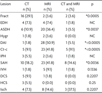
Table 1. Frequency and percentage of the patients whose lesion was diagnosed in each exam separately (CT or MRI); in both ex-ams simultaneously (CT and MRI); and value of P for the McNe-mar test.
Lesion CT n (%)
MRI n (%)
CT and MRI n (%)
p
Fract 16 (29.1) 2 (3.6) 2 (3.6) *0.0005 eDH 4 (7.3) 4 (7.4) 1 (1.8) NC ASDH 6 (10.9) 20 (36.4) 3 (5.5) *0.0037 Hygr 1 (1.8) 2 (3.6) 0 (0.0) NC DAI 1 (1.8) 28 (50.9) 1 (5.5) *<0.0005 Ct-c 5 (9.1) 23 (41.8) 5 (9.1) *<0.0005 IPH 3 (5.5) 2 (3.6) 1 (1.8) NC SAH 10 (18.2) 23 (41.8) 8 (14.6) *0.0036 IVH 1 (1.8) 5 (9.1) 1 (1.8) 0.1336 DCS 5 (9.1) 1 (1.8) 0 (0.0) 0.2207 HCS 3 (5.5) 0 (0.0) 0 (0.0) 0.25 Isch 4 (7.3) 8 (14.6) 3 (37.5) 0.2207
*Signiicant difference; NC, McNemar test may be faulty or not conclu-sive; Fract, skull fracture; eDH, extradural hematoma; ASDH, acute sub-dural hematoma; Hygr, subsub-dural hygroma; DAI, diffuse axonal injury; Ct-c, cortical contusions; IPH, intraparenchymal hematoma; SAH, subarach-noid hemorrhage; IVH, intraventricular hemorrhage; DCS, diffuse cere-bral swelling; HCS, hemispherical cerecere-bral swelling; Isch, ischemia.
Table 2. Frequency and percentage (chi-square test) to assess the relation between MRI diagnosis and the se-verity of TBI .
MRI (diagnosis) Chi-square test Mild TBI Moderate/severe TBI Total P ASDH yes
No Total
6 (37.5%) 10 (62.5%)
16
14 (35.9%) 25 (64.1%)
39
20 (36.4%) 35 (64.6%)
55 0.911 DAI yes
No Total
2 (12.5%) 14 (87.5%)
16
26 (66.7%) 13 (33.3%)
39
28 (50.9%) 27 (49.1%)
55 *<0.0001 Ct-c yes
No Total
7 (43.8%) 9 (56.3%)
16
16 (41.0%) 23 (59.0%)
39
23 (41.8%) 32 (58.2%)
55 0.105 SAH yes
No Total
4 (25.0%) 12 (75.0%)
16
19 (48.72) 20 (51.28%)
39
23 (41.8%) 32 (58.2)
55 0.105
with parameters that are adequate for the demonstration of soft parts (brain) the image of the hematoma may be confused with the one of the bony structures; 3) hemato-mas located below the temporal or occipital lobes, below the tentorium cerebelli and in the convexity, when small, tend to be darkened by the partial volume with the bone.
Inter-hemispheric acute subdural hematomas occur in adults who suffer trauma involving the whip mechanism (whiplash) or children who suffer lesions by a mechanism that is similar to spanking occasions. Although the CT is useful in the initial assessment of these patients, the MRI is more sensitive to detect small subdural collections that indicate the possibility of child beating and domestic vio-lence16,17. The most sensitive sequences to detect this le-sion are FLAIR and T2*.
For “laminate” acute subdural hematoma, the CT de-tected only 6 (10.9%) and the MRI 20 (36.4%) patients. Through McNemar’s test, the difference was signiicant. The MRI was more sensitive and detected this lesion in 86.95% of the patients, using the association of the se-quences T1, T2, FLAIR, T2* and diffusion.
Diffuse axonal injury: intra-axial lesion that along with the cortical contusions, represent the most impor-tant cause of morbid-mortality in patients with traumatic brain lesions13,15. The diagnosis is based upon the clinical condition and on the imaging exams (CT and MRI). Clini-cally, DAI is deined as an estate of immediate coma to the impact lasting more than 6 hours, that does not re-sult in expansive lesion (hematomas) or anoxic-ischemic lesions18. Its severity is graded according to the duration of the coma and the presence of signs of the impairment of the brainstem 19.
At the present study, the DAI was identiied by the CT in only 1 (1.8%) patient and in 28 (50.9%) by the MR. Through the McNemar’s test there was a statistically sig-niicant difference between the CT and the MRI, showing that MRI is more indicated when it comes to detecting and characterizing DAI20.
The initial CT on the patient who suffered DAI is usu-ally normal despite the severity of the clinical condition, only 25 to 50% of the patients with DAI showed abnor-malities on this exam13. Computed tomographies per-formed on subsequent dates may show lesions that were not visible on the initial exam. These lesions, on the acute phase, are seen as little hyper-dense petechial hemor-rhages, particularly at the point of convergence of white substance/grey substance at the rostral portion of the midbrain and at the corpus callosum8,20.
Showing the value of the MRI on the acute TBI, Mittl et al.21 investigated 20 patients with light TBI in which
the CT had not detected any abnormality. The MRI was performed in the sequences T2 and T2* for 4 days, show-ing abnormality compatible to a diffuse axonal injury in 6 (30%) patients.
In this investigation, the presence of diffuse axonal le-sion on the MRI was related to a moderate or severe TBI (Gl<14), where 26 patients (66.7%) out of the 39 with this score presented DAI. within those with mild TBI, only 2 (12.5%) presented DAI through MRI. These results are con-cordant with the ones obtained by Tokutomi et al.22, who studied 120 patients victims of closed, mild, moderate, and severe TBI with MRI using the sequences T1, T2, and FLAIR. The authors veriied that in 44 patients with mild TBI, only 1 (2%) presented signal alteration on the corpus callosum; only 3 patients (10%) out of the 31 with mod-erate TBI presented this alteration; 17 patients (38%) out of 45 with severe TBI had signal alteration on the corpus callosum compatible with DAI.
Paterakis et al.23 performed studies with an MRI in the sequences T1, T2, T2*, and FLAIR with 33 patients with acute and closed TBI in which the CT had not detected abnormalities. In this study, the DAI was found by the MRI in 24 patients, where 19 have severe TBI (Gl<8) and 5 have moderate TBI (Gl=9–12). They show that MRI is more sen-sitive than the CT in the detection of hemorrhagic and non-hemorrhagic axonal lesions, whereas the presence of hemorrhage is related to the worst prognostics. These re-sults are according to those obtained at the present study, especially those related to the highest occurrence of DAI in victims of severe and moderate TBI.
The diffuse axonal lesion is the most frequently ana-tomic-pathological inding detected in about 80% of the patients who were fatal victims of trafic accidents24,25. when studied by the CT, only larger hemorrhagic lesions were seen9. The magnetic resonance imaging has shown more sensitivity in the detection and characterization of these lesions9,21,22. By using MRI in the present study it was possible to evidence the DAI in about 51% of the patients, while the CT detected it in only 2%, with MRI being su-perior in relation to the majority of the projects consult-ed9,21,22. Furthermore, a direct correlation of the presence of this lesion with the severity of the GCS was established. Similar indings were found in the literature9,26.
typically increase in the irst days after the trauma and then decrease gradually over time. Cortical contusions may impregnate themselves through the contrast27.
The MRI is much more sensitive than the CT when de-tecting the presence and extension of the cortical contu-sions. It is observed the presence of multiple supericial areas that are poorly delimited and appear frequently heterogeneous, as on the images weighed in T1 and T28,16. In the acute phase, the FLAIR sequence is the one which best demonstrates the hyper-signal of the swelling on the cortical and the T2* the hypo-signal of the focuses of hemorrhages of the contusion24.
In relation to the cortical contusions, the MRI was more sensitive when detecting, characterizing and quanti-fying the primary traumatic lesions. Moreover, the results showed there was no relationship of dependency between the Ct-m and the severity in the GCS score. These results are similar to those found by other researchers3,8,9,15,16,20.
Subarachnoid hemorrhage appears with a thin collec-tion of blood on the subarachnoid space between the pia mater and the arachnoid mater. It is usually focal along the area of contusion, ASDH, laceration, fracture or diffuse in the arachnoid space and basal cisterns (interpeduncular cisterns). This lesion was found by CT in 10 (18.2%) patients and in 23 (41.8) by MRI. The superiority evidenced by MRI is due to the performance of the FLAIR sequence that is more sensitive when detecting the traumatic SAH24,28,29.
on CT, SAH appears as hyper dense images in relation to the brain tissue along the furrows and cisterns. This method is considered to be superior to the conventional MRI (T1, T2) in detecting this lesion. However, with the development of new sequences such as FLAIR, that sup-presses the cerebrospinal luid of the subarachnoid space, it was possible to better visualize SAH than with CT care-fully performing the exam with oxygen at 40 to 50% more room air or with nitrous oxide, because the oxygen at 100% would leave the subarachnoid space whitish, with the pos-sibility of super-estimating the diagnostic of the SAH30. on MRI, the subarachnoid hemorrhage may be better detected than by CT, mainly on FLAIR sequences through signal hyper-intensity and on ecoplanar gradient through signal hypo-intensity.
There was no statistical difference on the intraventric-ular hemorrhage, diffuse and hemispherical brain swelling and on the ischemia diagnosis. Moreover, it was not con-clusive for extradural hematoma, subdural hygroma, in-traparenchymal hematoma, due to the size of the sample and the number of patients with these lesions.
TBI cause great morbidity producing deiciencies on the information process when assessed by
neurophysi-ologic tests. The severity of the brain lesion should not be evaluated exclusively by the extension of the loss as de-termined on the neuropsychological tests; methods of di-agnosis through imaging must also be used to detect ana-tomic and physiological abnormalities of the brain tissue31. Many different MRI sequences have been applied to the assessment of closed TBI. The ecoplanar gradient se-quence at the T2* weighing contributed to demonstrate chronic and acute intra-axial hemorrhage32. The utility of FLAIR images in diagnosing swelling and subarachnoid hemorrhage was studied by many researchers28,29,33. New discoveries of images through diffusion ( diffusion-weight-ed, apparent diffusion coeficient, diffusion tensor imaging) besides demonstrating diffuse axonal and ischemic lesions better, also indicate the prognostic more precisely34-37.
In the present study, the magnetic resonance imaging was superior to the computerized tomography in diagnos-ing the diffuse axonal injury, subarachnoid hemorrhage, multiple contusions and laminate acute subdural hema-toma, though inferior to diagnosing fractures.
The magnetic resonance imaging detected a greater number of traumatic lesions and its greatest utility is di-agnosing the diffuse axonal injury. The presence of DAI is associated with greater severity of the acute traumatic brain injury.
REFERENCES
1. Kraus JF, McArthur DL, Silverman TA, Jayaraman M. Epidemiology of brain lesion In: Narayan RK, Wilberger JE, Povishock JT (Eds). Neu-rotrauma. New York: McGraw-Hill; 1996;13-30.
2. Comitê de Trauma do Colégio Americano de Cirurgiões. Suporte Avan-çado de Vida no Trauma (SAVT - ATLS®),6ª Ed. Illinois: American Col-lege of Surgeons; 1997.
3. Teasdale G, Jennett B. Assessment of coma and impaired consciousness. Lancet 1974;2:81-83.
4. Gentry LR. Imaging of closed head lesion. Radiology 1994;191:1-17. 5. Toyama Y, Kobayashi T, Nishiyama Y, Satoh K, Ohkawa M, Seki K. CT
for acute stage of closed head lesion: review. Radiation Med 2005;23:309-316.
6. Andrade AF, Almeida AN, Bor-Seng-Shu E, Lourenço L, Mandel M, Marino R Jr. The value of cranial computed tomography in high-risk, midly head-injured patients. Surg Neurol 2006;65:10-13.
7. Gean AD. Traumatic brain lesion: imaging update 2004; http://www. nordictraumarad.com/Syllabus04/gean.pdf.
8. Gentry RL, Godersky JC, Thompson B, Dunn VD. Prospective
compar-ative study of intermediate-ield MR and CT in the evaluation of closed
head trauma. AJR 1988;150:673-682.
9. Gentry RL, Thompson B, Godersky JC. Trauma to the corpus callosum: MR features. AJNR 1988;9:1129-1138.
10. Stein SC, Ross SE. Moderate head lesion: a guide to initial management. J Neurosurg 1992;77:562-564.
11. Zar JH. Biostatistical analysis. 4th Ed. New Jersey: Prentice Hall 1999. 12. Minitab, lnc. Minitab statistical softwareTM , Release 14.13.
Copy-right©2004.
13. Osborn AG. Traumatismo craniencefálico. In Osborn AG (Ed). Diag-nóstico neurorradiológico. Rio de Janeiro: Revinter 1999:199-247. 14. Macpherson BC, Macpherson P, Jennett B. CT evidence of intracranial
contusion and hematomas in relation to the presence, site and type of skull fracture. Clin Radiol 1990;42:321-326.
16. Kelly AB, Zimmerman RD, Snow RB, Gandy SE, Heier LA, Deck MD. Head trauma: comparison of MR and CT-experience in 100 patients. AJNR 1988;9:699-708.
17. Zimmerman RA. Computed tomography of craniocerebral lesion in the abused child. Radiology 1979;10:687-690.
18. Gentry RL. Primary neuronal lesions. Neuroimag Clin N Am 1991;1: 411-432.
19. Gennarelli TA. Cerebral concussion and diffuse brain lesions. In: Coo-per PR (Ed). Head lesion. 3ª Ed. Baltimore: Williams & Wilkins, 1993: 137-158.
20. Gentry RL, Godersky JC, Thompson B. MR imaging of head trauma: review of the distribution and radiopathologic features of traumatic le-sions. AJNR 1988;9:101-110.
21. Mittl RL, Grossman RI, Hiehle JF, et al. Prevalence of MR evidence of diffuse axonal lesion in patients with moderate head lesion and
nor-mal head CT indings. AJNR 1994;15:1583-1589.
22. Tokutomi T, Hirohata M, Miyagi T, Shigemori M. Posttraumatic edema in the corpus callosum shown by MRI. Act Neurochir 1997;70:80-83. 23. Paterakis K, Karantana AH, Komnos A, Volikas Z. Outcome of patients
with diffuse axonal lesion: the signiicance and prognostic value of MRI
in the acute phase. J Trauma 2000;49:1071-1075.
24. Osborn AG. Primary effects of CNS trauma. In Osborn AG (Ed). Diag-nostic imaging brain, 4ª Ed. Washington: AMIRSYS 2004:4-41. 25. Gusmão SN, Silveira RL, Arantes A. Lesão axonal difusa In Pereira CU
(Ed). Neurotraumatologia. Rio de Janeiro: Revinter 2000:170-175.
26. Kampl A, Franz G, Aichner F, et al. The persistent vegetative state af
-ter closed head lesion: clinical and magnetic resonance imaging ind -ings in 42 patients. J Neurosurg 1998;88:809-816.
27. Andrade AF. Considerações sobre a classiicação do traumatismo leve
e monitoração da pressão intracraniana no traumatismo craniencefáli-co moderado e grave [Tese]. São Paulo, 2004.
28. Parizel PM, Van Goethem JW, Özsarlak Ö, Maes M, Philips CD. New
developments in the neuroradiological diagnosis of craniocerebral trau-ma. Eur Radiol 2005;15:569-581.
29. Rocha AJ, Silva CJ, Gama HP, et al. Comparison of magnetic resonance imaging sequences with computed tomography to detect low-grade
subarachnoid hemorrhage: role of luid-attenuated recovery sequence.
J Comput Assit Tomogr 2006;2:295-303.
30. Braga FT, Rocha AJ, Hernandez G Filho, Arikawa RK, Ribeiro IM, Fon -seca RB. Relationship between the concentration of supplemental
oxy-gen and signal intensity of CSF depicted by luid-attenuated inversion
recovery imaging. AJNR 2003;9:1863-1881.
31. Marshall LF, Marshall SB, Klauber MR. A new classiication of head le
-sion based on computerized tomography. J Neurosurg 1991;75(Suppl):
S14-S20.
32. Yanagawa Y, Tsushima Y, Tokumaru A, et al. A quantitative analysis of head lesion using T2*-weighted gradient-echo imaging. J Trauma 2000;49:272-277.
33. Lee B, Newberg A. Neuroimaging in traumatic brain imaging: Neu-roRx® 2005;2:372-383.
34. Huisman TAGM, Schwamm LH, Schaefer PW, et al. Diffusion tensor imaging as potential biomarker of white matter lesion in diffuse axo-nal lesion. AJNR Am J Neuroradiol 2004;25:370-376.
35. Liu AY, Maldjian JA, Bagley LJ, Sinson GP, Grossman RI. Traumatic
brain lesion: diffusion-weighted MR imaging indings. Am J Neurora -diol 1999;20:1636-1641.
36. Ducreux D, Huynh I, Fillard P, et al. Brain MR diffusion tensor
imag-ing and ibre trackimag-ing to differentiate between two diffuse axonal le -sions. Neuroradiology 2005;47:604-608.