9
Abstract – Exposure to aluminium during gestation causes changes in mammalian brain development and behavior. Our aim was to perform spectral analysis on electrocortical activity of Sprague Dawley male pups (30±3 days of age) whose mothers were treated with aluminium during gestation and lactation. There was a higher presence of power spectra in the delta range of parietal electrocortical activity, a lower presence in the theta range and increased values of the parameter DT as the ratio of delta to theta range in pups indirectly exposed to aluminium (whose mothers were drinking a 0.5% water solution of aluminium chloride during the gestation and lactation periods), compared to controls.
Key words:Maternal aluminium exposure, young rats, electrocortical activity, power spectra
UDC 612.82.08:59:546.62
INTRODUCTION
Many studies have shown the association between oral aluminium exposure and brain development in early life and have pointed out the high toxicity of alu-minium on the growth and development of fetuses and sucklings both in humans and experimental animals (Bishop et al., 1997; Yumoto et al., 2000; Colomina et al., 2005 ). Animal studies have documented central nervous system impairments following aluminium exposure, including neuropathological, neurochemical, neurophysiological and neurobehavioral changes (Wang et al., 2002; Walton, 2007; Ali et al., 2008).
MATERIALS AND METHODS
Experimental animals and aluminium treatment
Experiments were performed on young male Sprague Dawley rats after the lactation period. All procedures were done in accordance with the European Council Directive (86/609/EEC) and the rules for the care and use of laboratory animals at the Institute for Biological
Research, University of Belgrade. All animals were maintained in a 12 h light-dark cycle and a tem-perature-controlled (22-25°C) animal room. Female rats were placed in cages for time mating with males. After two days, during the gestation and lactation periods, each female rat (at the time – with offspring) was housed in a separate cage. There were two feeding regimens of the female rats: the control and with aluminium. Thus, the control animals were exposed
Surgical procedure
Experiments examining acute exposure employed surgical procedures performed on young male rats at 30±3 days of age under Zoletil (Virbac S.A., Carros, France) anesthesia at an initial dose of 60
mg/kg and a subsequent supplement of ~15 mg/kg, when necessary to obtain stable anesthesia through-out the experiment. Each animal was mounted in a stereotaxic apparatus. Partial round-shaped cranio-tomies were made over the parietal cerebral cortex (P: 2-2.5mm; L/R: 2-2.5 mm).
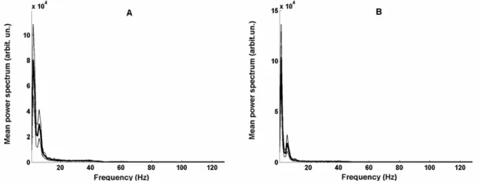
Fig. 2. Mean power spectra (±SD) of electrocortical activity from biosignals showed on Fig. 1.
recorded sequence lasted 121 s; there were at least 10 recorded sequences with interruptions of 5-10 min during 150 min of total acute experimentation. We analyzed digitized electrocortical signals at a sampling rate of 256 samples/s, filtered to avoid artifacts at 61, 107 and 121 Hz.
Data analysis
Spectral analysis of the recorded signals during sequences lasting 80 to 120 s (divided into epochs of 8 s), was obtained by Fast Fourier Transformation algorithm. Mean power spectra (of 10 to 15 epochs) of cerebrocortical activity were obtained in frequency ranges of 4 Hz up to 128 Hz. The parameter DT as the mean of at least five values of ratio of delta to theta power spectra of electro-cortical activity in each animal was established. Statistical t-test was used to test the difference of parameter DT between two groups of young animals.
RESULTS
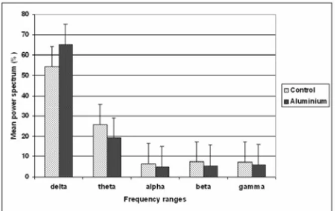
The anesthetized state of the investigated young rats was characterized by a predominance of slow delta range (0.1- 4.0 Hz) in the mean total power spectra at the parietocortical level, as was the case in adult animals (Culic et al., 2005). However, the slower rhythms were more pronounced in young rats with maternal aluminium treatment. The typical activities (Fig. 1) and spectral characteristics of recorded signals in one control rat and one rat indirectly treated with aluminium are shown in
Figs. 2, 3 and 4 (power spectra, frequency ranges histogram and the DT histogram of these two rats).
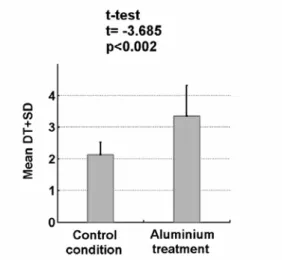
A statistical t-test showed that maternal aluminium treatment during gestation and lactation induced significant changes in the power spectral characteristics. The mean value of parameter DT was significantly increased (p<0.002) from 2.130 (control rats) to 3.348 (rats indirectly treated by aluminium) (Fig. 5).
DISCUSSION
Most behavioral studies have shown that chronic aluminium exposure at certain doses can cause behavioral morphological impairments in animals. In our study, results show that AlCl3 consumption (even the values from 45 mg/kg/day during ge-station to 160 mg/kg/day) during lactation, induce significant changes in the spectral characteristics of parietal cortical activity of offspring. There was an increase of power spectra in the delta range and a decrease in the theta range and particularly the established parameter DT as the ratio of delta and theta spectral powers was markedly greater in young rats with maternal consumption of alumi-nium during gestation and lactation, compared with the control rats. There could be some electro-physiological changes at some other level of the central nervous system but a greater sample should be studied, as we have already mentioned in our preliminary study (Podgorac et al., 2008) on young
rats with maternal aluminium treatment. The study on the relationship between aluminium exposure of female Wistar rats and the memory of their off-spring showed that consumption of not less than 800 mg/kg/day of AlCl3 during lactation can impair short-term and long-term memory of their off-spring (Ali et al., 2008). It seems that changes in brain activity could denote aluminium neuroto-xicity at lower doses than some behavioral tests. We have already shown that chronic aluminium chlo-ride parenteral administration may induce an in-crease of slow power spectra of the electric brain ac-tivity in adult rats (Martac et al., 2006).
The evaluated parameter DT as the ratio of relative delta and theta spectral powers of brain activity increased in young rats indirectly treated by aluminium during maternal exposure to aluminium chloride in gestation and lactation.
These trends of growth intensity in the delta range and decrease in the theta band could be detected in brain disorders by wavelet analysis. According to recent experimental results from Berkeley (Barbaro et al., 2006), slow frequency ranges (delta and theta) have a key role in the coordination of activities between the various centers in the brain, which, roughly speaking, exchange information at higher frequencies. From this point of view, a decrease of activities in the
theta range may indicate a neurotoxic effect of aluminium.
Acknowledgment – This work was supported by the
Minis-try of Science of the Republic of Serbia (Contract No. 143021).
REFERENCES
Ali, M. A., Simin, E. V., and C. Rahi (2008). Effect of Oral Aluminium Chloride Administration During Lactation on Short- and Long-Term Memory of Their Offspring. J. Biol. Sciences 8, 767–772.
Barbaro, N.M., Berger, M.S., Canolty, R.T., Dalal, S.S., Edwards,
E., Kirsh, H.E., Knight, R.T., Nagarajan, S.S., and
M.Soltani (2006). High Gamma Power Is Phase-Locked
to Theta Oscillations in Human Neocortex. Science313, 1626–1628.
Bishop, N. J., Morley, R., Chiz, B., Day, J. P., Lucas, A., andN.
Engl (1997). Aluminium neurotoxicity in preterm
infants receiving intravenous feeding solution. Medicine 336, 1557–1562.
Colomina, M.T., Roig, J. L., Torrente, M., Vicens, P., and J. L.
Domingo (2005). Concurrent exposure to aluminum
and stress during pregnancy in rats: Effects on postnatal development and behavior of the offspring. Neuro-toxicology and Teratology, 27, 565–574.
Ćulić, M., Martać-Blanuša, Lj., Grbić, G., Spasić, S., Janković, B., andA.Kalauzi (2005). Spectral analysis of cerebellar activity after acute brain injury in anesthetized rats. Acta Neurobiol. Exp. 65, 11–17.
Fig. 5. The mean value of parameter DT in the group of control young rats (N=10) and the group of young rats with maternal aluminium treatment (N=10).
СПЕКТРАЛНЕ ПРОМЕНЕ МОЖДАНЕ АКТИВНОСТИ КОД МЛАДУНАЦА ПАЦОВА ИЗЛОЖЕНИХ ДЕЈСТВУ АЛУМИНИЈУМА ТОКОМ ГЕСТАЦИЈЕ И ЛАКТАЦИЈЕ
Љ. МАРТАЋ1, Г. ГРБИЋ1, Г. КЕКОВИЋ1, Ј. ПОДГОРАЦ2, M. ЋУЛИЋ1, С. СЕКУЛИЋ3, Д. ЛАЛОШЕВИЋ3, и И. ЧАПО3
1 Институт за биолошка истраживања “Синиша Станковић”,
Универзитет у Београду, 11000 Београд, Србија
2Медицински факултет, Универзитет у Крагујевцу, 34000 Крагујевац, Србија 3Медицински факултет, Универзитет у Новом Саду, 21000 Нови Сад, Србија
Излагање дејству алуминијума током геста-ције и лактагеста-ције може довести до промена у развићу мозга и понашања код сисара. Наш циљ је био да се уради спектрална анализа електро-кортикалне активности младих Sprague Dawley
пацова мушког пола (старости 30±3 дана) чије су мајке биле третиране алуминијумом током гес-тације и лакгес-тације. У поређењу са контролом,
код младих пацова који су били индиректно тровани алуминијумом (чије су мајке пиле 0,5 % водени раствор AlCl3 током гестационог и