326
Despite optimized medical therapy, patients with chronic obs-tructive pulmonary disease (COPD) tend towards progressive dysp-nea, low tolerance to exercise, and an increase in morbidity and mortality when compared with those in cohorts of similar ages. When the forced expiratory volume in the first second (FEV1) is lower than 0.75 L or 30% of that predicted, the mortality rate is 40% to 50% in 3 years. Age > 65 years also contributes to increased mortality 1,2, and pulmonary hypertension, the major
com-plication of the disease, predisposes to the development of cor pulmonale, which, by itself, is associated with a poor prognosis 3.
The study of the ventricular function through conventional methods, such as echocardiography and computed tomography, is difficult 4,5.
Therefore, magnetic resonance imaging has been used as a more reliable method for analyzing the right ventricle (RV). In recent years, due to the difficulty in performing lung or heart-lung trans-plantation, lung volume reduction surgery (LVRS) has become an option to patients in an advanced stage of chronic obstructive pulmonary disease 6,7. From the cardiovascular point of view, LVRS
has been contraindicated to those with right and/or left ventricular dysfunction and to those with severe pulmonary hypertension 8.
Recently, some authors 9 have reported the possibility of reverting
right ventricular function with LVRS. If better ventricular function can be observed in some cases after pulmonary resection, why not expand its indication? What can truly suggest that the ventricular function is irreversible? Other questions regarding the left ventricle (LV) still persist. Is left ventricular dysfunction consequent to hypo-xia? Is coronary artery disease associated with or secondary to right ventricle dysfunction itself? 10–13
In the present study, the right and left ventricular functions of a group of patients with moderate to severe chronic obstructive pulmonary disease were carefully analyzed, aiming at, through the precise knowledge of biventricular function, understanding the mechanisms involved in the deterioration of the functions of 1 or both ventricles.
Methods
Twenty-seven patients with chronic obstructive pulmonary di-sease and 11 healthy individuals (control) were studied. The diag-nosis was based on the definition of the American Thoracic Society (ATS) 14. The inclusion criteria of the control group were the
presence of a normal physical examination, chest radiography, electrocardiography, and exercise testing, in addition to the absence
Original Article
Assessment of the Ventricular Function of Patients
with Advanced Chronic Obstructive Pulmonary
Disease by Using Magnetic Resonance Imaging
Nazareth de Novaes Rocha, Rafael Stelmach, Alberto Cukier, José Rodrigues Parga,
Luiz Francisco Ávila, Márcia Caldas, Paula Buck, Charles Mady
São Paulo, SP - Brazil
Instituto do Coração of the Hospital das Clínicas of the FMUSP Mailing address: Nazareth de Novaes Rocha - Rua Inhangá 7/601 – 22020-060 - Rio de Janeiro, RJ, Brazil
E-mail: [email protected] Received for publication: 8/12/03 Accepted for publication: 1/26/04 English version by Stela Maris Costalonga
Objective
To assess the mechanisms that may be involved in the evolu-tion of right and left ventricular dysfuncevolu-tion in patients with chronic obstructive pulmonary disease (COPD).
Methods
Magnetic resonance imaging was used in 11 control patients (group C) and 27 patients with COPD, who were divided into 2 groups, COPDc and COPDs, according to the presence or absence of right ventricular dysfunction, respectively. Doppler echocardiogra-phy was used for assessing the degree of pulmonary hypertension.
Results
The right ventricular diameter was similar in the 3 groups,
COPDs, COPDc and C (29±8 mm; 31±7 mm; and 30±6 mm;
respectively, P=NS). Right ventricular hypertrophy was observed only in the COPD groups (8±2 mm and 9±3 mm vs 5±1 mm; P<0.01). The percentage of systolic right ventricular lateral wall thickening (%RVLWT) in the 3 groups were as follows: 86±82% vs 41±35% vs 86±89%; P=NS). Different left ventricular ejec-tion fracejec-tions were observed in the groups as follows: 69±9% vs 55±16% vs 76±6%; P < 0.01. A positive and significant linear correlation was observed between the left ventricular (LV) diasto-lic diameter and the LV systodiasto-lic volume (r = 0.72; P < 0.01). No correlation was observed between the pulmonary volumes, arte-rial blood gas analysis, and ventricular function.
Conclusion
No correlation was observed between the severity of pulmo-nary function and the degree of ventricular function impairment. Whether a preserved %RVLWT means the possibility of reversi-bility of right ventricular function remains to be elucidated. However, the presence of the phenomenon of ventricular inter-dependence was confirmed.
Keywords
327
of a history of smoking. All participants signed a written informed consent after approval of the protocol by the local committee on ethics.
The patients with chronic obstructive pulmonary disease un-derwent pulmonary function tests and assessment of the ventricular function and the degree of pulmonary hypertension in a period of 10±3 days. After a 30-minute rest, an arterial blood sample was obtained for gas analysis. The pulmonary volumes and capacity of pulmonary diffusion were measured with adequate spirometric devices (Collins/GS; Milwaukee, USA) according to the recom-mendations of the ATS 15,16.
Pulmonary hypertension was quantified by using Doppler echo-cardiography (ATL – HDI 3000 or 5000 model; Bothell, WA, USA). The mean pulmonary artery pressure (MPAP) was quantified through the time of acceleration of the pulmonary flow (TAC) on Doppler 17-19. The systematic calculation of the pulmonary artery
systolic pressure through acquisition of the velocity of tricuspid regurgitation was not routinely used, due to the difficulty of obtai-ning the echocardiographic window in patients with pulmonary hyperinflation.
Coronary artery disease, as a cause of left ventricular dysfunction, was excluded due to the lack of history of precordial pain and of alterations in myocardial scintigraphy and in the pharmacological dobutamine stress test.
A previous history of systemic arterial hypertension and the presence of right bundle-branch block on the electrocardiogram, which could influence the interpretation of ventricular function, were recorded.
Considering that the irregular rhythm may hinder the measu-rement of cardiac chambers on magnetic resonance imaging, pa-tients with atrial fibrillation or frequent ectopic beats were excluded from the study protocol.
The 1.5T Horizon (GE) model was used for magnetic resonance imaging. To determine the anatomy and future planning of the images, 3 spin-echo sequences were obtained in the coronal, transverse, and sagittal planes. The gradient-echo technique was used with the following parameters: mean repetition time of 9 ms, echo time of 4 ms, cut thickness of 10 mm, field of view of 320°, tilt angle of 35 mm, and matrix of 128x256. The temporal resolution was 12 to 16 phases per cardiac cycle.
The following parameters were analyzed for each ventricle: ejection fraction (Simpson formula), systolic volume, cardiac output, thickening of the interventricular septum, percentage of septal thi-ckening during systole, thickness of the lateral ventricular wall, percentage of thickening of the lateral ventricular wall during sys-tole, diameter of the long and short ventricular axes during diastole and systole, and the ratio between the diameters of the long and short ventricular axes during both diastole and systole 20,21.
The percentage of systolic parietal thickening was obtained based on the equation 22:
% parietal thickening = (ST – DT) . 100% DT
where, ST = systolic parietal thickness, DT = diastolic parietal thickness.
The ratio between the short axis diameter and the long axis diameter (SA/LA) of the left ventricle during diastole and systole is used for observing the change in ventricular geometry during the cardiac cycle. The difference between the SA/LA ratios during
diastole and systole was used as a method for quantifying, in absolute values, the systolic motion of the interventricular septum towards the left ventricle. Therefore, the percentage of interven-tricular septal motion towards the left ventricle during systole (%LSD) was obtained based on the equation:
% LSD = (SA/LAdiastole – SA/LAsystole) . 100% SA/LA diastole
According to the literature 23,24, the patients with right
ventri-cular ejection fraction (RVEF) below 45% on magnetic resonance imaging were classified as having right ventricular dysfunction. Therefore, the patients with COPD were divided into the following 2 groups: COPDs group = without RV dysfunction; COPDc group = with RV dysfunction; group C = control (healthy).
The characteristics of the groups studied were compared using the Fisher exact test and the Student t test. The ANOVA and the Kruskal-Wallis test were used for assessing the differences between the parameters of the LV and RV obtained on magnetic resonance imaging. The Pearson test assessed the existence of linear corre-lations between the ejection fractions of the LV and RV, between the LV diameter during diastole and its systolic volume, and between the pulmonary parameters and MPAP. All calculations were per-formed using a system of statistical analysis (SSA). A P value < 0.05 was considered significant.
Results
The control, COPDs, and COPDc groups comprised 11, 16,
and 11 individuals, respectively. The characteristics of the 3 groups are shown in table I. Fifteen patients with chronic obstructive pulmonary disease were continuous users of O2 according to the national and international consensus of continuous O2 inhalation therapy 25-28.
Table II shows the results of arterial gas analysis and of the pulmonary function tests of the COPDs and COPDc groups. Statistical differences were not observed between both groups (P = NS). A linear correlation was observed between the predicted percentage of the forced expiration volume in the first second (%FEV1) and the partial pressure of arterial oxygen (PaO2) (r = 0.32; P<0.01). Quantification of the pulmonary artery pressure was technically possible in 20 patients with COPD (74%). The systolic pulmonary artery pressure (SPAP) and MPAP of the COPDs and COPDc groups were similar, 57±14 mmHg versus 59±17 mmHg, and 40± 9 mmHg versus 40±11 mmHg, respectively (P = NS). An MPAP
≥ 35 mmHg was observed in 13 patients with COPD, 8 in the
COPDs group, and 5 in the CPODc group. A linear and significant
correlation was observed between PaO2 and MPAP (r=0.45;
P < 0.01) (fig. 1). No correlation was observed between %FEV1 and MPAP. Moreover, no correlation was observed between MPAP and the RV ejection fraction (RVEF), and between PaO2 and the LV ejection fraction (LVEF).
Table III shows the parameters derived from the analyses of the right ventricle on magnetic resonance imaging in the 3 groups. The RVEF of the control, COPDs, and COPDc groups were, respec-tively, 54±8%, 53±6%, and 32±8%. These differences between
the RVEF were already expected, because the COPDc group was
328
systolic volume (RVSV) than the COPDs group (40±21 mL vs
66±19 mL; P = 0.006). No statistical difference was observed for right ventricular systolic volume (RVSV) between the COPDs and control groups. Although the COPDc group had a lower RVSV, the diameter of the right ventricle short axis during diastole (RVSA) was similar for the 3 groups. Patients with COPD had a greater right ventricular lateral wall thickness (RVLWT), which were
com-parable in the COPDs and COPDc groups (8±2 mm and 9±3 mm,
respectively); group C had a lower mean value (5±1 mm) (P<0.004, C vs COPDs, and P<0.004, C vs COPDc). No difference regarding the percentage of systolic right ventricular lateral wall thickening (%RVLWT) was observed in the 3 groups: C (86±89%),
COPDs (86±82%), and COPDc (41±35%) (P = NS).
Table IV shows the results derived from the assessment of left ventricular function on magnetic resonance imaging. The left ven-tricular ejection fraction (LVEF) was different in the 3 groups. The lowest LVEF was observed in the COPDc group.
A reversible perfusion defect in the inferoseptal area was obser-ved in 5 patients in the COPDc group and in 3 patients in the COPDs group on thallium myocardial scintigraphy and a pharma-cological dobutamine stress test. The LVEF values for individuals with and without these reversible perfusion defects were 52±15% and 66±11%, respectively (P < 0.01). The RVEF values for those with and without inferoseptal ischemia were 38±14% and 47±11%, respectively (P=0.07). The percentages of septal thi-ckening during systole (%ST) for those with and without infero-septal ischemia were 14±10% and 24±17%, respectively
(P=NS). During the study, 2 patients in the COPDc group with
Table I – Characteristics of the control, COPDs, and COPDc groups
Group C COPDs group COPDc group p
(n = 11) (n = 16) (n = 11)
Age (years)* 60±11 65±8 57±11 NS
Male sex (%) 9 (82) 13 (81) 7 (73) NS
BSA (m2)* 1.63±0.20 1.78±0.25 1.59±0.19 NS
Arterial hypertension (%) - 7 (44) 4 (36) NS
Right bundle-branch block (%) 0 (0) 4 (25) 2 (18) NS
Continuous use of O2 (%) - 8 (50) 7 (64) NS
* Results presented as mean ± standard deviation. BSA = body surface area. NS = P > 0.05; COPD = chronic obstructive pulmonary disease.
Table II – Arterial gas analysis and pulmonary function of the COPD* groups
COPDs group COPDc group p
PaO2 (mmHg) 66±14 63±17 NS
PaCO2 (mmHg) 48±13 50±07 NS
FEV1 (previewed %) 39±20 27±12 NS
RV (previewed %) 191±61 204±59 NS
TLC (previewed %) 115±21 104±29 NS
RV/TLC (previewed %) 167±33 185±26 NS
DLCO (previewed %) 42±29 53±16 NS
* Results presented as mean ± standard deviation. PaO2 = partial pressure of arterial oxygen; PaCO2 = partial pressure of carbon dioxide; FEV1 = forced expiration volume in the first second; RV = residual volume; TLC = total lung capacity; DLCO = diffusing lung capacity for carbon monoxide; COPD = chronic obstructive pulmonary disease. NS = P > 0.05.
Table III – Parameters of right ventricular function*
Group C COPDs group COPDc group p
RVEF (%) 54±8 53±6 32±8
RVSV (mL) 58±28 66±19 40±21 †
RVCO (L/min) 04±2 05±2 03±2 NS
RVLWT (mm) 05±1 08±2 09±3 ‡, §
%RVLWT 86±89 86±82 41±35 NS
RVSA (mm) 30±6 29±8 31±7 NS
* Results presented as mean ± standard deviation. †P < 0.01, COPDs
versus COPDc; ‡P < 0.01, group C versus COPDs group; §P < 0.01, group C versus COPDc group. RVEF = right ventricular ejection fraction; RVSV =
right ventricular systolic volume; RVCO = right ventricular cardiac output; RVLWT = right ventricular lateral wall thickness; %RVLWT = percentage of systolic right ventricular lateral wall thickness; RVSA = diameter of the right ventricular short axis at the end of diastole; COPD = chronic obstructive pulmonary disease. NS = P > 0.05.
Fig.1 - Graph of dispersion showing the linear correlation between the partial pressure of arterial oxygen (PaO2)and mean pulmonary artery pressure (MPAP).
Table IV – Parameters of left ventricular function*
Group C COPDs group COPDc group p
LVEF (%) 76±6 69±9 55±16 †, ‡, §
LVSV (ml) 70±22 50±17 52±20 †, xx
LVCO (L/min) 05±2 04±1 05±1 NS
ST (mm) 11±2 10±4 10±1 NS
%ST 46±63 38±44 24±20 NS
LVLWT (mm) 10±2 12±4 12±2 NS
%LVLWT 83±72 58±31 65±64 NS
LVSA (mm) 44±5 40±8 44±7 NS
329
inferoseptal ischemia underwent cine coronary angiography. The coronary arteries were normal.
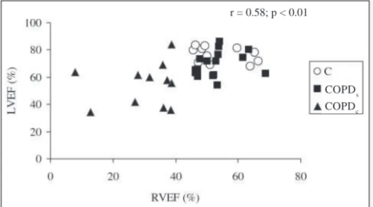
A positive and significant linear correlation was observed between LVEF and RVEF (r = 0.58) and between the diameter of the left ventricular short axis during diastole (LVSA) and its systolic volume (LVSV) (r = 0.72), figures 2 and 3, respectively.
The percentage of septal motion of the left ventricle during systole (%LSD) was similar in the 3 groups, C, COPDs, and COPDc, 7±12, 13±13, and 11±13, respectively (P = NS).
Discussion
Our population sample comprised patients with the advanced form of chronic obstructive pulmonary disease characterized by
the following parameters: %FEV1, 34±18; %RV/TLC (TLC = total
lung capacity), 175±31; and %DLCO (DLCO = diffusing lung
capacity for carbon monoxide), 47±24. Although PaO2 of 65±
15 mmHg may be considered high for such patients, it is worth noting that the arterial blood sample was obtained under continuous use of O2 in some patients. No correlation was observed between the degree of pulmonary impairment and the presence or absence of right ventricular dysfunction.
No differences in %RVLWT were observed between the groups.
Although a lower %RVLWT was observed in the COPDc group, it
did not reach statistical significance. On the other hand, the number of patients studied may have been too small to evidence differences between the groups. In addition, the fact that the RVLW thickens during systole suggests that right ventricular dys-function may still be reverted in those with RVEF < 45% after
lung transplantation or lung volume reduction surgery (LVRS). This may be an advantage of magnetic resonance imaging over the other methods, such as echocardiography and computed tomo-graphy, which do not provide a detailed analysis of the right ventri-cular lateral wall. Moreover, pulmonary hyperinflation, which is usually present in these patients, hinders echocardiographic per-formance 28-31. Measurement of the regional ventricular function
is important information for the analysis of myocardial feasibility. Certainly, a myocardial region that thickens during systole contains, at least, some viable tissue. The contrary, however, is not true – the absence of systolic parietal thickening does not necessarily implicate the absence of myocardial viability. However, still in these cases, the presence of viability may be assessed by using the contractile reserve test with dobutamine 31. Both the COPD
s
and the COPDc groups showed right ventricular hypertrophy with preserved %RVLWT.
The COPDs group had a greater RVSV than that in the COPDc group; however, the end-diastolic diameters of the right ventricle were similar. One explanation could be the restriction to right ventricular filling due to pulmonary hyperinflation.
Pulmonary artery pressure (PAP) in the COPDs and COPDc
groups were similar on Doppler echocardiography. Considering that PAP is the result of the product of RVSV by pulmonary vascular resistance, one may infer that pulmonary vascular resistance is greater in the COPDc group. Therefore, patients with the advanced form of chronic obstructive pulmonary disease may or may not have right ventricular dysfunction, depending on the degree of pulmonary vascular resistance. In 120 patients with emphysema who participated in the National Emphysema Treatment Trial, no correlation was observed between the ventilatory parameters and PAP 32. In our study, no correlation was observed between %FEV
1
and MPAP. Only a linear correlation was observed between PaO2
and MPAP. This, however, does not explain why patients with similar PaO2 may develop different values of pulmonary vascular resistance. Certainly, factors other than PaO2 and pulmonary func-tion seem to be involved, and these factors may be partially related to the individual genetic expression of pulmonary receptors for endothelin. The lungs account not only for the production, but also for the extraction of circulating endothelin, a peptide with potent vasoconstricting and proliferative action. In animals with pulmonary hypertension, a reduction in the expression of ETB recep-tors has been observed 33,34. Endothelin, when bound to ET
B
recep-tors, causes pulmonary vasodilation, while in the presence of only ETA receptors, it causes vasoconstriction.
Patients with chronic obstructive pulmonary disease showed lower LVEF as compared with that of controls. Factors considered risky for coronary artery disease, such as arterial hypertension and smoking, were observed in both COPD groups. Therefore, the possibility of coronary artery disease as a cause of left ventricular dysfunction had to be eliminated. Thus, all patients underwent myocardial scintigraphy with dobutamine, which showed reversible defects of radioisotope uptake in the inferoseptal region in 8 pa-tients. This finding may be a common artifact obtained in the per-fusion images, occurring in approximately 25% of the patients 35.
These defects are usually caused by attenuation of the radioisotopic activity of the inferior wall due to elevation of the left diaphragmatic cupula, as observed in obese patients.
Fig. 2 - Graph of dispersion showing the linear correlation between the right ventricular ejection fraction (RVEF) and left ventricular ejection fraction (LVEF) of all groups. Circle = control group; square = COPDs group; triangle = COPDc group.
COPD
s
COPDc
r = 0.58; p < 0.01
Fig. 3 - Graph of dispersion showing the linear correlation between the diameter of the left ventricular short axis (LVSA) and left ventricular systolic volume (LVSV) of all groups. Circle = control group; square = COPDs group; triangle = COPDc group.
COPD
s
COPDc
330
During our study, 1 patient underwent LVRS. That patient had severe left and right ventricular dysfunction prior to surgery. As that patient was 1 of those with defective inferoseptal radioisotopic uptake in myocardial scintigraphy, he underwent a complete hemo-dynamic study, and the absence of coronary artery disease was confirmed. After LVRS, the patient significantly improved his pul-monary function and remained out of the protocol of continuous O2 inhalation therapy. The major data of pulmonary and ventricular function before and 3 months after LVRS were, respectively: %FEV1,
18% vs 42%; %VEF1/FVC (FVC = forced vital capacity), 39% vs
43%; RVEF, 27% vs 64%; LVEF, 42% vs 53%; and MPAP, 50 mm Hg vs 33 mm Hg. As LVRS was performed in only 1 patient in our population sample, no inferences about the result of this surgery may be performed with the results of the present study.
Recently, Mineo et al 9 reported the effect of LVRS on ventricular
function. All patients underwent hemodynamic study before surgery, and 9 of the 12 patients had RVEF below 40% at rest. Those authors attributed the improvement in right ventricular function observed in the postoperative period to the Frank-Starling mecha-nism, because the increase in the right ventricular end-diastolic volume was accompanied by a parallel increase in RVSV. Those authors concluded that the reduction in the intrathoracic pressure may cause an increase in venous return, and, consequently, in ventricular filling. Other mechanisms include the decrease in pul-monary vascular resistance in the pulpul-monary regions that had previously undergone compression by the hyperinflated alveoli and improvement in the pulmonary elastic recoil 36-38.
Although LVEF was different in the 3 groups, LVSV was similar in the COPD groups. The increase in intrathoracic pressure seems to reduce left ventricular transmural pressure and increase cardiac output in patients with heart failure, while in those with normal ventricular function, the increase in intrathoracic pressure is asso-ciated with a reduction in cardiac output 39.
The contribution of interventricular septal motion towards the left to the phenomenon of ventricular interdependence remains controversial. The present study, showed no difference in the %LSD in the groups. However, it is worth noting that %LSD was assessed at the end of systole. Some studies 12 have reported the presence
of septal motion and its importance in restricting left ventricular
filling during the beginning of diastole. The presence of right ventri-cular hypertrophy and its smaller compliance cause a sudden in-crease in the right ventricular diastolic pressure with interventricular septal motion towards the left.
The presence of right bundle-branch block may contribute to loss of synchronism between the right and left ventricles and to a lower LVEF 40. In our study, a similar distribution of right
bundle-branch block was observed in the COPDs and COPDc groups, which cannot explain the differences observed in LVEF.
Our results are similar to those reported by Marcus et al 41,
who found left ventricular filling restricted in patients with primary pulmonary hypertension.
A limitation of the present study is the noninvasive measure-ment of PAP. However, some studies have already shown a good correlation between MPAP obtained on Doppler echocardiography and the direct measurement through hemodynamics, and between TAC also obtained by use of Doppler and the invasive measurement of pulmonary vascular resistance 42-44. However, future advances
in magnetic resonance imaging may facilitate the noninvasive mea-surement of PAP. Recently, Saba et al 45 reported a noninvasive
way of estimating PAP based on the ventricular mass index, and compared their results with those obtained on Doppler echocar-diography and invasive measurements. The confidence interval for the ventricular mass index was shorter than that for echocar-diography. The sensitivity and specificity for detecting pulmonary hypertension on magnetic resonance imaging were greater than those of echocardiography.
Another limitation was the fact that the diastolic function of the ventricles was not assessed, which could have provided addi-tional data regarding ventricular compliance.
In conclusion, patients with the advanced form of chronic obstructive pulmonary disease may have a preserved %RLWT, re-gardless of the presence or absence of right ventricular dysfunction. Left ventricular function depends, in most cases, on RVSV. In the present study, no correlation was observed between pulmonary and ventricular functions. Whether a preserved %RLWT means a possible reversion to right ventricular dysfunction is still to be defined and requires further studies.
1. Anthonisen NR. Prognosis in chronic obstructive pulmonary disease: results from multicenter clinical trials. Am Rev Respir Dis 1989; 14: 595-9.
2. Meyers BF, Yusen RD, Lefrak SS, Patterson GA, Pohl MA, Richardson VJ. Outcome of medicare patients with emphysema selected for, but denied, a lung volume reduction operation. Ann Thorac Surg 1998; 66: 331-6.
3. MacNee W. Pathophysiology of cor pulmonale in chronic obstructive pulmonary disease. Part two. Am J Respir Crit Care Med 1994; 150: 1158-8.
4. Global Strategy for the Diagnosis, Management and Prevention of Chronic Obs-tructive Lung Disease-GOLD. National Heart, Lung and Blood Institute/World Health Organization Workshop Report. www.goldcopd.com
5. Biernacki W, Flenley DC, Muir AL, MacNee W. Pulmonary hypertension and right ventricular function in patients with COPD. Chest 1988; 94: 1169-75. 6. Geerstma A, Tenvergert EM, Bonsel GJ, Boer WJ, van der Bij W. Does lung
trans-plantation prolong life? A comparison of survival with and without transplanta-tion. J Heart Lung Transplant 1998; 17: 511-6.
7. Maurer JR, Frost AE, Estenne M, Higenbottam T, Glanville AR. International gui-delines for the selection of lung transplant candidates. J Heart Lung Transpl 1998; 17: 703-9.
8. Yusen RD, Lefrak SS, Trulock EP. Evaluation and preoperative management of lung volume reduction surgery candidates. Clins Chest Med 1997; 18: 199-224.
References
9. Mineo TC, Pompeo E, Rogliani P et al. Effect of lung volume reduction surgery for severe emphysema on right ventricular function. Am J Respir Crit Care Med 2002; 165: 489-94.
10. Steele P, Ellis JH, Van Dyke D, Sutton F, Creagh E, Davies H. Left ventricular ejec-tion in severe chronic obstructive airways disease. Am J Med 1975; 59: 21-8. 11. Lazar JM, Flores AR, Grandis DJ, Orie JE, Schulman DS. Effects of chronic right
ventricular pressure overload on left ventricular diastolic dysfunction. Am J Cardiol 1993; 72: 1179-82.
12. Jessup M, Sutton MSJ, Weber KT, Janicki JS. The effect of chronic pulmonary hypertension on left ventricular size, function, and interventricular septal motion. Am Heart J 1987; 113: 1114-22.
13. Kun SL, Santamore WP. Contribution of each wall to biventricular function. Car-diovasc Research 1993; 27: 792-800.
14. American Thoracic Society. Standards for the diagnosis and care of patients with chro-nic obstructive pulmonary disease. Am J Respir Crit Care Med 1995; 152: S77–S120. 15. American Thoracic Society. Standardization of spirometry: 1987 update. Am Rev
Respir Dis 1987; 136: 1285-98.
331
17. Vazquez de Prada JA, Ruano J, Martin-Duran R et al. Noninvasive determination of pulmonary arterial systolic pressure by continuous wave doppler. Intern J Cardiol 1987; 16: 177-84.
18. Yock PG, Popp RL. Noninvasive estimation of right ventricular systolic pressure by Doppler ultrasound in patients with tricuspid regurgitation. Circulation 1984; 4: 657-62.
19. Chan KL, Currie PJ, Seward J, Hagler DJ, Mair DD, Tajik AJ. Comparison of three doppler ultrasound methods in prediction of pulmonary artery pressure. J Am Coll Cardiol 1987; 9: 549-54.
20. Feigenbaun H. Echocardiographic evaluation of cardiac chambers. Echocardiogra-phy. 5 ed. Baltimore, Lea & Febiger, 1999; 139-41p.
21. Peshock RM, Hundley WG, Willet D, Sayad DE, Chwialkowski MP. Quantitative magnetic resonance imaging of the heart. In: Skorton DJ, Schelbert HR, Wolf GL et al. Marcus Cardiac Imaging. A Companion to Braunwald’s Heart Disease. 2 ed. Philadelphia, Saunders, 1996; 759-83p.
22. Lima JAC, Jeremy R, Guier W et al. Accurate systolic wall thickening by nuclear magnetic resonance imaging with tissue tagging: correlation with sonomicrometers in normal and ischemic myocardium. J Am Coll Cardiol 1993; 21: 1741-51. 23. Rumberger JA, Behrenbeck T, Bell MR et al. For the Cardiovascular Imaging
Wor-king Group. Determination of ventricular ejection fraction: a comparison of avai-lable imaging methods. Mayo Clin. Proc 1997; 72: 860-70.
24. Turnbull LW, Ridgeway JP, Biernack W et al. Assessment of the right ventricle by magne-tic resonance imaging in chronic obstructive lung disease. Thorax 1990; 45: 597-610. 25. ATS Statement: Comprehensive outpatient management of COPD. Am J Resp Crit
Care Med 1995; 152 (Suppl): S84 – S96.
26. Nocturnal Oxygen Therapy Trial Group. Continuous or nocturnal oxygen therapy in hypoxemic chronic obstructive lung disease: a clinical trial. Ann Intern Med 1980; 93: 391-8.
27. Medical Research Council Working Party. Long term domiciliary oxygen therapy in chronic hypoxic cor pulmonale complicating chronic bronchitis and emphysema. Lancet 1981; 1: 681-6.
28. Morrison DA, Henry R, Goldman S. Preliminary study of the effects of low flow oxy-gen on oxyoxy-gen delivery and right ventricular function in chronic lung disease. Am Rev Respir Dis 1986; 133: 390.
29. Reiter SJ, Rumberger JA, Feiring AJ, Stanford A., Marcus ML. Precision of measu-rements of right and left ventricular volume by cine computed tomography. Circu-lation 1986; 74: 890-900.
30. Vigneswaran WT, Mc Dougall JC, Olson LJ, Breen JF, Mc Gregor CGA, Rumberger JA. Right ventricular assessment in patients presenting for lung transplantation. Transplantation 1993; 55: 1051-5.
31. Danchin N, Cornette EA, Henriquez A et al. Two dimensional echocardiography
assessment of the right ventricle in patients with chronic obstructive lung disease. Chest 1987; 92: 229-33.
32. Koebe HG, Kugler C, Dienemann H. Evidence-based medicine: lung volume reduc-tion surgery (LVRS). Thorac Cardiovasc Surg 2002 Oct; 50: 315-22.
33. Dupuis J, Stewart DJ, Cernacek P, Gosselin G. Human pulmonary circulation is an important site for both clearance and production of endothelin-1. Circulation 1996; 94: 1578-84.
34. Dupuis J, Cernacek P, Tardif JC et al. Reduced pulmonary clearance of endothelin-1 in pulmonary hypertension. Am Heart J endothelin-1998; endothelin-135: 6endothelin-14-20.
35. Wackers FJTH. Myocardial perfusing imaging. In Sandler MP, Coleman RE, Wackers FJTh, Gottschalk A. Diagnostic Nuclear Medicine, 3 ed. Baltimore, Williams and Wilkins, 1995, 443p.
36. Scharf SM, Iqbal M, Keller C, Criner G, Lee S, Fessler HE. Hemodynamic charac-terization of patients with severe emphysema. Am J Resp Crit Care Med 2002; 166: 314-22.
37. Noodergraaf AV, Marcus JT, Roseboom B, Postmus PE, Faes TJ, Vries PM. The effect of right ventricular hypertrophy on left ventricular ejection fraction in pulmo-nary emphysema. Chest 1997; 112: 640-5.
38. Louie EK, Lin SS, Reynertson SI, Brumdage BH, Levitaky S, Rich S. Pressure and volume loading of the right ventricle have opposite effects on left ventricular ejection fraction. Circulation 1995; 819-24.
39. Buda AJ, Pinsky MR, Ingels NB, Daughters GT II, Stinson EB, Alderman EL. Effect of intrathoracic pressure on left ventricular performance. N Engl J Med 1979; 310: 453-9.
40. Yasui H, Yoshitoshi M, Komori M et al. Cardiodynamic effects of experimental right bundle branch block in canine hearts with normal and hypertrophied right ventricles. Am Heart J 1985; 109: 69-77.
41. Marcus JT, Vonk Noordegraaf A, Roeleveld RJ et al. Impaired left ventricular filling due to right ventricular pressure overload in primary pulmonary hypertension. Chest 2001; 119: 1761-5.
42. Dabestani A, Mahan G, Gardin JM et al. Evaluation of pulmonary artery pressure and resistance by pulsed-wave doppler echocardiography. Am J Cardiol 1987; 59: 662-8.
43. Laaban JP, Diebold B, Zelinski R, Lafay M, Raffoul H, Rochemaure J. Noninvasive estimation of systolic pulmonary artery pressure using doppler echocardiography in patients with chronic obstructive pulmonary disease. Chest 1989; 96: 1258-62. 44. Marchandise B, De Bruyne B, Delaunois L, Kremer R. Noninvasive prediction of
pulmonary disease by doppler echocardiography. Chest 1987; 91: 361-5. 45. Saba TS, Foster J, Cockburn M, Cowan M, Peacock AJ. Ventricular mass index