www.jcol.org.br
Journal of
Coloproctology
☆ This study was conducted at Hospital Federal dos Servidores do Estado do Rio de Janeiro. * Corresponding author.
E-mail: [email protected] (L.H. Tauil).
2237-9363/$ - see front matter. © 2014 Sociedade Brasileira de Coloproctologia. Published by Elsevier Editora Ltda. All rights reserved. http://dx.doi.org/10.1016/j.jcol.2014.02.002
Original article
Immunohistochemistry expression of TCF4 protein on
carcinoma, adenoma and non neoplastic colorectal mucosa
☆Leonardo Huber Tauil
a,*
, Ana Maria Amaral Mader
b, Thamires Huber Tauil
c, Andrea Pires
d,
Lidia Maria Magalhães Rezende
e, Jaques Waisberg
aa Hospital Federal dos Servidores do Estado de São Paulo, São Paulo, SP, Brazil b Universidade de São Paulo, São Paulo, SP, Brazil
c Faculdades Souza Marques, Rio de Janeiro, RJ, Brazil d Universidade Federal Fluminense, Niterói, RJ, Brazil e Instituto Nacional do Câncer, Rio de Janeiro, RJ, Brazil
a r t i c l e i n f o
Article history:
Received 17 September 2013 Accepted 4 February 2014
Keywords:
Colorectal neoplasms TCF transcription factors Wnt proteins
Immunohistochemistry
a b s t r a c t
Purpose: To detect and quantify the immunoreactivity of TCF4 protein in colorectal carci-noma, colorectal adenoma and non-neoplasic colorectal epithelium.
Methods: We studied 129 individuals: 40 with colorectal cancer, 52 with colorectal ad-enoma and 37 with non-neoplastic colorectal epithelium. The colorectal adad-enoma and carcinoma samples were obtained from patients who underwent surgical procedures, and colonoscopies and samples of non-neoplastic colorectal epithelium were taken from patients who died from cardiovascular diseases, without diseases of the large intestine. Samples of different tissues were included in parafi n blocks, and the immunohistochem-ical expression of protein TCF4 was analyzed using the technique of tissue microarray (TMA) with polyclonal antibody TCF4. The immunoreactivity was analyzed and classii ed as positive and negative.
Results: The immunohistochemical expression of TCF4 protein was signii cantly higher (p < 0.01) in colorectal carcinoma than in the non-neoplastic colorectal epithelium and adenoma. There was no difference (p = 0.76) between TCF4 protein immunohistochemical expression in colorectal adenoma and non-neoplastic colorectal tissue.
Conclusions: TCF4 protein showed a more intense expression in colorectal carcinoma than in non-neoplastic colorectal epithelium and adenoma, indicating that this protein is in-volved in colorectal carcinogenesis.
Palavras-chave:
Neoplasias colorretais Fatores de transcrição TCF Proteínas Wnt
Imuno-histoquímica
r e s u m o
Expressão imuno-histoquímica da proteína TCF4 no carcinoma, no adenoma e na mucosa não neoplásica colorretal
Objetivos: Detectar e quantiicar a imunoexpressão da proteína TCF4 no carcinoma e no adenoma colorretal e no epitélio colorretal não neoplásico.
Método: Foram estudados 129 indivíduos: 40 com carcinoma colorretal, 52 com adenoma colorretal e 37 com epitélio colorretal não neoplásico. Os tecidos de adenoma e carcinoma colorretais foram representados por amostras da lesão retirada de doentes submetidos a procedimentos cirúrgicos e colonoscópicos, e as amostras de epitélio colorretal não neo-plásico foram retiradas de doentes falecidos por afecções cardiovasculares e sem compro-metimento do intestino grosso. As amostras dos diferentes tecidos foram incluídas em blo-cos de paraina e submetidas ao estudo da imunoexpressão da proteína TCF4 pela técnica do tissue microarray (TMA) com o anticorpo policlonal anti-TCF4. A imunorreatividade foi analisada e classiicada como positiva e negativa.
Resultados: A imunoexpressão da proteína TCF4 foi signiicantemente maior (p < 0,01) no carcinoma colorretal do que nos adenomas e no epitélio colorretal não doente. Não houve diferença signiicante (p = 0,76) entre a imunoexpressão da proteína TCF4 no adenoma co-lorretal e no epitélio coco-lorretal não doente.
Conclusão: A maior expressão da proteína TCF4 no carcinoma colorretal em relação ao ade-noma e ao epitélio não doente sugere que esta proteína possui participação na carcinogê-nese colorretal.
© 2014 Sociedade Brasileira de Coloproctologia. Publicado por Elsevier Editora Ltda. Todos os direitos reservados.
Introduction
With the increase in the average age of the world popu-lation in recent years, an increase of causes of death by neoplastic diseases was observed. Among these diseases, colorectal cancer (CRC) become prominent, especially in the western hemisphere population. About 50% of individ-uals will develop colorectal adenoma until the age of 70, and one in ten adenomas will progress to carcinoma.1 In
Brazil, 30,140 new cases of CRC will emerge, according to estimates: 14,180 in men and 15,960 in women, and half of these people will die from the disease.2
The sequence non-neoplastic colorectal tissue-ade-noma-carcinoma was irst described by Fearon and Vol-geistein,3 in 1978, and prompted studies to determine the
genetic and epigenetic alterations of this transformation. However, the molecular changes involved in this process are still not fully understood.
The Wnt signalling pathway plays an important role in the development of the human embryo, participating in the formation and homeostasis of organs and tissues. The improper functioning of this pathway participates in the carcinogenesis of human tissues, including colorectal tis-sue.4 When the Wnt pathway is activated, in most cases by
mutations of proteins members of the pathway itself, the usual result is an increase of the transcription of genes im-portant for growth, proliferation, differentiation, apoptosis, genetic stability, migration and angiogenesis.5 About 200
genes that are inluenced by this pathway, including genes c-myc, cyclin D1,6 VEGF (vascular endothelial growth factor)
and endothelin, were described.
The TCF/LEF family controls a great number of genes ac-tivated by Wnt. In this family, TCF4 gene is the component most commonly expressed in human colorectal tissue and is responsible for the activation of several genes linked to colorectal carcinogenesis, acting either in the more early stages (transformation of normal colorectal tissue to ad-enoma) as in later phases (distant metastases).7,8 Recently,
studies consistently showed evidence of the inluence of this gene in colorectal carcinogenesis. This gene exhibits a mutation 2.8 times more frequent in the CRC than in non-neoplastic colorectal tissue.9 Another study showed that
when overexpression of anti-TCF-4 occurred, colorectal tumours did not present a satisfactory response to neoad-juvant therapy,10 and when TCF4 is inhibited using
inter-ference RNA, an increased sensitivity of CRC to treatment occurs.11
Because of this evidence, it becomes important to deine patterns of expression of this protein in various types of colorectal tissue through a simpler, cheaper and more af-fordable method.
The aim of this study is the detection, by immunohis-tochemical method, of TCF4 protein, a member of the Wnt signalling pathway in colorectal adenoma and carcinoma and in non-neoplastic colorectal epithelium.
Methods
This is a retrospective study conducted at the Serviço de Co-loproctologia, Hospital Federal dos Servidores do Estado do Rio de Janeiro (HFSE), in which 163 parafi n blocks were analyzed in three groups: group 1 (carcinoma) with 46 blocks of patients with CRC operated, group 2 (adenoma) with 67 blocks of pa-tients who underwent endoscopic or surgical polypectomy, and group 3 (control) with 50 blocks of patients who died from heart disease and without indicative picture of digestive disease, and necropsied at the Serviço de Verii cação de Óbitos do Município de São Paulo (SVO - Universidade de São Paulo - USP).
For the formation of groups 1 and 2, patients treated in HFSE-RJ and who underwent colonoscopy or surgical proce-dures and were in outpatient follow-up during the study period were included.
Subjects under 18 years of age, patients with inl ammatory bowel disease or with non-adenomatous polyps and those who refused to participate in the study were excluded; 13 of 50 sam-ples from non-neoplastic colorectal tissue were excluded due to non-recognition of the colorectal mucosa or due to autolysis found in these samples; of 67 cases of colorectal adenomas, 15 were excluded due to the observation of only hyperplasic epi-thelium; and, i nally, of 46 carcinoma blocks 6 were excluded, due to samples that, on analysis, did not include areas with car-cinomatous transformation.
From the parafi n blocks obtained by the procedures above, blocks of tissue micro array (TMA) were prepared. The prepara-tion of TMA blocks followed the technique described by Pires et al.12 Three blocks were prepared : the i rst with the cases of
colorectal carcinoma, the second with colorectal adenomas and the third block with non-neoplastic colorectal tissue.
For the immunohistochemistry method, histological sec-tions of 3 mm thick were obtained; they were deposited on si-lanized slides and subsequently treated by streptavidin-biotin method.
Incubation with primary antibody was performed in a moist chamber at 4°C for a minimum of 16-18 hours (overnight), at a 1:10 dilution. The primary antibody used was polyclonal TCF-4 (NBP1-88633) obtained from rabbit (Novus Biologicals, Littleton, CO, USA).
The immunoreactivity of TCF4 protein was analyzed and classii ed as positive or negative.
The following data were recorded: clinical features of sam-ple (age and gender) for all groups of individuals. In the group of patients with colorectal adenoma, the morphological charac-teristics of the lesion (location, size of the longer axis, histologi-cal type, degree of cellular atypia) were recorded. In the group of patients with colorectal carcinoma, macroscopic lesion char-acteristics (location, appearance, size of the longer axis), mi-croscopic features (lymph node involvement, grade of cellular differentiation, lymphatic, venous and neural ini ltration), TNM classii cation (UICC, 2010),13 presence of synchronous
metasta-ses and tissue immunoexpression of the antibodies used (nega-tive or posi(nega-tive) were registered.
Statistical analysis of the quantitative results was reported as mean and standard deviation. The qualitative data were described as frequency. To analyze categorical variables, the Fisher exact test or the chi-square test was used; and for con-tinuous variables, the Student's t test was used, after testing for its normality.
The level of signii cance was set at 5% (p < 0.05) in all tests.
Results
This is a retrospective study in which three groups of sub-jects were studied, of which data of two groups were col-lected at HFSE-RJ, and data of the third group at SVO-USP. In both institutions the data were collected sequentially, with no attempt to matching among the groups with disease and the control group. No signii cant differences between groups in relation to clinical characteristics were found (Fig. 1).
Fifty-two subjects with colorectal adenoma were studied. These patients were classii ed according to location, size of the longest axis, histological type, degree of cellular atypia and TCF4 protein expression of colorectal adenoma. Most of the cases studied were of adenomas with < 10 mm and with low-grade cellular atypia and tubular shape (Fig. 2).
Expression of TCF4 protein in 7 (13%) cases was observed. In adenoma subtypes larger than 30 mm, with severe atyp-ia and villous histology, higher percentage of expression of TCF4 protein (42.9%, 25% and 33.3%, respectively) was noted, but the signii cance of this difference could not be demon-strated (Fig. 3).
Fig. 1 – Mean age of subjects, according to the group: carcinoma, adenoma and non-neoplastic colorectal tissue.
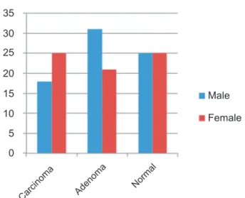
Fig. 2 – Distribution by gender of subjects according to the group: adenoma, carcinoma and non-neoplastic colorectal tissue.
90
80
70
60
50
40
30
20
10
0
Carcinoma Adenoma Normal
35
25
15
5
30
20
10
0
Carcinoma Adenoma
Normal
Male
Of the 40 cases of patients with colorectal carcinoma, most were located in the rectum, classii ed as II and III stag-es, greater than 50 mm, moderately differentiated, without vascular compromise and without metastases.
There was expression of TCF4 protein in 23 (57%) of sub-jects with colorectal carcinoma. The expression of TCF4 protein was compared, without significant difference with respect to gender (p = 0.52), age (p = 0.80), size (p = 0.20), grade of differentiation (p = 1.0), presence of metastasis (p = 1.0), vascular invasion (p = 1.0) and location (p = 0.80). The mean age of patients with carcinoma who expressed TCF4 protein was 65.5 ± 11.9 years; and of those who did not express the protein, the mean age was 66.4 ± 8.4 years, without significant difference between these values (Fig. 4).
The analysis of non-neoplastic colorectal tissues showed expression of the protein in 6 (16%) cases (Fig. 5).
We compared the expression of TCF4 protein between the group of patients with non-neoplastic colorectal tissue and the group of patients with colorectal adenoma. This compari-son showed no signii cant difference (p = 0.76) between the two groups.
The comparison of the group with colorectal carcinoma versus colorectal adenoma and versus non-neoplastic colorec-tal tissue showed signii cant difference (p < 0.01) (Table 1).
Discussion
Genes stimulated by β-catenin/TCF4 complex act both in
car-cinogenesis as in the progression and formation of metasta-sis of CRC.8 In this series, a signii cant difference (p < 0.01)
was observed between TCF4 protein immunoexpression in colorectal adenomas and carcinomas, suggesting a role of this protein in carcinogenesis. No signii cant difference was found between non-neoplastic colorectal tissue and colorectal ad-enoma. This i nding indicates that the activity of this protein may occur in later stages of colorectal carcinogenesis.
To evaluate the role of TCF4 protein in the progression and formation of metastasis of CRC, we compared, in the cases of colorectal carcinoma studied, the expression of TCF4 protein in relation to the diameter, depth of invasion of the intestinal wall, vascular, lymph nodal and neural invasion, degree of
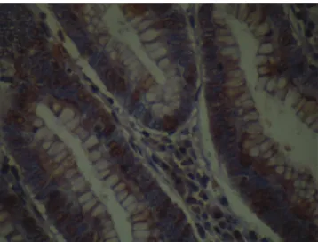
dif-Fig. 3 – Photomicrograph of colorectal adenoma with positive immunoexpression for the TCF4 antibody, represented by a brownish colour in the cell nucleus (immunohistochemistry ×200) (photo by the author).
Fig. 4 – Photomicrograph of colorectal adenoma with positive immunoexpression for the TCF4 antibody, represented by a brownish colour in the cell nucleus (immunohistochemistry ×200) (photo by the author).
Table 1 – Comparison of the expression of TCF4 protein in colorectal carcinoma, colorectal adenoma and non-neoplastic colorectal tissue.
TCF-4 protein expression
Colorectal carcinoma
Colorectal adenoma
Non-neoplastic colorectal
tissue
Negative 17 (42,5%) 45 (86,5%) 31 (83,8%) Positive 23 (57,5%) 7 (13,5%) 6 (16,2%)
ferentiation, lymph node metastasis, and distant metastasis; in none of them a signiicant difference was found.
Despite evidence of the participation of TCF4 protein in the homeostasis of normal colorectal tissue and in colorec-tal carcinogenesis, we found only one trial in literature that compared the expression of TCF4 protein in adenomas and colorectal carcinomas by immunohistochemistry studies.14
The authors of this trial studied 68 cases of colorectal tu-mours (19 adenomas, 14 adenomas with intraepithelial neo-plasia and 35 carcinomas), and compared these tumours with the mucosa adjacent to the lesion. These authors found a dif-ference between expression of TCF4 protein in the adenoma and in the adjacent normal mucosa, but no difference be-tween the expression in the adenocarcinoma and in the ad-jacent mucosa. This result may have been obtained by these authors thanks to the comparison made with the mucosa from the same individual and not from individuals with non-neoplastic colon epithelium. Other reasons that may explain this difference may be linked to the smaller number of cases analyzed, as well as the different types of antibodies used, including with different dilutions (1:200).
In the present study, we observed that the non-neoplastic colorectal tissue and the colorectal adenoma expressed TCF4 protein in less than 20% of cases. Although without signii-cant difference, the adenomas with higher probability of ma-lignant transformation (larger ones, with a higher degree of atypia, and of histologic villous type) tended to express more frequently the protein. Possibly due to the high percentage of cases that did not express TCF4 protein among adenomas (> 80%), we were unable to establish this difference. These data suggest that a future study with a larger sample may establish signiicant differences with respect to the expression of TCF4 protein among tumours with low probability of neoplastic transformation and with high probability of transformation.
Kriegl et al.15 correlated the survival of patients operated
on for colorectal carcinoma with expression of TCF4 protein. They identiied the expression of TCF4 protein in 46% of cases, whose survival was worse. In the current series, although the prognosis has not been studied, there was expression of the protein in 57% of cases of carcinoma, a result similar to that obtained by Kriegl et al.15 Comparing the positivity of
immu-noexpression of TCF4 protein in relation to depth of invasion of the intestinal wall, lymph node invasion and presence of metastasis – data that may indicate a worse prognosis –, there was no signiicant difference. Such data can corroborate the fact that this protein is linked more to colorectal carcinogene-sis, not participating in the processes considered as occurring in a later period in the progression of neoplasia, for instance, the invasion of adjacent tissue and occurrence of metastasis.
The difference in the expression of TCF4 protein between colorectal adenomas and carcinomas suggests that this pro-tein participates in colorectal carcinogenesis, which is in agreement with the indings of van de Wetering et al.16 These
authors stated that the β-catenin/TCF4 complex is the main
regulator of proliferation and differentiation of the non-neo-plastic colorectal tissue and in colorectal carcinomas; these authors also showed that the activation of TCF4 target genes, induced by mutations in the Wnt signalling pathway, consti-tute the transforming event of the normal colorectal mucosa in neoplastic mucosa.
Kendiziorra et al.33 studied colorectal carcinoma cell lines
and found that tumour cells that showed overexpression of TCF4 protein did not respond satisfactorily to radiotherapy. Moreover, the authors reported that the silencing of this overexpression renders these tumour cell lines more radio-sensitive. The conirmation of these data by other studies can conirm the clinical importance of evaluating the ex-pression of this protein for planning the treatment of CRC.
Chen et al.17 published studies on resveratrol, a substance
found in plants, which could suppress the Wnt signalling pathway. In vitro analysis showed that this action occurs due to rupture of the β-catenin-TCF4 complex, suggesting that this mechanism of action may be a potential target for therapy of CRC. The reported studies show promise for the use of TCF4 protein as a therapeutic target in CRC. Consid-ering that immunohistochemistry is a cheap and affordable method, it is important to determine patterns of TCF4 pro-tein immunoexpression in non-neoplastic colorectal tissue and in colorectal adenomas and carcinomas.
The results reported in this study may help in the under-standing of TCF4 protein activity and Wnt signalling path-way in colorectal carcinogenesis, and thereby increase the prospect to inding new therapeutic targets for drug devel-opment for the treatment of colorectal carcinoma.
Conclusions
Under the conditions of this study, the results showed that:
1) The immunoexpression of TCF4 protein in colorectal car-cinoma was superior versus in adenoma and non-neo-plastic colorectal tissue.
2) There was no difference of immunoexpression of TCF4 protein in adenoma and in non-neoplastic colorectal epi-thelium.
Conlicts of interest
The authors declare no conlicts of interest.
R E F E R E N C E S
1. Kinzler RJ, Volgenstein B. Lessons from hereditary colorectal cancer. Cell. 1996; 87:159–70.
2. Instituto Nacional de Câncer José Alencar Gomes da Silva. Estimativa 2012; incidência de câncer no Brasil [estimativa na Internet]; 2011; Rio de Janeiro [cited 06 September 2013]. Avaible from: http://.inca.gov.br/estimativa/2012
3. Fearon ER, Vogelstein B. A genetic model for colorectal tumorigenesis. Cell.1990;61(5):759-67.
4. Nusse R. Wnt signaling in disease and in development. Cell research. 2005;15(1):28-32.
5. Arce L, Yokoyama NN, Waterman ML. Diversity of LEF/ TCF action in development and disease. Oncogene. 2006;25(52):7492–504.
7. Barolo S. Transgenic Wnt/TCF pathway reporters: all you need is Lef? Oncogene. 2006;25(57):7505-11.
8. Bright-Thomas RM, Hargest R. APC, β-Catenin and hTCF4; an
unholy trinity in the genesis of colorectal cancer. European journal of surgical oncology. 2003;29(2):107-17.
9. Sjöblom T, Jones S, Wood LD, Parsons DW, Lin J, Barber TD, et al. The consensus coding sequences of human breast and colorectal cancers. Science. 2006;314:268-74.
10. Ghadimi BM, Grade M, Diilippantonio MJ, Varma S, Simon R, Montagna C, et al. Effectiveness of gene expression proiling for response prediction of rectal adenocarcinomas to preoperative chemoradiotherapy. J.Clin.Oncol.2005; 23(9):1826–38.
11. Kendziorra E, Ahlborn K, Spitzner M, Rave-Fränk M, Emons G, Gaedcke J, et al. Silencing of the Wnt transcription factor TCF4 sensitizes colorectal cancer cells to (chemo-) radiotherapy. Carcinogenesis. 2011;32(2):1824-31. 12. Pires AR, Andreiuolo FM, de Souza SR. TMA for all: a new
method for the construction of tissue microarrays without
recipient parafin block using custombuilt needles. Diagnostic Pathology 2006;1:14.
13. Sobin L, Gospodarowicz M, Wittekind C, (editors). TNM classiication of malignant tumors. 7th ed. [monograph on the Internet]; 2009 [cited 2013 Sept. 6]. Available from: www.uicc. org/uicc_old/resources/tnm.
14. Takeda K, Kinoshita I, Shimizu Y, Ohba Y, Itoh T, Matsuno Y, et al. Clinicopathological signiicance of expression of p-c-Jun, TCF4 and beta-Catenin in colorectal tumors. BMC Cancer, 2008;8:328. 15. Kriegl L, Horst D, Reiche JA, Engel J, Kirchner T, Jung A. LEF-1 and
TCF4 expression correlate inversely with survival in colorectal cancer. Journal of Tranlational Medicine. 2010;8:123.
16. van de Wetering M, Sancho E, Verweij C, de Lau W, Oving I, Hurlstone A, et al. The β-catenin/TCF-omplex imposes a
crypt progenitor phenotype on colorectal cancer cells. Cell. 2002;111(2):241–50.
17. Chen HJ, Hsu LS, Shia YT, Lin MW, Lin CM. The β-catenin/TCF
complex as a novel target of resveratrol in the Wnt/β-catenin