Research Article
Chemical Composition, Leishmanicidal and
Cytotoxic Activities of the Essential Oils from
Mangifera indica
L. var. Rosa and Espada
Eduardo H. S. Ramos,
1Marcílio M. Moraes,
2Laís L. de A. Nerys,
1Silene C. Nascimento,
1Gardênia C. G. Militão,
3Regina C. B. Q. de Figueiredo,
4Cláudio A. G. da Câmara,
2and Teresinha Gonçalves Silva
11Departamento de Antibi´oticos, Universidade Federal de Pernambuco, 50670-420 Recife, PE, Brazil
2Departamento de Ciˆencias Moleculares, Universidade Federal Rural de Pernambuco, 52171-900 Recife, PE, Brazil 3Departamento de Fisiologia e Farmacologia, Universidade Federal de Pernambuco, 50670-420 Recife, PE, Brazil 4Departamento de Microbiologia, Centro de Pesquisa Aggeu Magalh, FIOCRUZ, 50670-420 Recife, PE, Brazil
Correspondence should be addressed to Teresinha Gonc¸alves Silva; teresinha100@gmail.com
Received 27 February 2014; Revised 17 June 2014; Accepted 18 June 2014; Published 20 July 2014
Academic Editor: Silvia R. B. Uliana
Copyright © 2014 Eduardo H. S. Ramos et al. his is an open access article distributed under the Creative Commons Attribution License, which permits unrestricted use, distribution, and reproduction in any medium, provided the original work is properly cited.
he essential oils fromMangifera indicavar. Rosa and Espada latex were obtained by hydrodistillation and analyzed using
GC-FID and GC-MS. Twenty-seven components were identiied. he main compound in the essential oil fromM. indicavar. Espada
(EOMiE) was terpinolene (73.6%). he essential oil ofM. indicavar. Rosa (EOMiR) was characterized by high amounts of�-pinene
(40.7%) and terpinolene (28.3%). In the test for leishmanicidal activity against promastigotes forms ofL. amazonensis, EOMiR and
EOMiE showed IC50(72 h) of 39.1 and 23.0�g/mL, respectively. In macrophages, EOMiR and EOMiE showed CC50of 142.84 and
158.65�g/mL, respectively. However, both were more speciic to the parasite than macrophages, with values of selectivity index of
6.91 for EOMiE and 3.66 for EOMiR. he essential oils were evaluated for their cytotoxicity against the human tumor cells
HEp-2, HT-29, NCI-H29HEp-2, and HL-60. he EOMiR and EOMiE were most efective against the HL-60, with IC50values of 12.3 and
3.6�g/mL, respectively. he results demonstrated that the essential oils ofM. indicacan destroyL. amazonensisand inhibit tumor
cell growth. hese indings contribute to the knowledge of the Brazilian biodiversity as a source of potential therapeutic agents.
1. Introduction
Mangifera indicaL. is a perennial arboreal tree belonging to the family Anacardiaceae. Native to southeastern Asia, this plant had been domesticated for centuries before spreading to other parts of the tropical world. In Brazil, this tree is known commonly as “manga,” and it was irst introduced into the Northeast Region in the eighteenth century by the Portugue-ses. Manga-Espada and manga-Rosa are the most commonly varieties cultivated in the Pernambuco state, Brazil, where their leaves are used in folk medicine to treat backaches and bronchitis [1]. Previous investigations of the biological
properties ofM. indicahave found that this plant has antiviral, antibacterial, analgesic, anti-inlammatory, immunomodula-tory, and antioxidant activities [2, 3]. he composition of the essential oils of M. indica fruits, including the Espada and Rosa varieties, has been extensively investigated [4–12], but, to our knowledge, this is the irst report on the volatile components of latex fromM. indicavar. Espada and Rosa and their leishmanicidal and cytotoxicity activities.
Leishmaniasis is a disease caused by protozoan para-sites of the genus Leishmania, a protozoa transmitted by
Phlebotomussp., commonly called sand lies. Leishmaniasis is one of the leading causes of morbidity and mortality
worldwide and, as such, has been found to be a major global health problem, with 1.5 to 2 million humans afected by the disease annually. About 350 million people in 88 countries are estimated to be threatened by the disease [13].Leishmania (Leishmania) amazonensis is one of the causative agents of human cutaneous leishmaniasis in the Amazon region, Brazil, which is associated with both the simple and difuse forms of the disease [14]. Available chemotherapy still relies on pentavalent antimonials, amphotericin B, or pentamidine. However, the use of these compounds is limited by toxicity to the host and the development of resistance by the parasites [15–18]. Hence, a search for a new active compound with potential leishmanicidal property remains essential for the development of a new antileishmanial therapy. Extracts from medicinal plants are being widely tested for leishmanicidal activity [19].
he research for new antileishmanials and anticancer drugs is imperative and has been promoted by World Health Organization, which endorses the use of traditional medicine when appropriate health services are inaccessible [20]. More-over, due to their broad spectrum of reported biological activities [21–23], essential oils have been of interest, includ-ing leishmanicidal [24–27] and anticancer [28] activities. In addition, their hydrophobic nature makes these oils more permeable to the cell membranes, which is a very important feature for developing agents against intracellular pathogens, or act by destroying vital intracellular components of tumor cells leading to apoptosis [29]. It is also well established that antitumor drugs may also display antileishmanial activity [30].
As part of a systematic study of the chemical composition and biological properties of aromatic plants that grow in the portion of the Atlantic forest in the state of Pernambuco, the aim of the present study was to determine the chemical composition of the essential oils obtained from the latex of the fruits of M. indica var. Rosa and Espada, evaluate of leishmanicidal activity against promastigote forms of the
L. amazonensis, and the cytotoxicity of these oils against HEp-2 (human larynx carcinoma), HT-29 (human colon adenocarcinoma), NCI-H292 (human lung carcinoma), and HL-60 (human leukemia) cell lines.
2. Materials and Methods
2.1. Reagents. Fetal bovine serum was purchased from Gibco (Madrid, Spain), and RPMI-1640, Dulbecco’s modiied Eagle medium (DMEM), dimethyl sulfoxide (DMSO), 3-(4,5-dimethylthiazol-2-yl)-2,5-diphenyltetrazolium bromide (MTT), and doxorubicin were purchased from Sigma Chem-ical Co. (Madrid, Spain). he monoterpenes and sesquiter-penes used to identify the components of the essential oils of the latex of fruits fromM. indicavarieties were purchased from Sigma-Aldrich Co. he monoterpenes terpinolene,
�-pinene, �-pinene, and �-3-carene were purchased from Sigma-Aldrich Co.
2.2. Plant Material. he latex of fruits ofM. indicaL. var. Rosa and Espada was collected in December 2010 on the campus
of the Federal Rural University of Pernambuco (UFRPE), Recife, Pernambuco. he plants were identiied by a specialist, Ladjane C. Gomes, and voucher specimens of these species were deposited in the Vasconcelos Sobrinho Herbarium-PEURF of the UFRPE under numbers 364 forM. indica L. var. Espada and 363 forM. indicaL. var. Rosa.
2.3. Isolation of the Essential Oil from M. indica. he fruits were harvested with the pedicels (approximately 2 inches long) intact. Subsequently, the pedicel was detached from the fruit at the abscission zone. he fruit was immediately inverted over a glass tube, and the latex was allowed to low into the same tube. All latex in the glass tube was stored under refrigeration at 4∘C until essential oil isolation was achieved. he latex samples extracted from the two fruit varieties were treated separately. he samples were subjected to hydrodistillation for 2 h in a Clevenger-type apparatus. he oil layers were separated and dried over anhydrous sodium sulfate, stored in hermetically sealed glass containers, and refrigerated at 4∘C until the chemical analysis and cytotoxicity and leishmanicidal assays occurred. he total oil yield was expressed as a percentage (g/10 g of fresh plant material).
2.4. Gas Chromatography and Gas Chromatography-Mass Spectrometry (GC/MS). Quantitative GC analyses were per-formed with a Hewlett-Packard 5890 Series II GC apparatus equipped with a lame ionization detector (FID) by using a nonpolar DB-5 fused silica capillary column (30 m×0.25 mm
×0.25 mm ilm thickness) from J & W Scientiic. he oven temperature was programmed from 60 to 24∘C at 3∘C/min for integrating purposes. he injector and detector temperatures were set at 260∘C. Hydrogen was used as a carrier gas with a 1 mL/min low rate and a 30 psi inlet pressure. he system was operated in split mode (1 : 30). he injection volume was 0.5�L from the solution (1 : 100 of the oil in hexane). he amount of each compound was calculated from the GC peak areas in the order of elution from the DB-5 column and is expressed as a relative percentage of the total area of the chromatogram. Analyses were performed in triplicate, and the results were submitted to descriptive statistical analysis.
Qualitative GC/MS analyses were performed using a Varian GC/MS (CG: Varian 431/CG-MS: Varian 220-MS) system operating in EI mode at 70 eV. he system was operated with the same column and temperature program as those used for the GC experiments. he carrier gas was helium with a 1 mL/min low rate, and split mode (1 : 30) was used. he injection volume was 1�L of 1 : 100-diluted oil in hexane.
2.5. Leishmanicidal Activity Assay on Promastigotes. L. ama-zonensis amastigotes forms (MHOM/77BR/LTB0016) were isolated from Balb/c mice lesions and maintained as pro-mastigote forms at 26∘C in Schneider’s medium (Sigma-Aldrich, St. Louis, MO, USA) containing 10% inactivated fetal bovine serum (FBS) (Sigma-Aldrich, St. Louis, MO, USA), 100�g streptomycin/mL, and 100 U/mL penicillin (Sigma-Aldrich, St. Louis, MO, USA). he parasites (106 par-asites/mL) were incubated at 26∘C in Schneider’s Drosophila medium supplemented with 10% FBS in the absence, or presence, of the diferent concentrations of the essential oils. he cell density was determined using a Neubauer count-ing chamber [33]. he concentration that inhibited culture growth by 50% (IC50) was determined ater 72 h by regression analysis using the SPSS 8.0 sotware. All experiments were performed in triplicate.
2.6. Cytotoxicity Assay on Macrophage. Peritoneal macro-phages from Swiss (Mus musculus) mice were plated at 5×10⁴ cells/well in 96-well plate with RPMI medium, supplemented with 10% inactivated FBS, and incubated for 2 h at 37∘C in 5% CO2. Nonadherent cells were then removed, and the adhered macrophages were washed twice with PBS and grown for 72 h in RPMI in the absence, or presence, of the diferent concentrations of EOMiR and EOMiE (100 to 6.25�g/mL). Treated and untreated cells were washed and incubated in fresh culture medium containing 5 mg/mL of 3-(4,5-dimethylthiazol-2-yl)-2,5-diphenyl tetrazolium bromide (MTT) for 3 h at 37∘C under the same conditions. Ater incubation, the cells were solubilized in DMSO (100�L/well) and the formazan precipitates derived from MTT reduction were determined spectrophotometrically at 540 nm. he 50% cytotoxic concentration (CC50) was determined by regression analysis using the SPSS 8.0 for Windows. he selectivity index was determined as the ratio of CC50 for the macrophages to IC50 for the protozoa [33]. Each assay was carried out in triplicate in three independent experiments.
2.7. Cytotoxicity Assays on Tumoral Cell Line. he cell lines used for the cytotoxicity assays were HEp-2 (human larynx carcinoma), HT-29 (human colon adenocarcinoma), NCI-H292 (human lung mucoepidermoid carcinoma), and HL-60 (human promyelocytic leukemia), and these cells were obtained from the Cell Bank of Rio de Janeiro (Rio de Janeiro, Brazil). he HEp-2, NCI-H292, and HT-29 cells were maintained in DMEM and supplemented with 10% fetal bovine serum, 2 mM glutamine, 100 U/mL penicillin, and 100�g/mL streptomycin at 37∘C with 5% CO2. he HL-60 cells were cultured in RPMI-1640 medium under the same conditions.
Cell counts and viability were determined by trypan blue staining. he cell concentration was adjusted to 3 × 10⁵ cells/mL for HL-60 cells and 2×10⁵ cells/mL for the other cell lines. Approximately 100�L of cell suspension with the above concentration was cultured in each well of 96-well plates for 24 h at 37∘C in a humid atmosphere containing 5% CO2. Ater 24 h, EOMiR, EOMiE, or their major components (50.0, 25.0, 12.50, 6.25, 3.12, 1.56, or 0.78 mg/mL) “dissolved in
DMSO” were added to each well of the 96-well plates, and the plates were incubated again at 37∘C for 72 h. Control groups received DMSO (0.1%). Doxorubicin was used as a positive control. he growth of the tumor cells was quantiied by the ability of the living cells to reduce yellow tetrazolium MTT (3-(4,5-dimethylthiazolyl-2)-2,5-diphenyltetrazolium bromide) to a blue formazan product [34]. Ater 72 h of incubation, MTT (5.0 mg/mL) was added to the plate. Ater three hours for the suspended cells or two hours for the adherent cells, the formazan product from the reduction of MTT was dissolved in DMSO, and the absorbance was measured by using a multiplate reader. he efect of the oils was quantiied as the percentage of control absorbance of the reduced dye at 450 nm (Multiplate Reader hermoplate).
3. Results and Discussion
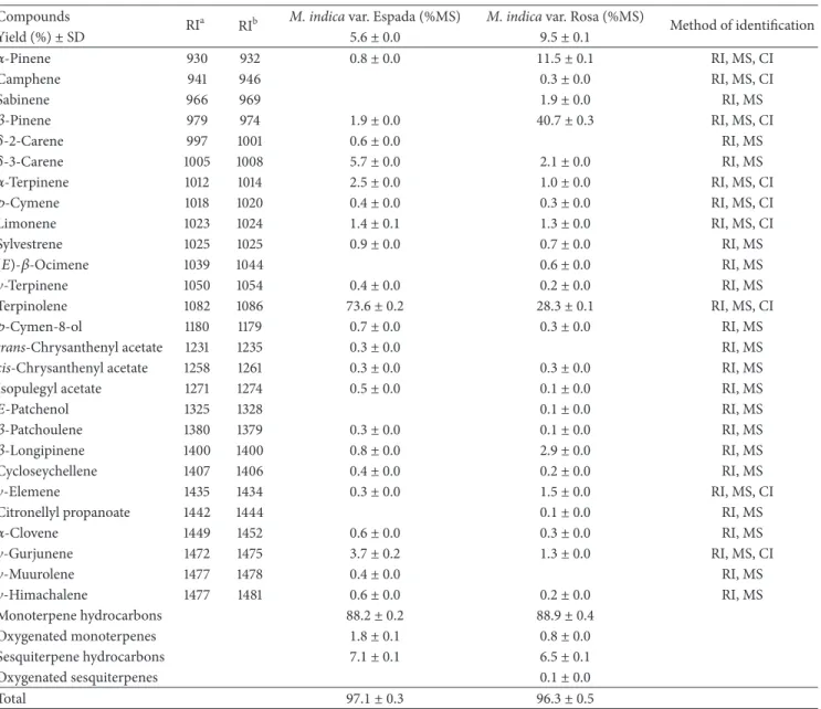
3.1. Chemical Composition of the Essential Oils. he latex samples collected from fruits of theM. indicavar. Espada and Rosa were viscous clear liquids with an aroma characteristic of the fresh ripe fruit. Both latex samples were submitted to hydrodistillation and yielded colorless and pleasant-smelling oils. he best yield was obtained for the EOMiR (9.50%). All constituents identiied in the essential oils of the latex of the fruits of the two studied mango varieties are listed inTable 1
in the order of elution from the DB-5 column.
Twenty-seven components were identiied in the oils of both varieties, representing approximately97.0 ± 0.3% and
96.4± 0.5% of the EOMiE and EOMiR, respectively (Table 1).
Nineteen compounds were common to both varieties. his analysis revealed that both oils were characterized by a high content of monoterpene hydrocarbons. he main compound identiied in the EOMiE was terpinolene (73.6 ± 0.2%), with
�-3-carene (5.7±0.0%) being the second most abundant. he main compound in the EOMiR was�-pinene (40.7 ± 0.3%), followed by terpinolene (28.3 ± 0.1%) and�-pinene (11.5 ±
0.1%). Both oils had similar chemical proiles, difering only in the percentages of the constituents. In fact, terpinolene, a main component identiied in both oils, was found to be the major constituent of the EOMiE, but it was found at a lower percentage in the EOMiR. he same pattern was observed for
�- and�-pinene, which were major compounds identiied in the EOMiR, but they were detected at percentages lower than 2% in the EOMiE.
A study performed by Loveys et al. [35] reported the presence of terpinolene (83.7%) and car-3-ene (89.8%) as major components in the Australian Kensingtonand Irwin
varieties, respectively. hese compounds were also found in the present study, whereas they were the major components in only theM. indicavar. Espada.
Table 1: Chemical composition of the essential oils from the latex of two varieties ofM. indicavar. Rosa and Espada.
Compounds
RIa RIb M. indicavar. Espada (%MS) M. indicavar. Rosa (%MS) Method of identiication
Yield (%)±SD 5.6 ± 0.0 9.5 ± 0.1
�-Pinene 930 932 0.8 ± 0.0 11.5 ± 0.1 RI, MS, CI
Camphene 941 946 0.3 ± 0.0 RI, MS, CI
Sabinene 966 969 1.9 ± 0.0 RI, MS
�-Pinene 979 974 1.9 ± 0.0 40.7 ± 0.3 RI, MS, CI
�-2-Carene 997 1001 0.6 ± 0.0 RI, MS
�-3-Carene 1005 1008 5.7 ± 0.0 2.1 ± 0.0 RI, MS
�-Terpinene 1012 1014 2.5 ± 0.0 1.0 ± 0.0 RI, MS, CI
�-Cymene 1018 1020 0.4 ± 0.0 0.3 ± 0.0 RI, MS, CI
Limonene 1023 1024 1.4 ± 0.1 1.3 ± 0.0 RI, MS, CI
Sylvestrene 1025 1025 0.9 ± 0.0 0.7 ± 0.0 RI, MS
(�)-�-Ocimene 1039 1044 0.6 ± 0.0 RI, MS
�-Terpinene 1050 1054 0.4 ± 0.0 0.2 ± 0.0 RI, MS
Terpinolene 1082 1086 73.6 ± 0.2 28.3 ± 0.1 RI, MS, CI
�-Cymen-8-ol 1180 1179 0.7 ± 0.0 0.3 ± 0.0 RI, MS
trans-Chrysanthenyl acetate 1231 1235 0.3 ± 0.0 RI, MS
cis-Chrysanthenyl acetate 1258 1261 0.3 ± 0.0 0.3 ± 0.0 RI, MS
Isopulegyl acetate 1271 1274 0.5 ± 0.0 0.1 ± 0.0 RI, MS
�-Patchenol 1325 1328 0.1 ± 0.0 RI, MS
�-Patchoulene 1380 1379 0.3 ± 0.0 0.1 ± 0.0 RI, MS
�-Longipinene 1400 1400 0.8 ± 0.0 2.9 ± 0.0 RI, MS
Cycloseychellene 1407 1406 0.4 ± 0.0 0.2 ± 0.0 RI, MS
�-Elemene 1435 1434 0.3 ± 0.0 1.5 ± 0.0 RI, MS, CI
Citronellyl propanoate 1442 1444 0.1 ± 0.0 RI, MS
�-Clovene 1449 1452 0.6 ± 0.0 0.3 ± 0.0 RI, MS
�-Gurjunene 1472 1475 3.7 ± 0.2 1.3 ± 0.0 RI, MS, CI
�-Muurolene 1477 1478 0.4 ± 0.0 RI, MS
�-Himachalene 1477 1481 0.6 ± 0.0 0.2 ± 0.0 RI, MS
Monoterpene hydrocarbons 88.2 ± 0.2 88.9 ± 0.4
Oxygenated monoterpenes 1.8 ± 0.1 0.8 ± 0.0
Sesquiterpene hydrocarbons 7.1 ± 0.1 6.5 ± 0.1
Oxygenated sesquiterpenes 0.1 ± 0.0
Total 97.1 ± 0.3 96.3 ± 0.5
ahe retention indices were calculated from the retention times in relation to those of a series of n-alkanes on a 30 m DB-5 capillary column.bLinear retention
indices from the literature. RI: retention index; MS: mass spectrum; CI: coinjection with authentic standards.
Terpinolene has been found in signiicant amounts in the volatile fractions of the fruits of eight varieties of mango (Chana = 62.4%, Coquinho = 51.4%, Comum = 45.4%, Carlota = 52.0%, Bacuri = 57.0%, Cheiro = 66.1%, Camet´a = 56.3%, and Gorjoba = 54.8%) collected in the state of Par´a, Brazil [4]. he essential oils of the pulp from the Choak Anand, Ok-rong, and yellow Keaw varieties of mango grown in hailand contain primarily terpinolene [12]. A similar pattern was recently reported for the essential oil from the lowers of a specimen from China, which had a high content of terpinolene [37].
Contrary to the terpinolene proile found in our study, Andrade et al. [4] did not report the presence of terpinolene in the volatile fraction ofM. indicavar. Rosa and only found
it in low amounts (0.2%) in the variety Espada from Par´a (Brazil). According to the literature, the essential oils from the exudate of the investigated mango varieties have marked diferences in composition with respect to the latex essential oils from seven varieties collected in India [36]. Speciically, these essential oils from India did not contain terpinolene, and these oils had lower contents of �-pinene and high contents of �-myrcene, limonene, cis-ocimene, and trans -ocimene.
0 50 100 150
EOMiR EOMiE
G
ro
w
th inhib
itio
n
(%)
6.25 12.5 25 50 100
(�g/mL)
Figure 1: Efects of essential oils onLeishmania amazonensis
pro-mastigote forms. Each bar represents the mean±standard deviation
of three independent experiments in triplicate.
Table 2: Leishmanicidal and cytotoxic efects of essentials oils ofM.
indicaL.
Treatment Promastigote
IC50(�g/mL)
Macrophage
CC50(�g/mL)
Selectivity index
EOMiR 39.1 ± 5.6 142.8 ± 6.0 3.7
EOMiE 23.0 ± 2.7 158.6 ± 2.5 6.9
he leishmanicidal activity was expressed as the IC50and CC50by the linear
regression analysis, respectively. Values are mean±standard deviation from
at least three independent experiments in duplicate. Diferences were
con-sidered signiicant at a 0.05 level of conidence. SI: CC50macrophages/IC50
promastigote forms. IC50: concentration inhibiting 50% of the growth of
promastigotes; CC50: concentration capable of causing cytotoxic efects of macrophages by 50%.
�-pinene as the main compound [5].�-Pinene was found in an appreciable amount (12.5%) in a sample of fruits of the variety Qalmi collected in India [8].
3.2. Leishmanicidal Activity of EOMiR and EOMiE. Our results showed that both oils demonstrated similar efect on theL. amazonensispromastigotes, inhibiting the parasite growth in dose-dependent manner (Figure 1). Only at a concentration of 50�g/mL did the activity proile between the two oils difer, seeing that EOMiE presented an inhibition rate of growth better than EOMiR.
he IC50/72 h estimated for the oils were 39.06 and 22.96�g/mL for EOMiR and EOMiE, respectively (Table 2). the oils had low cytotoxic efect on macrophages, with a CC50 of 142.84 and 158.65�g/mL for EOMiR and EOMiE, respectively. However, both oils were more speciic to the parasite than the macrophages, with values of a selectivity index of 6.91 for EOMiE, demonstrating that it is six times more toxic to the parasite compared to macrophages.
Essential oils, as well as their components, have been found to possess a wide spectrum of pharmacological efects including antibacterial, antifungal, antiviral, antihelminthic,
and antiprotozoal activities [38,39]. hey are also known to have important biological activities against trypanosomatids asTrypanosoma brucei [40],Leishmania [24], and T. cruzi
[41]. hese activities are mainly attributed to the presence of terpenic, aromatic, and aliphatic constituents [42].
It is usually assumed that terpenic constituents are responsible for the hydrophobic feature of essential oils [43] which allows essential oils to freely permeate the cell membranes and kill the parasites by afecting their cytoplas-mic metabolic pathways or organelles [44]. On the other hand, essential oils themselves could interact with parasite membranes and cause drastic physiologic changes leading to the loss of membrane permeability that ultimately leads to cell death [45]. However, due to the great number of constituents and the synergistic or antagonistic interactions existing among them, it is likely that essential oils have other cellular targets besides the cellular membranes. In fact, interactions of essential oils with lipids and proteins have already been reported [45].
3.3. Cell Viability Assay. he cytotoxic activity of the essential oils fromM. indicavar. Rosa and Espada, and of the major terpenes in the Rosa variety (�-pinene, terpinolene, and
�-pinene) and the Espada variety (terpinolene and � -3-carene), was investigated against human tumor cell lines. he cytotoxic efects of the essential oils are presented in
Table 3. he IC50values were ranged from 12.3 to 38.9�g/mL
for the EOMiR and from 3.6 to 14.5�g/mL for the EOMiE, depending upon the cell line. hese values indicate that these cancer cell lines difer with respect to their sensitivity to the substances contained in the essential oil of M. indica
latex. hese diferences may be due to the diferent molecular characteristics of these cells. Generally, the mechanism of cell death induced ater treatment is dependent on several factors, such as the compounds used, their concentrations, and the cell line used for the study.
According to the criteria of the National Cancer Institute (NCI-USA) for considering a crude extract promising for further puriication, the IC50 must be ≤30�g/mL, and, for a pure substance to be considered promising for use as an antitumor agent, it should have an IC50 ≤4�g/mL, which corresponds to strong cytotoxic activity [46]. he oils tested in this study had IC50 values below the value determined by NCI-USA for all cell lines tested. he essential oil is classiied as a crude extract, and it has a complex composition, containing from a dozen to several hundred components, with terpenes being the major components.
Terpinolene and�-3-carene are the major components of the EOMiE, and both were separately evaluated to determine their cytotoxic activities. Terpinolene showed cytotoxic activ-ity against all cell lines tested but was more potent against HT-29, HEp-2, and NCI-H292 cells.�-3-Carene exhibited little activity against all lines.
Table 3: Cytotoxic activity against human tumor cells of the essential oils from theM. indicavar. Rosa andM. indicavar. Espada.
Essential oil
aIC
50(�g/mL)
HT-29 HEp-2 NCI-292 HL-60
EOMiR 28.7 25.6 38.9 12.3
20.3–30.5 20.5–31.9 31.3–48.4 9.5–15.9
EOMiE 9.2 6.2 14.5 3.6
6.0–14.0 4.4–8.6 11.4–18.4 2.8–4.8
Terpinolene 16.7 13.7 17.4 28.8
13.0–21.6 11.5–16.3 14.8–19.6 25.6–32.3
�-Pinene 10.3 11.0 11.8 11.8
8.7–12.2 7.7–15.6 9.1–13.5 10.0–14.0
�-Pinene 6.6 6.3 7.5 10.5
5.8–7.5 5.0–8.0 5.1–11.0 9.1–12.0
�-3-Carene 45.5 35.0 42.6 63.1
37.6–55.0 29.6–41.2 35.1–51.9 47.4–84.0
Terpinolene (74%) +�-3-carene (6%) 22.8 26.5 29.3 27.0
16.4–31.6 22.8–30.8 25.8–33.2 23.03–31.8
�-Pinene (41%) + terpinolene (28%) +�-pinene (11%) 22.3 30.5 40.1 7.8
16.1–31.1 25.6–36.4 20.6–47.9 5.6–10.7
Terpinolene (74%) +�-pinene (2%) 39.4 35.0 30.4
36.0–43.1 27.7–44.38 21.8–42.2
Doxorubicin 0.4 0.7 0.01 0.2
0.3–0.5 0.2–1.4 0.004–0.3 0.01–0.2
aData are presented as IC
50values and 95% conidence intervals obtained by nonlinear regression for all cell lines from two independent experiments. Exposure
time: 72 h. HEp-2: larynx carcinoma; NCI-H292: lung mucoepidermoid carcinoma; HT-29: colon adenocarcinoma; and HL-60: promyelocytic leukemia cells.
Doxorubicin was used as a positive control. EOMiR: essential oil ofM. indicavar. Rosa; EOMiE: essential oil ofM. indicavar. Espada.
were analyzed together [(terpinolene :�-3-carene (74 : 6)] at the ratio present in the essential oil, the mixture showed lower activity than terpinolene itself. hese results suggest that the cytotoxic activity displayed by the EOMiE is associated with complex interaction between the major component terpinolene and other minor components of the mixture.
he major components in the EOMiR yielded the fol-lowing results when analyzed separately: �-pinene (IC50 6.6–10.5�g/mL), terpinolene (IC5013.7–28.8�g/mL), and� -pinene (IC5011.0–11.8�g/mL). Both�- and�-pinene showed higher cytotoxicity than terpinolene. When a mixture of these three components was analyzed [�-pinene (41%) + terpino-lene (28%) +�pinene (11%)], signiicant activity against all cell lines, except NCI-H292, was observed. Nevertheless, this activity appeared selective for leukemia cells because the IC50 value was very close to the cut-of set by the NCI for pure substances (IC50 ≤4�g/mL), despite the test substance being a mixture of terpenes.
Because strong cytotoxic activity was observed for � -pinene and given that it is a component of the EOMiE, the cytotoxic activity of a mixture of�-pinene and terpinolene was assessed. he�-pinene + terpinolene mixture was less active than the separate compounds and the EOMiE, indicat-ing that there is antagonism between these two components. his result shows that the activity of the EOMiE is associated with possible synergism among all the compounds present in the oil, not just a particular component.
With respect to their biological properties, it must be kept in mind that essential oils are complex mixtures of numerous molecules, and one might wonder if their biological efects are the result of synergism among all molecules or relect only the activities of the main molecules present at the highest con-centrations according to the gas chromatographic analysis. Generally, the major components are found to relect quite well the biophysical and biological features of the essential oils from which they were isolated, with the amplitudes of their efects being dependent on their concentration when they are tested on their own or in essential oils [47].
Because of the great number of constituents, essential oils appear to have no speciic cellular targets [48], involving multiple targets at the same time [49]. As typical lipophilic compounds, these oils pass through the cell wall and the cytoplasmic membrane, disrupting the structure of the difer-ent layers of polysaccharides, fatty acids, and phospholipids and permeabilizing these structures. Cytotoxicity appears to involve such membrane damage [50].
permeable, resulting in the leakage of radicals, cytochrome c, calcium ions, and proteins, as in the case of oxidative stress and bioenergetic failure. he permeabilization of the outer and inner mitochondrial membranes leads to cell death by apoptosis and necrosis [52]. In general, the cytotoxic activity of essential oils is primarily due to the presence of phenols, aldehydes, and alcohols [53].
4. Conclusions
his study revealed the chemical composition and leish-manicidal and cytotoxic activities of the essential oils of the latex ofM. indica var. Espada and Rosa, thus contributing to the knowledge of the biological properties of the species. Little is known about the compounds of the latex of these two varieties. Monoterpenes were identiied as dominant compounds in the essential oils from both varieties. Others classes of compounds, including oxygenated monoterpenes, sesquiterpene hydrocarbons, and oxygenated sesquiterpenes, were present at lower concentrations. he essential oils present a low toxicity to mammalian cell associated with signiicant leishmanicidal activity. he anticancer activityin vitroof these oils was considered promising. Further studies are required to elucidate the mechanisms of parasite death induced by the most promissory essential oils and identify their putative intracellular targets.
Conflict of Interests
he authors declare that there is no conlict of interests regarding the publication of this paper.
Acknowledgments
he authors thank the Conselho Nacional de Desenvolvi-mento Cient´ıico e Tecnol´ogico (CNPq Proc. no. 476503/2009 -4) and the Fundac¸˜ao de Amparo `a Ciˆencia e Tecnologia do Estado de Pernambuco (FACEPE-APQ no. 0520-55.01/08) for scholarships and inancial support.
References
[1] U. P. de Albuquerque, P. M. de Medeiros, A. L. S. de Almeida et al., “Medicinal plants of the caatinga (semi-arid) vegetation of
NE Brazil: a quantitative approach,”Journal of
Ethnopharmacol-ogy, vol. 114, no. 3, pp. 325–354, 2007.
[2] I. Rodeiro, M. T. Donato, N. Jim´enez, G. Garrido, R. Delgado,
and M. J. G´omez-Lech´on, “Efects ofMangifera indicaL.
aque-ous extract (Vimang) on primary culture of rat hepatocytes,”
Food and Chemical Toxicology, vol. 45, no. 12, pp. 2506–2512, 2007.
[3] G. L. Pardo-Andreu, M. F. Barrios, C. Curti et al., “Protective
efects ofMangifera indicaL extract (Vimang), and its major
component mangiferin, on iron-induced oxidative damage to
rat serum and liver,”Pharmacological Research, vol. 57, no. 1, pp.
79–86, 2008.
[4] E. H. A. Andrade, J. G. S. Maia, and M. D. G. B. Zoghbi, “Aroma volatile constituents of Brazilian varieties of mango
fruit,”Journal of Food Composition and Analysis, vol. 13, no. 1,
pp. 27–33, 2000.
[5] O. J. Alwala, W. Wanzala, R. A. Inyambukho, E. M. Osundwa, and I. O. Ndiege, “Characterization and evaluation of repellent
efect of essential oil ofMangifera indicaL. from Kenya,”Journal
of Essential Oil-Bearing Plants, vol. 13, no. 1, pp. 85–96, 2010. [6] S. H. Ansari, M. Ali, A. Velasco-Negueruela, and M. J.
P´erez-Alonso, “Volatile constituents of the fruits of three mango
cultivars,Mangifera indicaL.,”Journal of Essential Oil Research,
vol. 11, no. 1, pp. 65–68, 1999.
[7] S. H. Ansari, M. Ali, A. Velasco-Negueruela, and M. J.
Perez-Alonso, “Volatile constituents of Mango (Mangifera indica)
fruits cultivar Bombay,”Journal of Medicinal and Aromatic Plant
Sciences, vol. 21, no. 1, pp. 931–933, 1999.
[8] S. H. Ansari, M. Ali, A. Velasco-Negueruela, and M. J. Perez-Alonso, “Characterization of volatile constituents of mango
“Qalmi” (Mangifera indica L.) fruit,”Journal of Essential Oil
Research, vol. 16, no. 5, pp. 417–419, 2004.
[9] M. R. B. Franco, D. Rodriguez-Amaya, and F. M. F. Lanc¸as,
“Compostos vol´ateis de trˆes cultivares de manga (Mangifera
IndicaL.),”Food Science and Technology, vol. 24, no. 2, pp. 165– 169, 2004.
[10] J. A. Pino, J. Mesa, Y. Mu˜noz, M. P. Mart´ı, and R. Marbot,
“Volatile components from mango (Mangifera indicaL.)
culti-vars,”Journal of Agricultural and Food Chemistry, vol. 53, no. 6,
pp. 2213–2223, 2005.
[11] J. A. Pino and J. Mesa, “Contribution of volatile compounds
to mango (Mangifera indicaL.) aroma,”Flavour and Fragrance
Journal, vol. 21, no. 2, pp. 207–213, 2006.
[12] H. Tamura, S. Boonbumrung, T. Yoshizawa, and W. Varanyanond, “Volatile components of the essential oils
in the pulp of four yellow mangoes (Mangifera indicaL.) in
hailand,”Food Science and Technology Research, vol. 6, no. 1,
pp. 68–73, 2000.
[13] WHO, “Control of leishmaniasis. Report of the expert commit-tee,” Tech. Rep. Ser 949, World Health Organization, 2010. [14] R. Lainson and J. J. Shaw, “Evolution, classiication and
geo-graphical distribution,” in he Leishmaniases in Biologyand
Medicine, W. Peters and E. Killick-Kendrick, Eds., pp. 1–120, Academic Press, London, UK, 1987.
[15] A. J. Davis, H. W. Murray, and E. Handman, “Drugs against leishmaniasis: a synergy of technology and partnerships,”
Trends in Parasitology, vol. 20, no. 2, pp. 73–76, 2004.
[16] H. Goto and J. A. L. Lindoso, “Current diagnosis and treatment
of cutaneous and mucocutaneous leishmaniasis,”Expert Review
of Anti-Infective herapy, vol. 8, no. 4, pp. 419–433, 2010. [17] T. Polonio and T. Eferth, “Leishmaniasis: drug resistance and
natural products (review),”International Journal of Molecular
Medicine, vol. 22, no. 3, pp. 277–286, 2008.
[18] J. Berman, “Current treatment approaches to leishmaniasis,”
Current Opinion in Infectious Diseases, vol. 16, no. 5, pp. 397– 401, 2003.
[19] R. Sen and M. Chatterjee, “Plant derived therapeutics for the
treatment of Leishmaniasis,”Phytomedicine, vol. 18, no. 12, pp.
1056–1069, 2011.
[20] L. G. Rocha, J. R. G. S. Almeida, R. O. Macˆedo, and J. M. Barbosa-Filho, “A review of natural products with
antileishma-nial activity,”Phytomedicine, vol. 12, no. 6-7, pp. 514–535, 2005.
onEscherichia coliandBacillus subtilis,”Journal of Essential Oil Research, vol. 15, no. 5, pp. 356–362, 2003.
[22] S. Kulevanova, A. Katandzieva, A. Dimitrovska, G. Stekov, T. Grdanoska, and N. Panovski, “Investigation of antimicrobial
activity of essential oils of several Macedonian hymus L.
species (Lamiaceae),”Bollettino Chimico Farmaceutico, vol. 139,
no. 6, pp. 276–280, 2000.
[23] M. Marino, C. Bersani, and G. Comi, “Antimicrobial activity
of the essential oils ofhymus vulgarisL. measured using a
bioimpedometric method,”Journal of Food Protection, vol. 62,
no. 9, pp. 1017–1023, 1999.
[24] M. D. G. F. de Medeiros, A. C. da Silva, A. M. D. G. L. Cit´o et al., “In vitro antileishmanial activity and cytotoxicity of essential oil fromLippia sidoidesCham,”Parasitology International, vol. 60, no. 3, pp. 237–241, 2011.
[25] M. D. S. S. Rosa, R. R. Mendonc¸a-Filho, H. R. Bizzo et al., “Antileishmanial activity of a linalool-rich essential oil from
Croton cajucara,”Antimicrobial Agents and Chemotherapy, vol. 47, no. 6, pp. 1895–1901, 2003.
[26] V. D. Zheljazkov, C. L. Cantrell, B. Tekwani, and S. I. Khan, “Content, composition, and bioactivity of the essential oils of
three basil genotypes as a function of harvesting,”Journal of
Agricultural and Food Chemistry, vol. 56, no. 2, pp. 380–385, 2007.
[27] J. Mikus, M. Harkenthal, D. Steverding, and J. Reichling, “In vitro efect of essential oils and isolated mono- and
sesquiter-penes onLeishmania majorandTrypanosoma brucei,”Planta
Medica, vol. 66, no. 4, pp. 366–368, 2000.
[28] R. A. Mothana, M. S. Al-Said, M. A. Al-Yahya, A. J. Al-Rehaily, and J. M. Khaled, “GC and GC/MS analysis of essential oil composition of the endemic Soqotraen Leucas virgata Balf.f.
and its antimicrobial and antioxidant activities,”International
Journal of Molecular Sciences, vol. 14, no. 11, pp. 23129–23139, 2013.
[29] J. F. Sanchez-Suarez, I. Riveros, and G. Delgado, “Evaluation of the leishmanicidal and cytotoxic potential of essential oils
derived from ten Colombian plants,”Iranian Journal of
Para-sitology, vol. 8, no. 1, pp. 129–136, 2013.
[30] M. A. Fuertes, P. A. Nguewa, J. Castilla, C. Alonso, and J. M. P´erez, “Anticancer compounds as leishmanicidal drugs:
challenges in Chemotherapy and future perspectives,”Current
Medicinal Chemistry, vol. 15, no. 5, pp. 433–439, 2008. [31] H. van den Dool and P. Dec. Kratz, “A generalization of
the retention index system including linear temperature
pro-grammed gas-liquid partition chromatography,” Journal of
Chromatography A, vol. 11, pp. 463–471, 1963.
[32] O. D. Sparkman, “Identiication of essential oil components by gas chromatography/quadrupole mass spectroscopy Robert P.
Adams,”Journal of the American Society for Mass Spectrometry,
vol. 16, no. 11, pp. 1902–1903, 2005.
[33] M. D. G. F. de Medeiros, A. C. da Silva, A. M. D. G. L. Cit´o et al., “In vitroantileishmanial activity and cytotoxicity of essential oil
from Lippia sidoides Cham,”Parasitology International, vol. 60,
no. 3, pp. 237–241, 2011.
[34] T. Mosmann, “Rapid colorimetric assay for cellular growth and survival: application to proliferation and cytotoxicity assays,”
Journal of Immunological Methods, vol. 65, no. 1-2, pp. 55–63, 1983.
[35] B. R. Loveys, S. P. Robinson, J. J. Brophy, and E. K. Chacko, “Mango Sapburn: components of fruit sap and their role in
causing skin damage,”Australian Journal of Plant Physiology,
vol. 19, no. 5, pp. 449–457, 1992.
[36] K. Saby John, L. Jagan Mohan Rao, S. G. Bhat, and U. J. S. Prasada Rao, “Characterization of aroma components of sap
from diferent Indian mango varieties,”Phytochemistry, vol. 52,
no. 5, pp. 891–894, 1999.
[37] H. W. Wang, Y. Q. Liu, S. L. Wei, Z. J. Yan, and K. Lu, “Compar-ison of microwave-assisted and conventional hydrodistillation
in the extraction of essential oils from mango (Mangifera indica
L.) lowers,”Molecules, vol. 15, no. 11, pp. 7715–7723, 2010.
[38] I. T. F. Macedo, C. M. L. Bevilaqua, L. M. B. de Oliveira et al., “Anthelmintic efect of Eucalyptus staigeriana essential oil
against goat gastrointestinal nematodes,”Veterinary
Parasitol-ogy, vol. 173, no. 1-2, pp. 93–98, 2010.
[39] A. O. Santos, A. C. Santin, M. U. Yamaguchi et al., “Antileish-manial activity of an essential oil from the leaves and lowers of
Achillea millefolium,”Annals of Tropical Medicine and
Parasitol-ogy, vol. 104, no. 6, pp. 475–483, 2010.
[40] K. Otoguro, M. Iwatsuki, A. Ishiyama et al., “In vitro
antit-rypanosomal activity of plant terpenes against Trypanosoma
brucei,”Phytochemistry, vol. 72, no. 16, pp. 2024–2030, 2011. [41] G. F. Santoro, M. G. Cardoso, L. G. L. Guimar˜aes, J. M. Freire,
and M. J. Soares, “Anti-proliferative efect of the essential oil of
Cymbopogon citratus(DC) Stapf (lemongrass) on intracellular amastigotes, bloodstream trypomastigotes and culture
epi-mastigotes ofTrypanosoma cruzi(Protozoa: Kinetoplastida),”
Parasitology, vol. 134, no. 11, pp. 1649–1656, 2007.
[42] Z. Schelz, J. Hohmann, and J. Molnar, “Recent advances in research of antimicrobial efects of essential oils and plant
derived compounds on bacteria,” inEthnomedicine: A Source of
Complementary herapeutics, D. Chattopadhyay, Ed., pp. 281– 304, Research Signpost, Kerala, India, 2010.
[43] S. A. Burt, R. Vlielander, H. P. Haagsman, and E. J. A. Veldhuizen, “Increase in activity of essential oil components carvacrol and thymol against Escherichia coli O157:H7 by
addition of food stabilizers,”Journal of Food Protection, vol. 68,
no. 5, pp. 919–926, 2005.
[44] K. Knobloch, A. Pauli, B. Iberl, H. Weigand, and N. Weis, “Antibacterial and antifungal properties of essential oil
compo-nentes,”Journal of Essential Oil Research, vol. 1, no. 3, pp. 118–119,
1989.
[45] F. Bakkali, S. Averbeck, D. Averbeck, and M. Idaomar,
“Bio-logical efects of essential oils—a review,”Food and Chemical
Toxicology, vol. 46, no. 2, pp. 446–475, 2008.
[46] M. Sufness and J. M. Pezzuto, “Assays for bioactivity,” in
Methods in Plant Biochemistry, K. Hostettmann, Ed., vol. 6, pp. 71–133, Academic Press, London, UK, 1991.
[47] E. Ipek, H. Zeytinoglu, S. Okay, B. A. Tuylu, M. Kurkcuoglu, and K. H. C. Baser, “Genotoxicity and antigenotoxicity of Origanum oil and carvacrol evaluated by Ames Salmonella/microsomal
test,”Food Chemistry, vol. 93, no. 3, pp. 551–556, 2005.
[48] C. F. Carson, B. J. Mee, and T. V. Riley, “Mechanism of
action ofMelaleuca alternifolia(tea tree) oil onStaphylococcus
aureusdetermined by time-kill, lysis, leakage, and salt tolerance
assays and electron microscopy,” Antimicrobial Agents and
Chemotherapy, vol. 46, no. 6, pp. 1914–1920, 2002.
[49] R. di Pasqua, N. Hoskins, G. Betts, and G. Mauriello, “Changes in membrane fatty acids composition of microbial cells induced by addiction of thymol, carvacrol, limonene, cinnamaldehyde,
and eugenol in the growing media,”Journal of Agricultural and
Food Chemistry, vol. 54, no. 7, pp. 2745–2749, 2006.
[50] J. Sikkema, J. A. M. de Bont, and B. Poolman, “Mechanisms of
membrane toxicity of hydrocarbons,”Microbiological Reviews,
[51] A. E. Vercesi, A. J. Kowaltowski, M. T. Grijalba, A. R. Meinicke, and R. F. Castilho, “he role of reactive oxygen species in
mitochondrial permeability transition,”Bioscience Reports, vol.
17, no. 1, pp. 43–52, 1997.
[52] J. S. Armstrong, “Mitochondrial membrane permeabilization:
the sine qua non for cell death,”BioEssays, vol. 28, no. 3, pp.
253–260, 2006.
[53] G. Sacchetti, S. Maietti, M. Muzzoli et al., “Comparative evaluation of 11 essential oils of diferent origin as functional
antioxidants, antiradicals and antimicrobials in foods,”Food
Submit your manuscripts at
http://www.hindawi.com
Pain
Research and Treatment
Hindawi Publishing Corporation
http://www.hindawi.com Volume 2014
World Journal
Hindawi Publishing Corporationhttp://www.hindawi.com Volume 2014
Hindawi Publishing Corporation http://www.hindawi.com
Volume 2014
Toxins
Journal of
Vaccines
Journal ofHindawi Publishing Corporation
http://www.hindawi.com Volume 2014
Hindawi Publishing Corporation
http://www.hindawi.com Volume 2014
Antibiotics
Toxicology
Journal of
Hindawi Publishing Corporation
http://www.hindawi.com Volume 2014
Stroke
Research and Treatment
Hindawi Publishing Corporation
http://www.hindawi.com Volume 2014
Drug Delivery
Journal ofHindawi Publishing Corporation
http://www.hindawi.com Volume 2014 Hindawi Publishing Corporation
http://www.hindawi.com Volume 2014
Advances in Pharmacological Sciences
Tropical Medicine
Hindawi Publishing Corporation
http://www.hindawi.com Volume 2014
Medicinal ChemistryInternational Journal of Hindawi Publishing Corporation
http://www.hindawi.com Volume 2014
Addiction
Journal ofHindawi Publishing Corporation
http://www.hindawi.com Volume 2014
Hindawi Publishing Corporation
http://www.hindawi.com Volume 2014
BioMed
Research International Emergency Medicine International
Hindawi Publishing Corporation
http://www.hindawi.com Volume 2014
Hindawi Publishing Corporation
http://www.hindawi.com Volume 2014
Diseases
Hindawi Publishing Corporation
http://www.hindawi.com Volume 2014
Anesthesiology Research and Practice
Scientifica
Hindawi Publishing Corporation
http://www.hindawi.com Volume 2014
Journal of
Hindawi Publishing Corporation
http://www.hindawi.com Volume 2014
Pharmaceutics
Hindawi Publishing Corporation
http://www.hindawi.com Volume 2014
MEDIATORS
INFLAMMATION