Abstract
Submitted: February 16, 2017 Modiication: July 3, 2017 Accepted: August 2, 2017
Determination of the effective
anticandidal concentration of denture
cleanser tablets on some denture
base resins
Objective: Although the effectiveness of chemical cleansing against
Candida albicans bioilm has been shown, the effective concentration of denture cleanser tablets has not been studied. The aim of this study was
to assess the effect of three denture materials against Candida albicans
bioilm and to determine effective concentrations of denture cleanser tablets. Material and methods: The surface-roughness of Acron-hi™, QC-20™ and Delex™ (n=45 per resin) resins was standardized by using a proilometer and their contact angle or surface free energy was calculated. C. albicans bioilm was formed on all three resins and were treated with Polident 3 min™, Corega™ and Fittydent™ cleanser solutions at various concentrations and both resin-bioilm and cleanser-bioilm interest were determined by using a MTT protocol according to the European Committee on Antimicrobial Susceptibility Testing’s antifungal susceptibility testing (AFST-EUCAST). Scanning electron microscopy was used to compare the eficacy of different
resin materials against C. albicans bioilm. Anticandidal activity and surface
free energy statistical parameters were calculated by using 3-way and 1-way ANOVA, respectively (p<0.05). Results: Polident 3 min™ and Corega™ tablets signiicantly inhibited (p<0.05) the proliferation of C. albicans against all denture resins at 27-37 mg/mL. Scanning electron microscopy results indicated that there was no signiicant difference among resin specimens regarding bioilm formation on dentures. We failed to ind a signiicant relationship between surface free energy and the anticandidal effect of resin types. However, the polarity value of the resins was statistically associated with their anticandidal activity. Conclusions: The polarity of the resins, the concentrations of tablets and the chemical content of the cleanser may directly
affect C. albicans bioilm formations. Polident 3 min™ and Corega™ tablets
should be suggested for patients who use any denture resin types, whereas the Fittydent™ tablet should only be proposed for those who use Delex™, when two tablets are dropped into 150 mL water.
Keywords: Candida albicans. Surface properties. Denture cleansers.
Denture bases. Yeliz HAYRAN1
Işıl SARIKAYA1 Ali AYDIN2 Yadel Hazır TEKIN1
1Gaziosmanpaşa University, Faculty of Dentistry, Department of Prosthodontics, Tokat, Turkey. 2Gaziosmanpasa University, Faculty of Science and Art, Department of Molecular Biology & Genetics,
Tokat, Turkey.
Introduction
Denture stomatitis is a common infection of the oral mucosa in denture wearers and Candida albicans
is the most signiicant etiological agent of denture stomatitis15,20. C. albicans is an obstinate infection agent which is dificult to eliminate once it has been colonized as a complex bioilm formation6,8,14,15. The
surface of acrylic resin denture base provides an ideal environment for microorganisms and bioilm formation, thus the development of C. albicans
in such places4,6,9,14,15,20,24,27,29. The risk of denture
stomatitis increases in the presence of poor oral and denture hygiene, misit prosthesis and night wear of removable dentures4,14,20,24. It has been found that
repeated inhalation and ingestion of microorganisms adhering to the mucosa and denture base can be a reason for various infections in patients with immune deiciency or in those receiving treatment20. Therefore,
oral and denture hygiene is very important to remove microorganisms. Two methods are recommended to remove denture bioilm: mechanical or chemical, or a combination of both. Although the eficiency of mechanical methods in removing denture bioilm or microorganisms has been clearly shown, some people do not have the ability to apply suficient denture hygiene14,21. This is especially the case for patients
with limited motor capacity who have dificulty in cleaning the prosthesis with mechanical methods. To use unsuitable toothbrush with a dentifrice may also lead to surface roughness, which allows more microbial colonization14. The effectiveness of chemical cleansing
to control C. albicans bioilm is shown in many studies,
and denture cleansers are recommended for reducing bioilm formation on the dentures for these patients4,6.
These cleansers are available as commercial products, and they usually include alkaline peroxides19,23, sodium
hypochlorite5,29, acids29, enzymes19, and neutral
enzymatic peroxides solution4-6,15,19. Effervescent
tablets yielding an alkaline peroxide dilution with water are the preferred denture cleansers3,7,13,16,18,22
because they can easily provide enough cleansing without causing damage to surface resins26. These
effervescent tablets act differently as mechanisms against microbial lora. For example, Polident 3 min™, one of the cleanser effervescent tablets, achieves chemical cleaning by using the release of oxygen from a neutral enzymatic peroxide solution4-6,15,19. However,
the bioilm layer often cannot be completely removed
from the resin surface and a number of viable cells
remain on resins29. According to our knowledge, three
signiicant factors – resin types with physicochemical features, cleanser types and cleanser concentrations – can be suggested to explain this situation.
PMMA, one of the resins, is the most commonly used denture base material due its favourable mechanical, physical and aesthetic properties. However, it has some disadvantages, such as low lexural and impact strength10. Therefore, alternative
materials, such as polyamide thermoplastic resin and chemical modiication of PMMA with high impact resin, have been developed to achieve better mechanical properties of denture base materials. High impact acrylic resin has a high resistance against unexpected falls25. Also, polyamide thermoplastic resin is more
elastic than PMMA. Polyamide resins are especially preferred for patients with tissue allergies to PMMA30.
Thus, polyamide thermoplastic resin and high impact resin are suggested for patients with a tendency to drop their prosthesis, such as elderly and handicapped denture wearers.
In addition, one of the signiicant physicochemical features of resin surface is the surface free energy (SFE) resulted from the asymmetry between the energies of the molecules at the surface and in the bulk of resin, since the molecules at the surface of a solid-phase material are under the pressure of a one-side force, whereas in the bulk material, molecules do not have net forces due to being under equal pressure from every direction. Surface free energy (SFE) and surface roughness (Ra) both have important roles in the irst adhesion of microorganisms2,17. Some studies showed that Ra and, to a lesser extent, SFE of resins, along with environmental conditions, are responsible for the
C. albicans bioilm formation on the resin surface2,17.
However, the effect of the Ra on bioilm formations can be minimized and standardized by polishing resin surfaces to see the net effect from the SFE.
The effectiveness of various denture cleanser
tablets in removing C. albicans bioilm formation on
denture acrylic resin surfaces has been evaluated in
other studies4,6,15,29. These studies showed a signiicant
decrease in the amount of C. albicans after exposure
to different cleansers4,6,14,29.
Cleanser concentrations may also play a signiicant role in the removal of C. albicans bioilm from resin
concentration and biofilm removal from denture
surface.
Thus, the purpose of this study was to evaluate the effect of different resin types on C. albicans bioilm
formation and to determine the effective concentrations
of commercial denture cleansing tablets to remove bioilm formation according to the manufacturers’ recommendation times. Null hypotheses were that 1) there is no signiicant difference between the amount of C. albicans inhibited by chemical cleanser tablets in MTT assay; 2) There is no signiicant difference between the amount of C. albicans bind to resin types in scanning electron microscope (SEM) analyser and MTT assay; 3) There is no signiicant interaction between the amount of C. albicans inhibited by resin types, tablet types and tablet concentrations in all assays; 4) There would be no differentiation among the concentration of chemical cleanser in decreasing
Candida levels; and 5) The amount of C. albicans
adhesion would not be associated with resin polarity.
Material and methods
Specimen preparation
Two types of heat-polymerized PMMA resin and one type of thermoplastic polyamide resin were used for the fabrication of specimens (n=45 per resin). All
denture base specimens were prepared according to the manufacturers’ instructions. Circular wax pattern discs with dimensions of 10 mm in diameter and 2 mm in thickness were prepared using a stainless steel mould5,6. Wax discs were invested in denture lasks
followed by a compression moulding technique for conventional heat-polymerized acrylic resin (Q-type) (QC-20, Dentsply, Addlestone, UK) and high-impact heat-polymerized acrylic resin (A-type) (Acron-hi, Kemdent, Swindon, UK); then, wax discs were invested in injection lasks followed by a rapid injection technique for polyamide thermoplastic resin (D-type) (Delex classic SR, Buenos Ares, AR) and afterwards melted with boiling water. The heat-polymerized acrylic resins were then packed into the mould, and the metal lasks were placed in a boiler unit for polymerization. The infection lask and thermoplastic polyamide resin cartridge were placed in the device, and the resin was injected into the mould. All lasks were allowed to cool down for 2 h. All specimens were immersed in distilled water for 24 h for residual monomer release28.
Following this, specimens were labelled on one surface. Respectively, one side of each specimen was ground wet with 600, 800 and 1,000 grit emery paper to standardize surface roughness, which was measured using a proilometer (Taylor Hobson, Surtronic 25, Leicester, UK). Evaluation length and range were calibrated at 1.25 mm and 100 µm, respectively. Three readings were made for each specimen, and a mean value was calculated. For all resins, surface roughness (Ra) was standardized at 0.3±0.02 µm. After surface roughness measurements were completed, the specimens were ultrasonically (Pro-Sonic 600, Sultan Healthcare, Hackensack, NJ) cleansed in sterilized distilled water at 50°C, at 28 kHz frequency for 10 min. Thus, any contaminants or artefacts from the surfaces were removed before the measurement of surface free energy (SFE).
Contact angle and surface free energy
measurements
For the contact angle and SFE calculation, three liquids with well-established polar and dispersive components of surface tension were chosen. Distilled water, diiodomethane and formamide were used with the sessile drop technique on the surface of the specimens for measurement. It is known that water and formamide are polar and that diiodomethane is nonpolar. Therefore, the water-formamide pair gave accurate values for polar components, and the water-formamide-diiodomethane combination gave better results in the calculations of both polar components and dispersive components. Dispersive, polar, acidic and basic components of the SFE and the SFE of denture resin surfaces were calculated using ive different methods: the acid-base approach; equation of state; OWRK/Fowkes; Wu; and Zisman. Contact angle measurements were obtained using a KSV Attension Theta Lite Optical Tensiometer (Helsinki, FI). Five drops were measured on each sample at room temperature. SFE was calculated using contact angle values.
Fungal cultures and growth conditions
The C. albicans (ATCC 1023) strain was maintained
35°C for 48 h in a shaker incubator at 150 rpm. The
C. albicans cell counting with the Neubauer chamber
was also standardized according to McFarland turbidity standards for experimental conirmation.
Test tablets
Tablets of the alkaline peroxide denture cleansers Corega™ and Fittydent™ and the neutral peroxide enzymatic denture cleanser Polident 3 min™ were obtained from the manufacturers and used as received. The tablet contents (Figure 1) were reconstituted by using distilled water, and the cleanser working solution was used immediately. One test tablet is approximately 2.5 g. For cleanser working solutions, tablets were dissolved in a concentration ranging from 1-5 tablets/150 mL of warm distilled water, corresponding to 16, 32, 48, 64 and 80 mg/ mL, respectively.
Susceptibility testing
An MTT assay gives an accurate estimate of the number of viable cells. Thus, we performed an MTT assay according to AFST-EUCAST guidelines. An MTT stock solution (5 mg of MTT/mL of distilled water) was ilter sterilized and kept at -20°C until use. First, the bioilm was grown as described previously. After a 48-h incubation period, the old medium in the wells was carefully removed, and the cells were treated with 200 μL of the cleanser solutions of Polident 3 min™, Corega™, and Fittydent™ for 3, 5, and 5 min, respectively, at inal concentrations of 16, 32, 48, 64, and 80 mg/mL. Afterwards, cleanser solutions were replaced with fresh RPMI-2% glucose liquid medium containing MTT (inal concentration, 0.5 mg/mL). The mixture was incubated for 4 h on a shaker incubator (150 rpm at 35°C). After the incubation period, 180 µL of the medium were removed, 30 µL of Sorenson’s buffer and 150 μL of DMSO were added to the well, and the plate was vortexed for 5 min. The optical density
of sample and blanks (DMSO with Sorenson’s buffer) was measured with a spectrophotometer at 560 nm, with 690 nm as a reference interval. The percentage of viability was calculated using the Excel software. Each experiment was repeated at least three times for each of the cleanser tablets.
Calculation of % inhibition and IC
50The half maximal inhibitory concentration (IC50) represents the required concentration of an agent to inhibit a biological process by 50% in vitro. The MTT assay results were reported as the percentages of viability of the test substances. The IC50 of the test compounds was calculated using these percentages of viability with the help of the XLit5 software (IDBS) and expressed in µg/mL at 95% conidence intervals.
Determination of the minimum inhibitory
concentration
The minimum inhibitory concentration (MIC) of tablets was examined using C. albicans growing in the
RPMI-2% glucose liquid medium. A 24-well microtiter plate was used for this measurement. Test compounds (16, 32, 48, 64, and 80 mg/mL) together with different resin types bearing C. albicans bioilm were incubated
at 20-25°C for 3-5 min in air. Any plate well showing no visible growth was recorded as an MIC value.
Scanning electron microscopy
Scanning electron microscopy (SEM) was conducted using bioilms of C. albicans formed on the surface of resins. Bioilms were treated with the test compounds with the IC50 concentrations for 3-5 min. Cells were
then washed twice with DPBS and ixed in 2.5% glutaraldehyde in a phosphate buffer for 16 h and, shortly after, reixed in 2% osmium tetroxide for 2 h. Then, they were dehydrated through ethanol rinses (30, 50, 90, 95, and 100%) and mounted and sputter-coated with gold. Sample surfaces were examined
Denture cleanser Manufacturer Ingredients
Corega™ GlaxoSmithKline Healthcare,
Istanbul, Turkey Sodium bicarbonate, citric acid, potassium monopersulfate, sodium carbonate, sodium carbonate peroxide, TAED, sodium benzoate, PEG-180, sodium lauryl sulfoacetate, VP/VA copolymer, aroma, subtilisin, CI 42090, CI 73015 Fittydent™ Fittydent International GmbH,
Pinkafeld, Austria Sodium perborate, sodium bicarbonate, potassium monopersulfate, trisodium phosphate, PEG-240, sulfamic acid, PVP, TAED, silica, sodium methyl oleoyl taurate, cellulose-lactose, colour C.I. 42090, aroma
Polident 3 min™ GlaxoSmithKline Healtcare,
Moon Township, PA Sodium bicarbonate, citric acid, potassium monopersulfate, sodium carbonate, sodium percarbonate, TAED, sodium benzoate, PEG-180, sodium lauryl sulfoacetate, VP/VA copolymer, aroma, blue 1 aluminum lake, blue 2, yellow 5
aluminum lake, yellow 5
using SEM (Zeiss LEO 440, Cambridge, UK).
Statistical analysis
The statistical significance of differences was determined by the three-way analysis of variance (three-way ANOVA) followed by Tukey’s test. Data that did not show homogeneity variance were analysed by the non-parametric Kruskal-Wallis test. The SPSS for Windows computer program was used for statistical analyses. Results of SFE were reported as mean values±SD of three independent assays, and differences among groups were considered to be signiicant at p<0.05.
Results
Anticandidal effects of the three cleansing tablets
against C. albicans bioilm were initially screened using
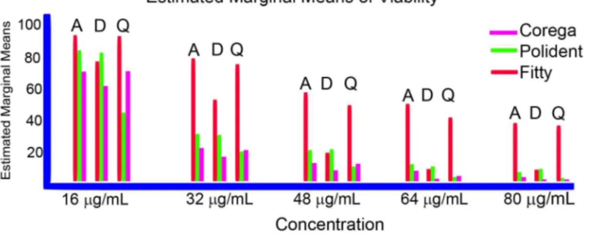
the MTT viability assay.According to MTT test results, Polident 3 min™ and Corega™ tablets exhibited strong anticandidal effects on C. albicans bioilm on all denture
resin at nearly 2 tablets/150 mL of water concentration (p<0.05), whereas Fittydent™ had only anticandidal
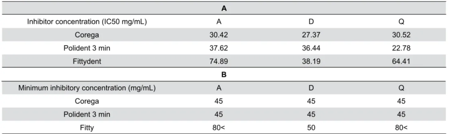
effects against the bioilm on D-type resin at nearly 2½ tablets/150 mL of water concentration (Figure 2) (Table 1). The anticandidal activity of the Corega™ was higher on A- and D-type resins compared to Polident™ and Fittydent™. The effective concentrations of Corega™ tablets were found to be 30.42 mg/mL, 27.37 mg/mL and 30.52mg/mL (approximately 2 tablets) on the A-, D- and Q-type resins, respectively (Table 1A). However, Polident 3 min™ anticandidal activity was strongest on the Q-type denture (22.78 mg/mL, approximately 1½ tablet). The effective concentrations of Polident 3 min™ tablets were 37.62 mg/mL, 36.44 mg/mL (approximately 2½ tablets), and 22.78 mg/mL (approximately 1½ tablet) on the A-, D-, and Q-type resins, respectively. The effective concentrations of Fittydent™ were 74.89 mg/mL (approximately 4½ tablets), 38.19mg/mL (approximately 2½ tablets) and 64.41mg/mL (approximately 4 tablets) on A-, D-, and Q-type resins, respectively. IC50 values to be used in subsequent studies were determined by performing the MTT assay, as indicated in Table 1A.
After an incubation time, the inhibition zone (optically clear) was produced by each cleansing solution, and the lowest concentration at which there
Figure 2- The effects of Corega, Polident 3 min or Fittydent tablets on the viability of Candida albicans bioilm on the surface A-, Q-, or
D-type resin specimens. Exponentially growing cells on denture surface were incubated with Corega, Polident 3 min or Fittydent cleansing solutions for 5, 3, and 5 min, respectively, and the cell viability was measured by the MTT assay. Percent viability was reported as mean values±SEM of three independent assays (p<.05)
A
Inhibitor concentration (IC50 mg/mL) A D Q
Corega 30.42 27.37 30.52
Polident 3 min 37.62 36.44 22.78
Fittydent 74.89 38.19 64.41
B
Minimum inhibitory concentration (mg/mL) A D Q
Corega 45 45 45
Polident 3 min 45 45 45
Fitty 80< 50 80<
was no visible zone of inhibition was taken as the MIC. The experiment was repeated three times, and the MIC values are presented in Table 1B. As shown in Table 1B, Corega™ and Polident 3 min™ had a higher inhibitory effect against bioilm.
Statistical analysis was achieved by using a three-way ANOVA test and showed signiicant difference in the mean values of resin types, tablet types, and tablet concentrations. The three-way ANOVA was run on a sample of 135 resins to examine the effect of resin type, tablet type or tablet concentrations against bioilm. There was a statistically signiicant three-way interaction between resin type, tablet type and tablet concentrations, F(16, 90)=18.81, p=.000 (Table 2A).
When calculating the two-way ANOVA,the resin types by tablet types (F(4, 90)=324.79, p=.000), resin types
by tablet concentrations (F(8, 90)=16.56, p=.000),
and tablet types by tablet concentrations (F(8,
90)=80.96, p=.000) are statistically signiicant (Table
2A). Application of Tukey’s HSD multiple comparisons test showed a statistically signiicant difference among all test groups (Tables 2B and 2C) (mean difference is “*” indicating signiicant difference among groups).
The plot of the mean “viability” score for each combination of groups of “resins” and “tablets” are plotted in a line graph at all concentrations, as shown in Figure 3.
The adhesion and the spreading of cells on surfaces
A
Source Type III Sum of
Squares df Mean Square F Sig.
Corrected Model a88651.08 44 2014.80 408.41 .000
Intercept 180547.92 1 180547.92 36597.55 .000
Resin type 2953.44 2 1476.72 299.34 .000
Tablet type 22468.55 2 11234.27 2277.22 .000
Concentration 51486.42 4 12871.60 2609.11 .000
Resin type * Tablet type 6409.1 4 1602.27 324.79 .000
Resin type * Concentration 653.45 8 81.68 16.56 .000
Tablet type * Concentration 3195.23 8 399.4 80.96 .000
Resin type * Tablet type *
Concentration 1484.90 16 92.81 18.81 .000
Error 444.00 90 4.93
a.R Squared = .995 (Adjusted R
Squared = .993)
B
(I) Resin type (J) Resin type Mean Difference
(I-J) Std. Error Sig.
A D 11,40* 0.468 .000
Q 6,69* 0.468 .000
D A -11,40* 0.468 .000
Q -4,71* 0.468 .000
Q A -6,69* 0.468 .000
D 4,71* 0.468 .000
C
(I) Tablet type (J) Tablet type Mean Difference
(I-J) Std. Error Sig.
Corega Polident -4,31* 0.468 .000
Fitty -29,27* 0.468 .000
Polident Corega 4,31* 0.468 .000
Fitty -24,96* 0.468 .000
Fitty Corega 29,27* 0.468 .000
Polident 24,96* 0.468 .000
were investigated using SEM. As shown in Figure 4, obvious cell spreading changes were not observed in the treated cells compared to the untreated cells. The bioilm exposed to the ive concentrations of cleansing tablets did not exhibit signiicantly greater adhesion strength. This situation was not consistent with the results of the above mentioned MTT assays.
Also, we found that surface characteristics among the thermoplastic polyamide resin (D-type) and the PMMAs (Q-type and A-type) may cause the formation of the C. albicans bioilm layer in different conluences
on resin types. In fact, we observed that the C. albicans bioilm layer effectively spread on the A-
and Q-type resins by penetrating into their notched surfaces, whereas this was not found in the D-type
resin (Figure 4, IV-VI).
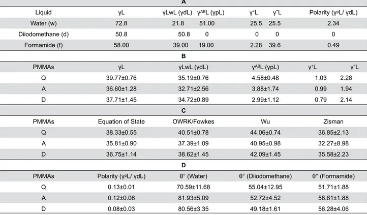
Results of SFE analysis and the analysis of its components are shown in Tables 3A-3D. The dispersive and polar components of Q-, A-, and D-type resins were found to be 35.19-4.58 mJ/m2 for distilled water,
32.71-3.88 mJ/m2 for diodomethane, and 34.72-2.99
mJ/m2 for formamide. According to the acid-base
approach, SFE values of Q-, A-, and D-type resins were 39.77, 36.60, and 37.71 mJ/m2, respectively.
Q-type resin exhibited the highest SFE value (p<0.05),
whereas the lowest values were found for A-type resin in all methods (p<0.05) (Table 3B and 3C). Generally,
differences in the SFE of these resins were identiied for all methods (p<0.05). Polarity values of Q-, A- and
D-type resins were 0.13, 0.12, and 0.08, respectively.
Figure 3- The graphics illustrated that an interaction effect is among resin*tablet at all concentration
According to the contact angle results, the wettability value of these resins was in the following order: Q-type (70.59) > D-type (80.56) > A-type (81.93) (Table 3D).
Discussion
The null hypothesis that denture base material type, chemical cleanser type, different concentration of chemical cleanser solution and polarity of resin would not interfere with C. albicans bioilm growth
was rejected.
First, we evaluated the anticandidal effect of two alkaline peroxide denture cleansers, Corega™ and Fittydent™, and one neutral enzymatic peroxide denture cleanser, Polident 3 min™, on three different resins so their surfaces were standardized to avoid surface imbalance. The SEM and surface analyses were used in this stage. It is known that polyamide resin surfaces generally exhibit a rougher texture compared to PMMA resins6,12 and the surface
structure may lead to increased microbial lora and the attenuated effect of cleansers1,24. However, we
utilised a simple process to obtain standardized surface roughness from three types of resin with different surface properties and applied a smoothing
method that adjusted their roughness to 0.32±0.02 µm through a proilometer. However, even though a standardized surface roughness was used, all cells on the polyamide resin were very weakly attached to the surface and spontaneously separated more easily from the surface compared to PMMAs. These results showed that the surface properties of resins are not the only factor governing the C. albicans adhesion,
and, at the same time, the chemical content of the material may affect the C. albicans adhesion (Table 1).
That is why the Fittydent™ tablet had only anticandidal effects against bioilm on the D-type resin with the same administrative concentrations. In addition, the MTT analysis indicated that the polyamide resin with low polarity (Table 4D) exhibited a high anticandidal effect against C. albicans cells, whereas the PMMAs
with high polarity had low anticandidal effect. The PMMAs, A-type resin, and Q-type resin exhibited approximately equal anticandidal effects, and one of the reasons may be that their polarity values were very close to one another. The bacterial attachment
to resins has not been fully revealed to be affected by its SFE and wettability propertybecause there are many inconsistent results from various studies8. For
example, some studies showed a linear correlation between SFE values and C. albicans adhesion2,17,
A
Liquid γL γLwL (γdL) γᴬᴮL (γpL) γ+L γˉL Polarity (γᵖL/ γdL)
Water (w) 72.8 21.8 51.00 25.5 25.5 2.34
Diiodomethane (d) 50.8 50.8 0 0 0 0
Formamide (f) 58.00 39.00 19.00 2.28 39.6 0.49
B
PMMAs γL γLwL (γdL) γᴬᴮL (γpL) γ+L γˉL
Q 39.77±0.76 35.19±0.76 4.58±0.48 1.03 2.28
A 36.60±1.28 32.71±2.56 3.88±1.74 0.99 1.94
D 37.71±1.45 34.72±0.89 2.99±1.12 0.79 2.14
C
PMMAs Equation of State OWRK/Fowkes Wu Zisman
Q 38.33±0.55 40.51±0.78 44.06±0.74 36.85±2.13
A 35.81±0.90 37.39±1.09 40.95±0.98 32.27±8.98
D 36.75±1.14 38.62±1.45 42.09±1.45 35.58±2.23
D
PMMAs Polarity (γᵖL/ γdL) θ° (Water) θ° (Diiodomethane) θ° (Formamide)
Q 0.13±0.01 70.59±11.68 55.04±12.95 51.71±1.88
A 0.12±0.06 81.93±5.09 52.72±4.52 56.81±1.88
D 0.08±0.03 80.56±3.35 49.18±1.61 56.28±4.06
Table 3- A) Acidic, Basic Components of Surface Free Energies of Test Liquids used in this work (mJ/m2). B) Surface Free Energy
Components of PMMAs Surface Calculated by Acid-Base Approach (mJ/m2). C) Surface Free Energy of PMMAs Surface Calculated by
whereas other studies reported no correlation at al9,27. Likewise, in this study, we failed to ind a strong
correlation between SFE and C. albicans adhesion. It is speculated that low polarity, low SFE value and low wettability may lead to a signiicantly increased anticandidal effect. However, we found that merely the polarity feature of resins may alter its anticandidal affect. The SEM images substantially conirmed our speculations about resin types used in this study. For example, C. albicans bioilm layers on A- and
Q-type resins were covered a much larger area and presented a higher level of growth than D-type resin. However, even though D-type resin showed rouger surface than the others, it can be pretty smooth and slippery following the surface deburring and polishing prosses. In addition, according to SEM presentations for all resins, we failed to ind a signiicant difference between C. albicans forms such as yeast and hyphal
formation.
Second, we conducted a MTT method to determine inhibitor concentration (IC50 mg/mL) and the minimum inhibitor concentration (MIC) values of these cleanser tablets. Results indicated that C. albicans viability was affected to alter the denture cleanser trademark, concentration, and resin type. Denture cleanser tablets act to bioilm layer in a concentration-dependent manner – that is, increasing the concentrations of denture cleanser tablets on bioilms layer lead to a gradual increase in the cell inhibition, showing a typical inhibitor effect. This means that the tablet concentrations are effective in terms of eliminating the bioilm layers. In addition, the concentration increasing effect reached maximum impact against cell viability at 64 mg/mL concentration (four 2.5 g effervescent tablets dissolved in 150 mL of water to prepare a 64 mg/mL solution). Regarding the eficacy of the tablets considered together with denture cleanser trademark and resin type, the Corega™ tablets should be advised to provide effective cleansing of A- and D-type resin (IC50, 30.42 and 27.37 mg/mL, respectively, correspond to approximately 2 tablets) and the Polident 3 min™ tablets are suggested for Q-type resin (IC50, 22.78 mg/mL, correspond to approximately 1½ tablet). However, Fitty™ tablets must be used in a more concentrated manner for the same effect on bioilm layer when compared withother types (IC50, 38.19 to 74.89 mg/mL, correspond to approximately 2½ to 4½ tablets). Other studies also showed that the type of resin of denture base affects
the amount of C. albicans bioilm layers colonization,
as observed in this study4,6. Murata, et al.19 (2010)
reported that the influence of neutral enzymatic denture cleanser on the surface properties was less than that of alkaline peroxide denture cleanser due to the neutral enzymatic denture cleanser containing less peroxide. However, none of the denture cleanser tablet concentrations were able to remove C. albicans
bioilm completely in up to 25 mg/mL concentrations (approximately 1½ tablet). Most studies were conducted to remove C. albicans bioilm formation on
the denture base resins of PMMAs via denture cleanser tablets15,29, whereas, to the best of our knowledge, a few studies evaluated the eficacy of denture cleansers on thermoplastic polyamide resin4,6. One of the
thermoplastic polyamide resin studies demonstrated smaller C. albicans growth on the PMMA surface than
on the thermoplastic polyamide resin4. They found that
the residual monomer was released from the PMMA, and they were putting this forward as a serious theory. Therefore, in this study, specimens were soaked in distilled water for 24 h after polymerization to reduce the residual monomer. The cytotoxic effects of the acrylic resins remained at high levels within the irst 24 h following polymerization28. The water immersion
method was suggested to reduce the level of residual monomer11 because this toxic effect is reduced in a
time-dependent manner29. Another study determined
that the cleanser tablets tested were more effective for PMMA resin than for thermoplastic polyamide resin6. This result was inconsistent with our indings. The reason we applied the surface roughness process to the resins using a proilometer was because of the varying study indings for both resins.
Conclusion
We have clearly demonstrated that the polarity of resins and the chemical content of the cleanser may
affect C. albicans bioilm adhesion. Also, the results
was shown that anticandidal activity appears to be a function of the nature of the resins, their roughness, the type of cleanser and the speciic concentrations of the cleanser.
Acknowledgments
The authors are thankful to Dr. Şaban Tekin and Dr. Isa Karaman for generous assistance, and to the Gaziosmanpaşa University Research Foundation (Grant 2014/90) for inancial support.
References
1- Berger JC, Driscoll CF, Romberg E, Luo Q, Thompson G. Surface roughness of denture base acrylic resins after processing and after polishing. J Prosthodont. 2006;15(3):180-6.
2- Busscher HJ, Weerkamp AH, van der Mei HC, van Pelt AW, de Jong HP, Arends J. Measurement of the surface free energy of bacterial cell surfaces and its relevance for adhesion. Appl Environ Microbiol. 1984;48(5):980-3.
3- Dills SS, Olshan AM, Goldner S, Brogdon C. Comparison of the antimicrobial capability of an abrasive paste and chemical-soak denture cleaners. J Prosthet Dent. 1988;60(4):467-70.
4- Fernandes FS, Pereira-Cenci T, Silva WJ, Ricomini AP Filho, Straioto FG, Del Bel Cury AA. Eficacy of denture cleansers on Candida spp.
bioilm formed on polyamide and polymethyl methacrylate resins. J Prosthet Dent. 2011;105(1):51-8.
5- Ferreira MA, Pereira-Cenci T, Vasconcelos LM, Rodrigues-Garcia RC, Del Bel Cury AA. Efficacy of denture cleansers on denture liners contaminated with Candida species. Clin Oral Investig. 2009;13(2):237-42.
6- Freitas-Fernandes FS, Cavalcanti YW, Ricomini AP Filho, Silva WJ, Cury AADB, Bertolini MM. Effect of daily use of an enzymatic denture
cleanser on Candida albicans bioilms formed on polyamide and poly (methyl methacrylate) resins: an in vitro study. J Prosthet Dent. 2014;112(6):1349-55.
7- Ghalichebaf M, Graser GN, Zander HA. The eficacy of denture-cleansing agents. J Prosthet Dent. 1982;48(5):515-20.
8- Hahnel S, Rosentritt M, Burgers R, Handel G, Lang R. Candida albicans bioilm formation on soft denture liners and eficacy of cleaning protocols. Gerodontology. 2012;29(2):e383-91.
9- Hahnel S, Rosentritt M, Handel G, Burgers R. In vitro evaluation
of artiicial ageing on surface properties and early Candida albicans adhesion to prosthetic resins. J Mater Sci Mater Med. 2009;20(1):249-55.
10- Jagger DC, Harrison A, Jandt KD. The reinforcement of dentures. J Oral Rehabil. 1999;26(3):185-94.
11- Jorge JH, Giampaolo ET, Machado AL, Vergani CE. Cytotoxicity of denture base acrylic resins: a literature review. J Prosthet Dent. 2003;90(2):190-3.
12- Kawara M, Iwata Y, Iwasaki M, Komoda Y, Iida T, Asano T, et al. Scratch test of thermoplastic denture base resins for non-metal clasp
dentures. J Prosthodont Res. 2014;58(1):35-40.
13- Kulak Y, Arikan A, Albak S, Okar I, Kazazoglu E. Scanning electron microscopic examination of different cleaners: surface contaminant removal from dentures. J Oral Rehabil. 1997;24(3):209-15. 14- Kulak-Ozkan Y, Kazazoglu E, Arikan A. Oral hygiene habits, denture cleanliness, presence of yeasts and stomatitis in elderly people. J Oral Rehabil. 2002;29(3):300-4.
15- Lucena-Ferreira SC, Ricomini-Filho AP, Silva WJ, Cury JA, Cury AA. Inluence of daily immersion in denture cleanser on multispecies bioilm. Clin Oral Investig. 2014;18(9):2179-85.
16- McCabe JF, Murray ID, Kelly PJ. The eficacy of denture cleansers. Eur J Prosthodont Restor Dent. 1995;3(5):203-7.
17- Minagi S, Miyake Y, Inagaki K, Tsuru H, Suginaka H. Hydrophobic
interaction in Candida albicans and Candida tropicalis adherence to
various denture base resin materials. Infect Immun. 1985;47(1):11-4. 18- Minagi S, Tsunoda T, Yoshida K, Tsuru H. Objective testing of the efficiency of denture-cleansing agents. J Prosthet Dent. 1987;58(5):595-8.
19- Murata H, Chimori H, Hong G, Hamada T, Nikawa H. Compatibility of tissue conditioners and denture cleansers: inluence on surface conditions. Dent Mater J. 2010;29(4):446-53.
20- Nikawa H, Hamada T, Yamamoto T. Denture plaque: past and recent concerns. J Dent. 1998;26(4):299-304.
21- Nikawa H, Hamada T, Yamashiro H, Kumagai H. A review of in vitro
and in vivo methods to evaluate the eficacy of denture cleansers. Int J Prosthodont.1999;12(2):153-9.
22- Nikawa H, Iwanaga H, Hamada T, Yuhta S. Effects of denture cleansers on direct soft denture lining materials. J Prosthet Dent. 1994;72(6):657-62.
23- Peracini A, Davi LR, Ribeiro NQ, Souza RF, Silva CH, Freitas Oliveira Paranhos H. Effect of denture cleansers on physical properties of heat-polymerized acrylic resin. J Prosthodont Res. 2010;54(2):78-83. 24- Radford DR, Sweet SP, Challacombe SJ, Walter JD. Adherence
of Candida albicans to denture-base materials with different surface inishes. J Dent. 1998;26(7):577-83.
25- Rodford R. The development of high impact strength denture-base materials. J Dent. 1986;14(5):214-7.
26- Sato S, Cavalcante MR, Orsi IA, Paranhos HF, Zaniquelli O. Assessment of lexural strength and color alteration of heat-polymerized acrylic resins after simulated use of denture cleansers. Braz Dent J. 2005;16(2):124-8.
27- Serrano-Granger C, Cerero-Lapiedra R, Campo-Trapero J, Del Rio-Highsmith J. In vitro study of the adherence of Candida albicans to acrylic resins: relationship to surface energy. Int J Prosthodont. 2005;18(5):392-8.
28- Sheridan PJ, Koka S, Ewoldsen NO, Lefebvre CA, Lavin MT. Cytotoxicity of denture base resins. Int J Prosthodont. 1997;10(1):73-7.
29- Silva FC, Kimpara ET, Mancini MN, Balducci I, Jorge AO, Koga-Ito CY. Effectiveness of six different disinfectants on removing ive microbial species and effects on the topographic characteristics of acrylic resin. J Prosthodont. 2008;17(8):627-33.