Analysis of the Breast Cancer Control Program
Information System (SISMAMA), with review of 1,000
mammograms in the cities of Barra Mansa and Volta
Redonda*
Análise do Sistema de Informação do Programa de Controle do Câncer de Mama (SISMAMA) mediante avaliação de 1.000 exames nas cidades de Barra Mansa e Volta Redonda
Sissy Bullos Lins dos Santos1, Hilton Augusto Koch2
OBJECTIVE: To analyze the Breast Cancer Control Program Information System (SISMAMA) implemented in 2009 by the Brazilian Health Ministry. MATERIALS AND METHODS: This was a retrospective study involving the analysis of 1,000 requisition forms and results of mammograms performed by SUS – Sistema Único de Saúde (the Brazilian unified public health system) in the cities participating in the present study, during the period from August to October/2009. The study covered the qualitative analysis of the information sent through the data processing and the deviations resulting from the failure or inappropriateness in the forms filling. RESULTS: The most frequent issue was data omission, particularly regarding data on previous surgeries, achieving 302 omissions (30.2%). CONCLUSION: Despite the necessity of adjustments because of the time elapsed between the system creation and implementation, such adjustments do not affect directly the system’s validity. Errors in data input in the Health Ministry database corresponded to the failure in the provision of information relevant for reports completion, and the lack of familiarity and capacity of professionals involved in this process and in the forwarding of data regarding mammography results.
Keywords: SISMAMA; SISMAMA analysis; SISMAMA review.
OBJETIVO: Fazer uma análise do Sistema de Informação do Programa de Controle do Câncer de Mama (SIS-MAMA), implantado em 2009 pelo Ministério da Saúde. MATERIAIS E MÉTODOS: Tratou-se de um estudo retrospectivo, feito mediante análise de 1.000 fichas de requisição e resultado de mamografias realizadas pelo Sistema Único de Saúde (SUS) nos municípios participantes desta pesquisa, no período de tempo com-preendido entre agosto e outubro de 2009. Foram analisados a qualidade das informações enviadas através do processamento desses dados e os desvios gerados pelo não preenchimento ou pelo inadequado preen-chimento dos dados nessas fichas. RESULTADOS: O problema mais frequentemente encontrado foi a omis-são de dados nas fichas, principalmente no quesito cirurgias anteriores, constatando-se 302 omissões (30,2%). CONCLUSÃO: Apesar do Sistema necessitar de alguns ajustes, pelo lapso temporal transcorrido entre sua criação até sua implementação, esses ajustes não afetam diretamente a validade do Sistema, encontrando-se como principal fator de erros na alimentação do banco de dados do Ministério da Saúde o não preenchi-mento de informações relevantes para o fechapreenchi-mento dos laudos, e a falta de familiarização e capacitação dos profissionais envolvidos nesse processo e no repasse de dados do resultado da mamografia.
Unitermos: SISMAMA; Análise do SISMAMA; Revisão SISMAMA. Abstract
Resumo
* Study developed at the Unit of Radiology and Post-graduation Prof. Dr. Hilton Koch, Santa Casa da Misericórdia do Rio de Ja-neiro, RJ, Brazil. Data collection in private clinical centers under contract to the SUS (the Brazilian unified health system) in the cities participating in the present study.
1. MD, Post-graduate in Radiology and Imaging Diagnosis, Pontifícia Universidade Católica do Rio de Janeiro (PUC-Rio), Rio de Janeiro, RJ.
2. PhD, Head for the Unit of Radiology, Santa Casa da Mise-ricórdia do Rio de Janeiro and Courses of Post-graduation at Pontifícia Universidade Católica do Rio de Janeiro (PUC-Rio), Rio de Janeiro, RJ, Brazil.
Mailing address: Dra. Sissy Bullos Lins dos Santos. Rua Cinco de Julho, 94/601, Copacabana. Rio de Janeiro, RJ, Brazil, 22051-030. E-mail: [email protected]
Sistema Único de Saúde – SUS (the Bra-zilian unified health system). Since the dis-closure in 2004 of the “Controle do Câncer de Mama: Documento de Consenso” (sensus Document on Breast Cancer Con-trol)(1), governmental actions have been
targeted at providing the population with the access to procedures for early detection of this disease, also called screening pro-cedures or screening mammography, with yearly clinical examination of the breast for Bullos SB, Koch HA. Analysis of the Breast Cancer Control Program Information System (SISMAMA), with review of 1,000 mammograms in the cities of Barra Mansa and Volta Redonda. Radiol Bras. 2010;43(5):295–301.
INTRODUCTION
Breast cancer control is a priority in the Brazilian health policy and has been in-cluded as one of the targets of the “Pacto pela Saúde (2006)” (Health Covenant), a program aimed at strengthening, integrat-ing and promotintegrat-ing resoluteness of the
all women above 40 years of age, mammog-raphy for women in the age range between 50 and 69 years at a maximum two-year interval between examinations, and yearly clinical examination and mammography for women above 35 years of age at high risk for developing breast cancer. Still, there are situations in which routine mammography is also recommended, such as before start-ing hormone replacement therapy, prior to plastic surgery and in the postoperative follow-up of mastectomy , for the study of the contralateral breast, or after conserva-tive surgery, and the so called diagnostic mammography performed in women with signs or symptoms of breast cancer.
For the year of 2010, 49,240 new cases of breast cancer are expected. Breast cer is the second most frequent type of can-cer in the world, and the most frequent among women. Every year 22% of the new cases of cancer are breast cancer(1).
With the objective of acting in a more practical and effective manner, based on data on breast cancer that could be regu-larly, safely and quickly evaluated, an elec-tronic information system was developed, the “Sistema de Informação do Controle do Câncer de Mama” (SISMAMA) (Breast Cancer Information and Control System), aimed at creating a national databank aimed at gathering data from the different regions of the country on the disease through the standardization of mammogra-phy requisition forms e respective reports. The SISMAMA started operating in 2009, after the previous capacitation of the professionals involved in this process both at regional and municipal levels, promoted in 2008 by the State Health Secretariats.
The SISMAMA system was jointly de-veloped by the Instituto Nacional de Câncer and DATASUS/MS as from 2000 and, af-ter an inaf-terruption, its development was resumed between 2005 and 2006, with the objective of obtaining data on breast can-cer screening in the country. SISMAMA is a subsystem of the SUS outpatient billing system, in which the collected data are uti-lized for the billing of mammography, cy-topathology and hiscy-topathology services and for the management of actions for breast cancer screening by the program coordina-tion at municipal, regional and state levels. The SISMAMA relied on the collaboration
of Colégio Brasileiro de Radiologia e Diagnóstico por Imagem (CBR) in the matters related to mammography(2).
This system automatically reproduces the Breast Imaging Reporting and Data System (BI-RADS®) categories, according to mammographic findings, allowing a re-liable reporting of such findings within their respective categories, since the system excludes antagonistic information. This is an essential tool in the management of the disease control.
MATERIALS AND METHODS
This was a retrospective study, devel-oped means of the analysis of
mammog-raphy request forms and respective
re-sults records issued by SUS in the cities participating in the present study between August and October of 2009.
Data were collected in a clinic located in the city of Barra Mansa, and in another in the city of Volta Redonda, RJ, Brazil. Both clinics are duly accredited and quali-fied by SUS, utilize the SISMAMA since 2009 and receive part of the patients in the region to be submitted to mammography.
The data collection was carried out through the analysis of the information in-cluded in the requisition forms (filled in by the patient’s assisting physician or nurse in the original health service) and in the mam-mography results records (filled immedi-ately after the examination, in part by the mammographers, and part by the radiolo-gist responsible for the preparation of the mammographic report).
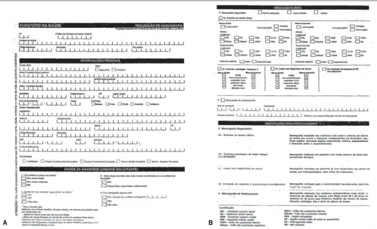
The first phase of the study comprised the analysis of the mammography request forms (Figure 1) including the patient´s personal data such as name, address, age, ethnicity, schooling, and anamnesis data such as breast nodule, high risk for breast cancer, previous breast evaluation by a health professional and previous mammo-gram. Finally, clinical indication, as fol-lows: screening or diagnosis; in the case of diagnostic mammography, marking of the affected breast (left or right) and type of finding (papillary lesion, papillary dis-charge, nodule, thickening or palpable lymph node).
The second phase comprised the analy-sis of the mammography results record
form (Figure 2) that is filled in, part by the mammographer with data regarding anam-nesis, menstrual background, use of hor-mones, information on pregnancy, radio-therapy treatment (if applicable, date of treatment), previous breast surgery and year. The other part is filled in by the radi-ologist with information on the number of films, radiological findings, BI-RADS cat-egory and respective recommendation. It is important to mention that the mammogra-phy results records defined by SUS/ SISMAMA consider the left and right breasts separately, also separating them by BI-RADS category.
For the purposes of the present study, the major category was always taken into consideration, following the appropriate recommendation for such category.
The main problems faced in the daily activities by the involved professionals as regards the deployment of the system and eventual failures or absence of data consid-ered as relevant by SISMAMA were evalu-ated through the analysis of both forms and the data digitization.
It is important to note that both forms were designed and distributed by the Min-istry of Health, in the case of the results record form, with the collaboration of CBR; and in none of them changes were made by the authors in the course of the present study. It should also be mentioned that, in the results of the present study, some of the data were not taken into con-sideration, either for not representing rel-evant information, or for not being statis-tically significant.
RESULTS
Figure 1. Mammography request form: front (A) and back (B).
A B
Figure 2. Mammography results record form: front (A) and back (B).
In some situations disagreement was observed between the System and BI-RADS, like that observed in the character-ization of microcalcifications shape. This occurred whenever microcalcifications where characterized as punctate, indepen-dently from their distribution and recom-mendation for radiological follow-up of lesion classified as category 3, because the System considered them as category 4, rec-ommending biopsy, and leading the report-ing radiologist to characterize them as round instead of punctate. Among the 1% of reported cases with microcalcifications, 0.6%, i.e., more than half of this total, were punctate microcalcifications described as rounded, so that biopsy was not recom-mended.
The inappropriateness or lack of any item on the forms designed by the Minis-try of Health generated “adaptations” by the reporting radiologists. The lack of space (restriction in the number of characters) in the field for remarks was also a factor caus-ing shortcomcaus-ing on the reports, particularly in cases of unforeseen situations on the forms.
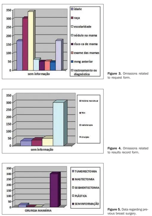
Omissions on the mammography request form (Figure 3)
Figure 3, regarding the first analysis phase, shows that the three major figures on omissions refer to schooling (342 pa-tients), ethnicity (305), purpose of the study – screening or diagnosis (174), age (172), presence of a mass or lump in the breast (65), risk for breast cancer (53), previous clinical examination of the breast by a health professional (54) and, finally, infor-mation on a previous mammography (48). Among the reported figures on omissions, the one causing greatest impact on the fi-nal conclusion of the examinations by the SISMAMA was the information regarding the reason for performing the mammogra-phy, whether screening or diagnosis. Le-sions previously classified as BI-RADS category 3 were not duly followed-up, be-sides complaints and clinical findings that could not be duly evaluated by the report-ing radiologist.
Additionally, the absence of data re-garding other items generated undernoti-fication.
Figure 3. Omissions related to request form.
Figure 5. Data regarding pre-vious breast surgery. Figure 4. Omissions related to results record form.
Omissions on the mammography results record form (Figure 4)
At the second analysis phase, the results record forms were evaluated, with the num-ber of omissions shown on Figure 4. One observed in this phase of the study that the item with the highest number of omissions refers to information on previous breast surgery (302 omissions), followed by ra-diotherapy (52), hormone replacement therapy (41) and finally, menstrual back-ground (35). The high number of omissions in the item regarding previous breast
sur-geries ended in error in the final version of the mammography reports, as the System excluded antagonistic data(2). This means
con-flicting results and hinders the report completion.
Data regarding previous breast surgeries (Figure 5)
On this Figure, a comparison is made, within the item regarding previous breast surgeries, demonstrating the most docu-mented types of surgeries. Nevertheless, the omissions represented the greatest part of the cases (302), followed by tumorectomy (23), plastic surgeries (9) mastectomy and segmentectomy (both with 4 cases). Other types of surgery were not found in the stud-ied sample.
DISCUSSION
BI-RADS is a system that classifies mammographic findings into categories and proposes clinical approaches according to each category. This system was developed by the American College of Radiology, with the objective of minimizing the differences in approaches inherent to due to interob-server variability (or disagreement)(3–6).
SISMAMA was deployed to standard-ize mammographic reports and avoid de-viations in images interpretation, and for that reason it adopts the BI-RADS, making data forwarding from SUS to the Ministry of Health, faster, easier and most reliable. Considering its recent implementation, it is important to analyze and document even-tual occurrences that may be negatively impact the data forwarding process, not only as a result of the necessity of adjust-ments in the System itself, but also because of incorrect filling or failure in filling out the forms by the involved professionals.
Omission of surgical data or inappropriate filling
On the mammography results record form, the 11th subitem (regarding data of the anamnesis), there is question on previ-ous breast surgeries and year, including different surgery modalities options (tumorectomy, segmentectomy, etc.). Ob-viously, it is appropriate that all patients’ data be accurately collected. However most of times the patients are unable to accu-rately report the procedure they were sub-mitted to, so the professional performing the anamnesis must deduct the required
information and hence the importance of a trained professional for the data collection. Most of times, the data collection is per-formed on the day of the examination by the mammographer. In the present study, this was the item with the highest number of omissions of data, totaling 302 (30.2% of the sample). This omission was also the one with greatest impact on the final result of the reports, as the reporting radiologist many times does not know that the System requires preliminary data to confirm what is described on the report. If the mammog-rapher responsible for the anamnesis does not report a previous surgery, a postopera-tive architectural distortion, for example, considered as BI-RADS category 2 by the physician, will be taken as category 4 by the System, as there is no record on previous surgery. Likewise, skin thickening or re-traction and architectural distortion will always be interpreted as a pathological finding if no data on previous surgery is available. Hence the importance of know-ing the system and workknow-ing in conjunction to use it properly.
Another problem regarding the item on previous surgery is related to patients that underwent mastectomy and breast recon-struction. In spite of the two options being comprised in the item, one observed the recurrence of certain situations: cases of breast reconstruction where the mammog-raphers report that the reconstruction was performed, but do not perform mammog-raphy for that side, generating the opening of a report for both breasts; or cases where in spite of the reconstruction, they only report mastectomy, performing bilateral mammography. In these cases, the System opens only the report for the preserved breast, disregarding the opening of the re-port for the reconstructed breast since it was not reported on the appropriate item.
Among the four mastectomies reported in the sample, one had reconstruction with-out mammography of the reconstructed breast, with the System being fed only with the data regarding mastectomy, so that re-porting on the reconstructed breast was not required, for which mammography was not performed, again causing wrong informa-tion to be entered into the System.
The above described situations clearly demonstrate that the absence of correct
information in the filling of anamnesis data is responsible for the greatest majority of errors in the interpretation of data by the System, and consequential failure in the data forwarding to the Ministry of Health.
Postoperative architectural distortion, surgical sequels and actinic lesions
In spite of the options for normal skin, skin thickening, and skin retraction (the two later ones being very common in cases of conservative surgery and actinic lesions) under sub-item “skin”, if one opts for skin retraction or skin thickening, and the field “anamnesis data” is not correctly filled out by the mammographer (as regards previous surgery), the system will automatically go to BI-RADS category 4, with the conse-quential recommendation for histopatho-logical study for typical sequel. On the other hand, as the sub-item “previous sur-geries” is correctly filled out, categories 2 or 3 will be accepted.
There is no sub-item covering the typi-cal breast volume reduction, a certain out-come in cases of conservative surgery.
Parenchymal distortion under the surgi-cal bed is a frequent occurrence, and most times it disappears within the first postop-erative year. Only 3% of all mammograms demonstrate some degree of architectural distortion two or three years after biopsy of benign lesions(7).
For breasts with distortion areas, the system provides two possibilities:
a) the first one refers to focal distortion and its location. Its selection leads the System to automatically select a biopsy category (BI-RADS 4), independently of previous breast surgery reporting. This sub-item was certainly created to anticipate situations with distortion not related to surgical procedures and with accurate in-dication for biopsy;
b) the second one refers to
postopera-tive architectural distortion, in which
there is no possibility of localizing the le-sion.
Comparison with previous studies and mammographic follow-up for lesions classified as BI-RADS category 3
The comparison with previous images is indispensable in cases of mammo-graphic follow-up for lesions classified as category 3.
On the back of the mammography
re-quest form there is item 5- diagnostic
mammography, which breaks down into 5-b radiological follow-up for category 3, and the professional responsible for filling in the form (nurse, assisting physician or duly trained personnel) is expected to pro-vide such information. Certain confusion is observed in the forms, as many times a single mammography was indicated as for diagnostic and screening purposes, without any justification in cases where the study was diagnostic.
In cases where the item radiological follow-up for category 3 is filled out, the System will only make the breast in ques-tion available for reporting. Nevertheless, the majority of such cases with incorrect filling of clinical indication in cases of fol-low-up of microcalcifications and nodules were not performed as follow-up, with re-peated bilateral mammography in the first follow-up after six months (2.4%), gener-ating unnecessary expenses with films and unnecessary radiation exposure for the pa-tient. Aguillar et al., in their description of mammographic reports according to BI-RADS, corroborate the recommendation for approaching lesions classified as cat-egory 3, with follow-up after six months (unilateral, only the breast with alteration), and bilaterally after 12, 24 and 36 months(7).
The mammography results record form does not include any provision for compari-son with previous images, i.e., even in the case of previous information being accu-rate, a follow-up for an appropriate period of time must be provided for, filling out the date of the previous study, and informing the patient as the mammography request form is filled - in the case of lesions requir-ing radiological follow-up (category 3) – , that she should always take the previous studies with her on the date scheduled for the new mammographic study, leaving them for comparison by the radiologist,
thus avoiding that the patient be left with inappropriate follow-up of the lesion, and without a forecast for the determination of its stability.
Microcalcifications
As the interpretation of the BI-RADS category by the System is concerned, it is automatically done based on certain data provided by the reporting radiologist, to-gether with other data collected by the mammographer.
In the presence of microcalcifications, there is the possibility of classifying them according to their location, shape and dis-tribution. In what regards to location, the options for breast quadrants and axillary extension are available in the System, and these topics have no impact on category classification. Subsequently, the System requests the shape, with the following op-tions: rounded, punctate, irregular and branching; the distribution is also requested with the following options: clustered, seg-mented and ductal. When the reporting ra-diologist characterizes the calcifications as rounded, the System automatically catego-rizes the findings as BI-RADS category 3; when the microcalcifications are described as punctate, the System categorizes the finding as BI-RADS category 4, and con-sequently recommending histopathological study.
Clustered, rounded or punctate calcifi-cations (clustered calcificalcifi-cations in a circu-lar or ovoid arrangement, suggestive of lobular origin) are classified as category 3, and clustered, monomorphic, predomi-nantly punctate or rounded calcifications; if absent in previous study, are classified as BI-RADS category 4a(7).
According to Kopans, very rounded, punctuate, regular calcifications are rarely associated with breast cancer. However, if they include, or are associated with calci-fications whose shapes are not rounded or smooth, but rather heterogeneous, they should be considered as suspicious(8).
In the present sample, 0.6% of all the microcalcifications, in spite of being actu-ally punctuate, were reported as rounded in order to avoid categorization for biopsy in findings with characteristics of benignity, again generating a distortion in the data forwarding.
Enlarged axillary lymph nodes
BI-RADS does not contemplate lymph node alterations. This is a controversial point for radiologists, as an occult carci-noma may present with lymph node alter-ation as its single manifestalter-ation.
For this reason, some radiologists con-sider biopsy in the presence of such alter-ations, as does the SISMAMA, consider-ing any lymph node alteration (enlarge-ment, density alteration and confluence) as category 4, with indication for biopsy.
Aguillar et al.(7) consider as probable
that the BI-RADS committee, on the next issue, will adopt the following classifica-tion for axillary adenopathy: if related to rheumatoid arthritis or sarcoidosis – cat-egory 2; related to lesion requiring further investigation with other imaging method (ultrasonography, magnetic resonance im-aging) – category 0; related to a lesion re-quiring biopsy – category 4; and, if related to other known disease (lymphoma, leuke-mia) – category 6.
Restricted space for general observations
In certain cases, the radiologist faces unusual situations that require a justifica-tion for the report or even informajustifica-tion to the assisting physician. In the case of SUS patients, accessing the requesting physician may be very difficult or not feasible at all because of the number of studies to be re-ported. So the space left in the field for general observations may be the single form of contact between the professionals. In seven cases of the present sample the space in the field for general observations was simply not enough to clarify points of the reports; five of these cases were from Barra Mansa, and two from Volta Redonda.
Unforeseen situations and findings
Findings observed outside the breast
parenchyma – During the forms analysis,
histopatho-logical diagnosis of infiltrating ductal breast carcinoma affecting the skin, classi-fied as BI-RADS category 5. Follow-up mammography was performed in August 2009, on which three small skin nodules were found and confirmed at clinical ex-amination. The nodules were located on the skin next to the surgical scar, and the need for biopsy could not be ruled out because of the previous histopathological result, which reported extension to the skin, with high probability for local recurrence.
The System does not comprise the pres-ence of skin nodules, only in the breasts. As a nodule and its location are identified, the possible options comprise quadrants, retroareolar region, and axillary extension, implying that that all mentioned nodular lesions are within the breast. In the sub-item “general observations”, it would not be possible to report the case because of the limited number of characters. In this spe-cific case, a telephone call was made to the assisting physician, explaining that the limitations inherent to the System did not allow an appropriate description of the case. In the present sample, this occurred with a single case (0.1%).
Steatonecrosis – Another common
postoperative finding refers to steatone-crosis. This is a common benign condition that may be asymptomatic or may be present as a palpable mass, with pain or associated findings, such as skin thicken-ing or nipple retraction. Steatonecrosis may present with different mammographic ap-pearances. Radiotransparent well-circum-scribed masses, mixed fat densities and soft tissue with or without calcified rim, known as lipid cysts constitute common and typi-cal findings(9). These findings may be
ob-served after any trauma in the breast, in-cluding surgery. Steatonecrosis is com-monly seen after a nodule resection and
radiotherapy for breast carcinoma and af-ter an extensive surgery; however such finding is not covered by the System. No report on these findings was observed in the sample.
Accessory breasts – Other unforeseen
situations of lesser relevance are the rela-tively common cases of accessory breasts. There is no mention of such alteration in the item regarding “other findings”, in which it could be mentioned.
Gynecomastia – Although the present
study is about female patients because they are the vast majority of those being submit-ted to mammography, it is important to mention that cases of gynecomastia are not comprised by the System.
CONCLUSION
It is obvious that no available computer system can anticipate all the situations and variables involved in a process, which also depend on the subjective analysis of the reporting radiologist. Adjustments are nec-essary in the System, and even more impor-tant, appropriate training of the profession-als involved in the process. Actually, what is intended is anticipating some relatively recurrent situations to improve the System with more accurate technical information. Undoubtedly, SISMAMA is a project that provides greater agility in the process-ing of data necessary for plannprocess-ing actions in health and better resources allocation. However, considering the short training time, the System is still underutilized by technicians, physicians and other involved professionals; not all of them know how it works and the importance of filling out the forms as accurately as possible to avoid the exclusion of antagonistic data. Physicians, technicians and those professionals respon-sible for the processing of such data should
know the operation of the System to allow that the statistical data generated by the System be accurately forwarded to the Ministry of Health, reducing undernoti-fication and erroneous notiundernoti-fications with consequential inappropriate allocation of financial resources. Reviews and adjust-ments in the System are in fact necessary, however proper training of involved pro-fessionals is the crucial point.
REFERENCES
1. Brasil. Ministério da Saúde. Instituto Nacional de Câncer. Controle do câncer de mama – documen-to de consenso. [acessado em 25 de maio de 2010]. Disponível em: www.inca.gov.br 2. SISMAMA Internet. [acessado em 10 de
novem-bro de 2009]. Disponível em: www.datasus.gov. br/siscam/siscam.php
3. Althoff FP, Petrelli ASC. Correlação dos achados mamográficos com o sistema BI-RADS. [Mono-grafia]. Rio de Janeiro, RJ: Curso de Pós-Gradua-ção da Santa Casa de Misericórdia do Rio de Ja-neiro; 2005.
4. American College of Radiology. ACR BI-RADS-Mammography. 4th ed. In: ACR Breast Imaging Reporting and Data System, Breast Imaging At-las. Reston, VA. American College of Radiology; 2003.
5. Berg WA, Campassi C, Langenberg P, et al. Breast Imaging Reporting and Data System: inter- and intraobserver variability in feature analysis and final assessment. AJR Am J Roentgenol. 2000; 174:1769–77.
6. Godinho ER, Koch HA. Submissão às recomen-dações do BI-RADS por médicos e pacientes: análise preliminar de 3.000 exames realizados em uma clínica particular. Radiol Bras. 2004;37:21– 3.
7. Aguillar V, Bauab S, Maranhão N. Relatório ma-mográfico e ultra-sonográfico segundo o BI-RADS. Guia e dúvidas. In: Aguillar V, Bauab S, Maranhão N. Mama – diagnóstico por imagem. 1ª ed. Rio de Janeiro, RJ: Revinter; 2009. p. 301–4. 8. Kopans DB. Analisando a mamografia. In: Ko-pans DB, editor. Imagem da mama. 2ª ed. Rio de Janeiro, RJ: Medsi; 1998. p. 332.