XIII
Radiol Bras. 2012 Nov/Dez;45(6):XIII–XIV
Hebert Ferro Monteiro1, Pedro Paulo Teixeira e Silva Torres2, Veronica Nogueira Garcia Edelhoff3, Pedro José de Santana Júnior4, Gustavo Ribeiro Fiori4, Kim-Ir-Sen Santos Teixeira5
0100-3984 © Colégio Brasileiro de Radiologia e Diagnóstico por Imagem
WHICH IS YOUR DIAGNOSIS?
Monteiro HF, Torres PPTS, Edelhoff VNG, Santana Júnior PJ, Fiori GR, Teixeira KISS. Which is your diagnosis? Radiol Bras. 2012 Nov/Dez;45(6):XIII–XIV.
Female, 11-year-old patient with lumbar pain and intermit-tent macroscopic hematuria for three months, sometimes with blood clots. The patient did not present any comorbidity and clini-cal examination was normal. All the laboratory blood tests results, including blood count, coagulation profile, protein electrophore-sis, FAN, C3, PCR and biochemistry were normal. Simple urine test revealed reddish color urine and presence of hemoglobin and Study developed in the Department of Radiology and Imaging Diagnosis, Hospital das Clínicas da Universidade Federal de Goiás (HC-UFG), Goiânia, GO, Brazil. 1. MD, Resident of Radiology and Imaging Diagnosis at Hospital das Clínicas da Universidade Federal de Goiás (HC-UFG), Goiânia, GO, Brazil. 2. Titular Member of Colégio Brasileiro de Radiologia e Diagnóstico por Imagem (CBR), Substitute Professor of Radiology at Hospital das Clíni-cas da Universidade Federal de Goiás (HC-UFG), Goiânia, GO, Brazil. 3. Titular Member of Colégio Brasileiro de Radiologia e Diagnóstico por Imagem (CBR), MD, Radiologist,
De-partment of Radiology, Hospital das Clínicas da Universidade Federal de Goiás (HC-UFG), Goiânia, GO, Brazil. 4. Titular Members of Colégio Brasileiro de Radiologia e Diagnóstico por Imagem (CBR), MDs, Radiologists at Clínica São Camilo, Goiânia, GO, Brazil. 5. PhD, Associate Professor, Department of Radiology, Hospital das Clínicas da Universidade Fe-deral de Goiás (HC-UFG), Goiânia, GO, Brazil. Mailing Address: Dr. Hebert Ferro Monteiro. Rua Longitudinal, Qd J, Lt 1-5, Ed. Condomínio dos Guaranis, ap. 501A, Setor Leste Vila Nova. Goiânia, GO, Brazil, 74633-300. E-mail: [email protected]
red blood cells. The tests for alcohol-acid-resistant bacilli and deformed red blood cells were negative. The 24-hour urine test demonstrated subtle proteinuria.
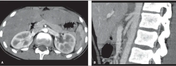
Abdominal multidetector computed tomography angiography (MDCT-angiography) was performed (Figure 1). Ureterocysto-scopy demonstrated intermittent bleeding and presence of blood clots throughout the left ureteral meatus.
Figure 1. Abdominal MDCT-angiography image in the axial plane (A) and MIP (maximum intensity projection) reformatted image in the sagittal plane (B).
XIV Radiol Bras. 2012 Nov/Dez;45(6):XIII–XIV Images description
Figure 1. Abdominal MDCT-angiogra-phy image in the axial plane (A) and MIP (maximum intensity projection) reformat-ted image in the sagittal plane (B) show compression of left renal vein (LRV) be-tween the aorta and the superior mesenteric artery (SMA) due to narrowing of the space between those two arteries, characterizing the nutcracker phenomenon. Both the dis-tance (A) and the mesoaortic angle (B) are reduced, measuring about 3 mm and 30°, respectively.
Diagnosis: Nutcracker syndrome.
COMMENTS
Nutcracker phenomenon (NCP) refers to compression of the LRV, generally be-cause of a narrowing of the angle between the aorta and the SMA. In such a situation, it is described as anterior nutcracker phe-nomenon, corresponding to the anatomy observed in the present patient. Posterior NCP is also described, where the retro-aortic or circumretro-aortic renal vein is com-pressed between the aorta and the vertebral body. In cases where there is association with clinical manifestations, nutcracker syndrome (NCS) occurs. The anatomy of NCP without clinical manifestations may represent a variant of normality(1,2).
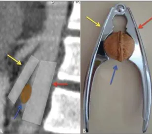
The use of the term nutcracker is gen-erally credited to de Schepper who, in 1972, reported the cases of two patients with hematuria and LRV compression. However, the term was first utilized by Chait et al. in 1971(3,4). Figure 2 illustrates
the analogy with a nutcracker.
The nutcracker syndrome is most fre-quently found in young women; and, be-cause of symptoms variability and lack of consensus about diagnostic criteria, its prevalence remains unknown, but probably it is an underdiagnosed entity. Most com-mon clinical manifestations include hema-turia, lumbar or abdominal pain, pelvic varices, varicocele and proteinuria in var-ied degrees. Hematuria occurs because of rupture of the thin walls of the venous collaterals within the adjacent calyceal sys-tem due to venous hypertension secondary to the LRV compression(1,5).
In the present case, the diagnosis of NCS was achieved after ruling out other
possible causes of hematuria, upon visual-ization of bleeding through the left ureteral meatus at ureterocystoscopy and character-ization of the anatomical findings of NCP by MDCT angiography.
Several imaging studies are utilized in the diagnostic workup of NCS, such as Doppler ultrasonography (US), MDCT an-giography, magnetic resonance angiogra-phy (MRA) and venograangiogra-phy. Although venography with measurement of the dif-ference of pressure between LRV and su-perior vena cava is considered as the most informative method in diagnostic terms, it is invasive and is not performed in patients presenting mild symptoms as well as in children. Additionally, it is not an entirely reliable method, since overlap of pressoric levels may occur between asymptomatic patients and those with NCS(1,5).
MDCT angiography with multiplanar reformatting allows the evaluation of the relationship between aorta, SMA and LRV, so it is easy to measure both the mesoaortic angle and the distance between the SMA and the aorta. This method has shown to be superior to venography and US in the iden-tification of LRV compression(6).
There is a great variability in the mea-surement of the mesoaortic angle, and small angles are associated with NCS tients as compared with asymptomatic pa-tients. Typically, the mesoaortic angle and the distance between the aorta and the SMA at the level of the LRV in symptomatic pa-tients are 39.3° ± 4.3° and 3.1 mm ± 0.2 mm, respectively. On the other hand, in asymptomatic patients, they are 90° ± 10°
and 12 mm ± 1.8 mm(5). The present patient
presented an angle of about 30° and dis-tance of 3 mm, in compatibility with the NCP anatomy.
The treatment for NCS is dependent on the symptoms severity. In patients under the age of 18 years, with mild to moderate he-maturia, the best option is a conservative approach with clinical follow-up, consid-ering that complete resolution is observed in 75% of cases over a two-year period(7).
Such approach was adopted in the present case.
One can conclude that MDCT-angiog-raphy can be utilized as a quite reliable, noninvasive method to characterize the NCP anatomy, allowing the diagnosis of NCS in patients with a compatible clinical presentation.
REFERENCES
1. Kurklinsky AK, Rooke TW. Nutcracker phenom-enon and nutcracker syndrome. Mayo Clin Proc. 2010;85:552–9.
2. Shin JI, Lee JS. Nutcracker phenomenon or nut-cracker syndrome? Nephrol Dial Transplant. 2005; 20:2015.
3. de Schepper A.“Nutcracker” phenomenon of the renal vein and venous pathology of the left kidney. J Belge Radiol. 1972;55:507–11.
4. Chait A, Matasar KW, Fabian CE, et al. Vascular impressions on the ureters. Am J Roentgenol Ra-dium Ther Nucl Med. 1971;111:729–49. 5. Fu WJ, Hong BF, Gao JP, et al. Nutcracker
phenom-enon: a new diagnostic method of multislice com-puted tomography angiography. Int J Urol. 2006; 13:870–3.
6. Shokeir AA, el-Diasty TA, Ghoneim MA. The nut-cracker syndrome: new methods of diagnosis and treatment. Br J Urol. 1994;74:139–43. 7. Shin JI, Park JM, Lee SM, et al. Factors affecting
spontaneous resolution of hematuria in childhood nutcracker syndrome. Pediatr Nephrol. 2005;20: 609–13.