http://dx.doi.org/10.1590/s2175-97902018000217149
Article
*Correspondence: G. P. Silveira. Departamento de Química Orgânica, Ins-tituto de Química, Universidade Federal do Rio Grande do Sul. Av. Bento Gonçalves, 9500, Agronomia, 91501-970 - Porto Alegre, Brasil. E-mail: [email protected]
In Vitro
additive effect on griseofulvin and terbinafine
combinations against multidrug-resistant dermatophytes
Aline Jacobi Dalla Lana
1, Bruna Pippi
1, Anderson Ramos Carvalho
1, Renata Cougo Moraes
1,
Samuel Kaiser
1, George Gonzalez Ortega
1, Alexandre Meneghello Fuentefria
1, Gustavo Pozza
Silveira*
21Faculdade de Farmácia, Universidade Federal do Rio Grande do Sul, Porto Alegre, Brazil, 2Departamento de Química Orgânica, Universidade Federal do Rio Grande do Sul, Porto Alegre, Brazil
Griseofulvin (GF) and terbinafine (TF) are commonly used drugs to treat dermatophytosis, a fungal infection of the skin. Today there is an increase in drug resistance to these antifungals which highlight the need for alternative synergistic therapies. Minimum Inhibitory Concentration (MIC) of GF and TF were determined against fungi clinical isolates from local hospitals with values ranging 0.03−2.0 µg mL-1 and 0.24−4.0 µg mL-1, respectively. A checkboard test was used to determine the combination
of GF:TF which could induce an additive effectagainst the fungi isolates Multidrug-resistant isolates showed susceptibility after treatment with 16:2 µg mL-1 GF:TF. An MTT assay further verified that GF
and TF combinations have greater additive effect against pathological and multidrug-resistant isolates than antifungals alone. Herein we disclose GF:TF combinations that could constitute as a possible new anti-dermatophyte therapy.
Keywords: Griseofulvin. Terbinafine. Dermatophyte. Multidrug-resistant fungi. Drug combination.
INTRODUCTION
Skin infections caused by dermatophytes fungus are common and widespread worldwide. The disease causing fungus result primarily from three genus Epidermophyton,
Microsporum, and Trichophyton. These infections are
typically limited to the stratum corneum of skin, nails,
and scalp, causing tinea-like mycoses and onychomycosis characterized by the symptoms of local irritation, scaling, redness, swelling, and inflammation (Patel, Schwartz, 2011). Although rarely fatal, these fungal diseases are considered difficult to treat, since their therapy is long and frequently recur (Molina de Diego, 2011).
Fungal resistance to conventional antifungals is on
the rise which further complicates the treatment (Grover, Arora, Manchanda, 2012). In addition, it is especially difficult to treat immunocompromised patients because of high toxicity associated with many antifungal agents
(Vandeputte, Ferrari, Coste, 2012). Griseofulvin (GF) and terbinafine (TF) are among the most commonly used drugs for the treatment of dermatophytosis but often not effective alone (Badali et al., 2015).
The current approach to treat dermatophytosis utilizes a combination of antifungals to overcome fungal resistance, especially in chronic cases (Tamura
et al., 2014). This strategy has been successful against
other diseases and is particularly attractive to the pharmaceutical industry since approved drugs gain extra
lifetime.
GF and TF act on different targets in the fungal cell. GF alters DNA synthesis inhibiting mitosis by interfering with microtubules function (Kathiravan et al., 2012).
Whereas, TF inhibits squalene epoxidase leading to ergosterol depletion and squalene accumulation (Campoy
et al., 2017; Scorzoni et al., 2017).
Co-administration of drugs of different mechanisms of action can often be synergist through inhibition of complementary targets inside fungal cells (Scorzoni et al.,
2017). This strategy has been shown to achieve a wider spectrum of antifungal activity (Mukherjee et al., 2005;
Herein, we present a study of the in vitro effects of
GF and TF combinations against multidrug-resistant fungi using fungi clinical isolates, which are simultaneously or individually resistant to GF and TF.
MATERIAL AND METHODS
Microorganisms
Clinical fungi isolates (ten strains of T. mentagrophytes, eleven of T. rubrum, eight of M. canis, and twelve of M. gypseum) were obtained from
the culture collections deposited on the Laboratory of Applied Mycological Research, Universidade Federal do Rio Grande do Sul, Brazil. PCR and direct sequencing, targeting the internal transcribed spacer (ITS) region of rDNA, were used in the identification of T. mentagrophytes
isolates employed in this study.
Antifungal solutions
Stock solutions of GF (Wallace Pharmaceuticals, Mumbai, India) and TF (terbinafine hydrochloride - Cristália, São Paulo, Brazil) were prepared by dilution with dimethyl sulfoxide (DMSO; Synth, São Paulo) at 1600 µg mL-1 and stored at −20 °C. For the experiments,
antifungal drugs were diluted with RPMI 1640 (Roswell Park Memorial Institute; Gibco) medium supplemented with L-glutamine, without sodium bicarbonate, and buffered at pH 7.0 with MOPS (morpholinepropansulfonic acid; Sigma-Aldrich) buffer 0.165 M. After dilution, the maximum final concentration of DMSO was 2%.
Antifungal susceptibility testing
The minimum inhibitory concentration (MIC) of each antifungal drug was determined by the broth microdilution method using RPMI 1640 medium, according to CLSI protocol M38-A2 (CLSI, 2008). GF and TF resistance was defined as MIC ≥ 3 µg mL-1 (Galuppi et
al., 2010) and MIC ≥ 4 µg mL-1 (Mukherjee et al., 2003),
respectively, since no breakpoints are available in the CLSI and EUCAST for these drugs. Sterility (without drugs and fungi) and cell viability controls were used respectively
and performed in triplicate.
Phenotypic Study of ATP-binding cassette (ABC) efflux pumps
MICs of GF and TF were determined to the multi-resistant isolates in the presence of verapamil (100 µM,
RPMI) into the culture medium and compared with the MIC values without the addition of this efflux pump inhibitor (Pinto e Silva et al., 2009). Incubation conditions
were the same as those used on antifungal susceptibility
tests.
Susceptibility assay of the combined antifungal drugs by the checkerboard method
Two-dimensional characterization of the GF:TF interaction was performed in quadruplicate by checkerboard technique as previously described by Lewis et al. (2002). Antifungal interactions were
evaluated by comparing the fractional inhibitory concentration index (FICI) expressed as the sum of the fractional inhibitory concentrations (FIC), as defined by the following equation:
where MICGF and MICTF are the MICs of GF and TF,
respectively (Mukherjee et al, 2005). Interactions were
defined as synergistic (FICI ≤ 0.5), additive (0.5< FICI < 1), indifferent (1 ≤ FICI < 4), or antagonistic (FICI ≥ 4) (Lewis et al., 2002).
Cell injury test of the combined antifungal drugs
After the Checkerboard time incubation, the hypha damage caused by GF:TF association was assayed by the colorimetric test using 3-(4,5-dimethylthiazol-2-yl)-2,5-diphenyltetrazolium bromide (MTT) (Sigma-Aldrich) (Chiou et al., 2001). The supernatant from each
microplate-well was substituted with 160 µL of MTT
(0.05 mg mL-1 – RPMI), followed by incubation at 35
°C for 24h. Next, the supernatant was replaced with 200 µl of isopropanol. Then, 100 µl from each well was transferred to another 96-well microplate for absorbance
(A) readings at 570 and 690 nm (EnVision 2104 Multilabel
Reader, PerkinElmer, USA). The cell damage (CD%) was calculated by the equation CD% = [1 - (A570nm − A690 nm with drug) / (A570nm− A690 nm without drug)]×100.
Statistical analysis
RESULTS
Antifungal susceptibility test
MIC and MIC50 values (Table I) showed that all
forty-one isolates tested were more susceptible to TF than GF. GF MIC values among susceptible isolates varied
from 0.25 to 2 µg mL-1. Seven isolates (MCA 36, MCA
40, MGY 58, TME 16, TME 34, TRU 25, and TRU 43) were classified as resistant (MIC ≥ 3 µg mL-1) to GF.
TF MIC results among susceptible isolates varied from 0.03 to 2.0 µg mL-1. Two isolates (TME 16 and TME 34)
were classified as resistant (MIC ≥ 4 µg mL-1). Finally,
two isolates (TME 16 and TME 34) were identified as multi-resistant to GF and TF. Statistical analysis also demonstrated higher presence of GF-resistant than TF-resistant (p<0.05) dermatophytes.
Antifungal activity of the GF:TF combination
Seven isolates characterized as resistant to either GF or TF, and sixteen isolates susceptible to both antifungals were selected for this study. The FICI values calculated revealed an additive effect in about 70% of these selected strains (Table II). Multidrug-resistant isolates TME 16 and TME 34 showed to be more susceptible to GF:TF mixtures (Table II - bold) which was unexpected but an
encouraging result.
Cell damage evaluation by the MTT test
Sixteen isolates, which presented additive effect in the qualitative checkerboard test (Table II) were then selected to have their its interaction substantiated by the
quantitative MTT test The MTT test corroborates to the
results obtained by the checkerboard assay because the GF and TF mixtures in all cases could inhibit greater amount of cell growth compared to both drugs alone. (Table III). The statistical analysis of cell damage verified that the combinations were significantly (p<0.05) more effective
in all isolates tested.
Cell viability was evaluated in the multidrug-resistant isolate TME 16 (Figure 1 - A) and the drug sensitive isolate TME 32 (Figure 1 - B) by the MTT assay. It was found that cell damage to TME 16 was decreased by 40% after treatment with GF (32 and 16 µg mL-1) and
TF (2 µg mL-1). In contrast, treatment with the GF:TF
mixtures (16:2 and 64:2 µg mL-1, respectively) resulted
in much greater cell damage (>90%) indicating possible hypha death due to synergy (Figure 1 – A). Similar trend in potency improvement was noted with to isolate TME 32
as cell damage could be enhanced to 80% or greater after treatment with GF:TF mixtures (Figure 1 – B).
Statistical analysis of the cell damage further validate that the GF:TF combinations were significantly more effective in all isolates tested (p<0.05).
Phenotypic study of ABC efflux pumps
Effect of ABC efflux-pumps on potency was checked by using Verapamil (Sigma), a known efflux pump inhibitor. Drug efflux is one of the main mechanisms responsible for decrease the MIC within drug resistant organism. It was found that the MICs of GF and TF were not changed with addition of verapamil suggesting that drug efflux is not responsible for drug resistance.
DISCUSSION
According to the critical points chosen from Table I, seven out forty-one isolates assayed were classified as resistant to GF (MIC values ≥ 3 µg mL-1) (Galuppi et al.,
2010). This finding is consistent with previously reported
results (Galuppi et al., 2010; Nardoni et al., 2013) as was
the prevalence of GF-resistant isolates compared to TF
(Andes et al., 2006).
Two T. mentagrophytes isolates (TME 16 and TME
32) showed resistance to GF and TF. This resistance to distinct antifungal drug classes is alarming, since the probability of therapeutic-treatment failure could be extremely high when these agents are given alone. The multi-drug resistance of dermatophytes to GF and TF is unusual, so further tests was performed focusing on these two multidrug-resistant isolates.
Literature has reported that one particular fungi strain (having resistance to GF, and tioconazole) which was related to the efflux pumps (Fachin, Maffei, Martinez-Rossi, 1996). Therefore, we hypothesized that TME 16 and TME 32 might be drug resistant due to the activity of efflux pumps. This was checked using verapamil to inhibit the ABC efflux pumps and MICs determined. Since the MICs were not improved upon addition of verapamil it is suggestive that the mechanism of resistance is not drug efflux. Additional studies will need to be conducted to further verify this result and uncover the mechanism of
drug resistance.
TABLE I - MICs to GF and TF against dermatophyte isolates
Isolates GF (µg/mL) TF (µg/mL)
MIC Mean MIC MIC50 MIC Mean MIC MIC50
Microsporum canis
MCA 01 1.0
8.38 0.5
0.03
0.30 0.125
MCA 29 0.5 0.125
MCA 32 0.5 0.125
MCA 36 >32.0 1.0
MCA W3 0.25 0.06
MCA 38 0.5 0.03
MCA 39 0.25 0.03
MCA 40 >32.0 1.0
Microsporum gypseum
MGY 42 1.0
3.91 1.0
0.03
0.24 0.06
MGY 45 1.0 0.03
MGY 46 1.0 0.03
MGY 48 2.0 0.125
MGY 49 1.0 0.03
MGY 50 2.0 0.125
MGY 51 2.0 0.25
MGY 52 1.0 0.125
MGY 53 2.0 0.06
MGY 54 1.0 0.06
MGY 57 1.0 0.03
MGY 58 >32.0 2.0
Trichophyton mentagrophytes
TME 16 >32.0
7.15 0.5
4.0
0.84 0.03
TME 18 2.0 0.06
TME 31 0.5 0.03
TME 32 1.0 0.06
TME 33 0.5 0.03
TME 34 >32.0 4.0
TME 38 2.0 0.03
TME 40 0.5 0.03
TME 44 0.5 0.03
TME 46 0.5 0.125
Trichophyton rubrum
TRU 20 1.0
4.45 1.0
0.06
0.12 0.03
TRU 23 1.0 0.03
TRU 25 >32.0 1.0
TRU 40 1.0 0.06
TRU 42 2.0 0.03
TRU 43 4.0 0.03
TRU 46 2.0 0.03
TRU 48 2.0 0.03
TRU 49 1.0 0.03
TRU 50 1.0 0.06
TRU 52 2.0 0.03
TABLE II - Fractional inhibitory concentration index (FICI) and type of interaction obtained for hyphae by testing GF:TF
combinations according to the checkerboard microdilution method (qualitative methodology)
Isolates MIC (µg/mL) MIC Combinations (µg/mL) FICI Interaction
GF TF GF TF
MCA 29 0.50 0.12 0.25 0.06 0.98 Add
MCA 32 0.50 0.12 0.25 0.06 0.98 Add
MCA 36 32.00 1.00 0.25 1.00 1.01 Ind
MCA 38 0.50 0.03 0.12 0.01 0.58 Add
MCA 39 0.25 0.03 0.06 0.01 0.57 Add
MCA 40 32.00 1.00 0.25 1.00 1.01 Ind
MGY 46 2.00 0.03 0.50 0.01 0.58 Add
MGY 48 2.00 0.12 0.12 0.06 0.54 Add
MGY 50 2.00 0.12 0.50 0.06 0.73 Add
MGY 53 2.00 0.06 0.50 0.03 0.75 Add
MGY 58 32.00 2.00 0.25 2.00 1.01 Ind
TME 16* 64.00 4.00 16.00 2.00 0.75 Add
TME 18 2.00 0.06 1.00 0.06 0.67 Add
TME 32 1.00 0.06 0.06 0.03 0.56 Add
TME 33 0.50 0.03 0.03 0.03 1.06 Ind
TME 34* 64.00 4.00 16.00 2.00 0.75 Add
TRU 20 1.00 0.06 0.125 0.03 0.63 Add
TRU 25 32.00 1.00 0.25 1.00 1.01 Ind
TRU 42 2.00 0.03 1.00 0.007 0.73 Add
TRU 46 2.00 0.06 0.50 0.03 0.75 Add
TRU 48 2.00 0.03 0.25 0.03 1.01 Ind
TRU 50 1.00 0.06 0.50 0.03 1.00 Ind
TRU 52 2.00 0.06 0.25 0.03 0.63 Add
Synergistic (Syn); additive (Add); indifferent (Ind). *multi-resistant isolates.
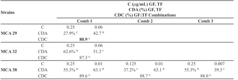
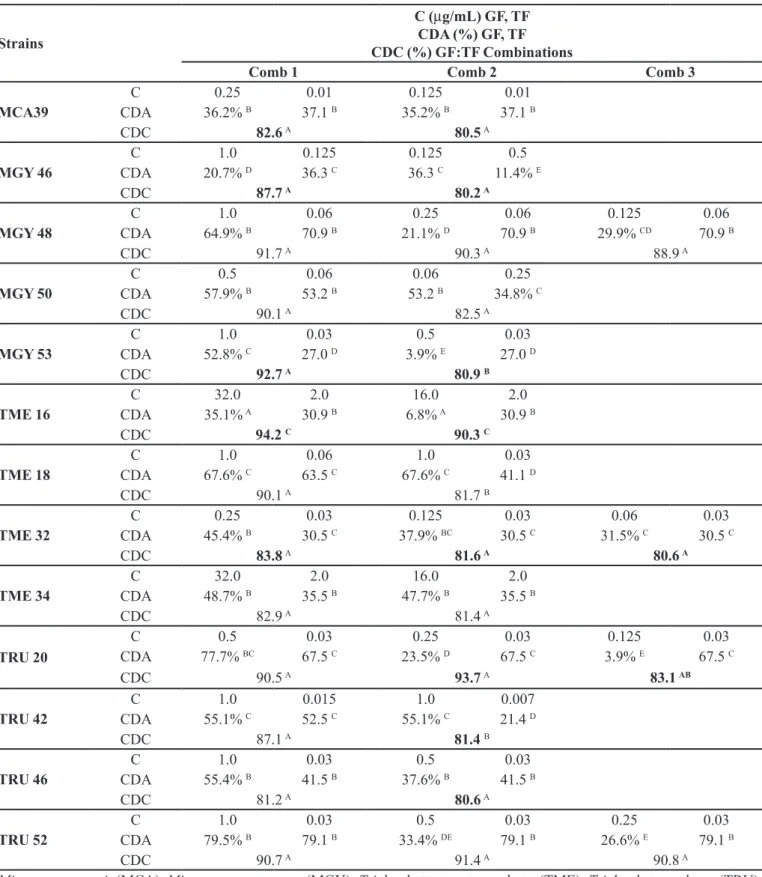
TABLE III - Cell damage after treatment with GF, TF, and GF:TF combinations (quantitative methodology)
Strains
C (µg/mL) GF, TF CDA (%) GF, TF CDC (%) GF:TF Combinations
Comb 1 Comb 2 Comb 3
MCA 29
C 0.25 0.06
CDA 27.9% C 42.7 B
CDC 80.9A
MCA 32
C 0.25 0.06
CDA 62.6% B 51.2 C
CDC 87.3 A
MCA 38
C 0.25 0.01 0.125 0.01 0.25 0.007
CDA 55.3% B 63.1 B 37.2% C 63.1 B 55.3% B 39.3 C
Strains
C (µg/mL) GF, TF CDA (%) GF, TF CDC (%) GF:TF Combinations
Comb 1 Comb 2 Comb 3
MCA39
C 0.25 0.01 0.125 0.01
CDA 36.2% B 37.1 B 35.2% B 37.1 B
CDC 82.6A 80.5A
MGY 46
C 1.0 0.125 0.125 0.5
CDA 20.7% D 36.3 C 36.3 C 11.4% E
CDC 87.7 A 80.2 A
MGY 48
C 1.0 0.06 0.25 0.06 0.125 0.06
CDA 64.9% B 70.9 B 21.1% D 70.9 B 29.9% CD 70.9 B
CDC 91.7 A 90.3 A 88.9 A
MGY 50
C 0.5 0.06 0.06 0.25
CDA 57.9% B 53.2 B 53.2 B 34.8% C
CDC 90.1 A 82.5 A
MGY 53
C 1.0 0.03 0.5 0.03
CDA 52.8% C 27.0 D 3.9% E 27.0 D
CDC 92.7 A 80.9 B
TME 16
C 32.0 2.0 16.0 2.0
CDA 35.1% A 30.9 B 6.8% A 30.9 B
CDC 94.2 C 90.3 C
TME 18
C 1.0 0.06 1.0 0.03
CDA 67.6% C 63.5 C 67.6% C 41.1 D
CDC 90.1 A 81.7 B
TME 32
C 0.25 0.03 0.125 0.03 0.06 0.03
CDA 45.4% B 30.5 C 37.9% BC 30.5 C 31.5% C 30.5 C
CDC 83.8A 81.6 A 80.6 A
TME 34
C 32.0 2.0 16.0 2.0
CDA 48.7% B 35.5 B 47.7% B 35.5 B
CDC 82.9 A 81.4 A
TRU 20
C 0.5 0.03 0.25 0.03 0.125 0.03
CDA 77.7% BC 67.5 C 23.5% D 67.5 C 3.9% E 67.5 C
CDC 90.5 A 93.7A 83.1 AB
TRU 42
C 1.0 0.015 1.0 0.007
CDA 55.1% C 52.5 C 55.1% C 21.4 D
CDC 87.1 A 81.4B
TRU 46
C 1.0 0.03 0.5 0.03
CDA 55.4% B 41.5 B 37.6% B 41.5 B
CDC 81.2 A 80.6A
TRU 52
C 1.0 0.03 0.5 0.03 0.25 0.03
CDA 79.5% B 79.1 B 33.4% DE 79.1 B 26.6% E 79.1 B
CDC 90.7 A 91.4 A 90.8 A
Microsporum canis (MCA); Microsporum gypseum (MGY); Trichophyton mentagrophytes (TME); Trichophyton rubrum (TRU).
C: concentration to each antifungal.CDA: percentage of cell damage to GF or TF. CDC: percentage of cell damage to GF:TF combinations (Ccomb=[GF]+[TF]). Comb 1-3: concentrations of GF:TF combinations to result in cellular damage over 80%. Bold:
indication of synergist effect. Two superscript letters represents significance p <0.05 between treatments. One letter relates to no significant difference.
merit for treatment of dermatophytosis (Scorzoni et al., 2017).
TF combined with amorolfine was checked in a randomized study of severe dermatophyte onychomycosis. Patients outcome were good and this combination
treatment demonstrated an improvement cost per cure ratio (Baran et al., 2000). Onychomycosis caused
by dermatophytes were also successfully treated by combination of oral terbinafine with ciclopyroxolamine, imidazole and other such topical antifungals. (Romano
et al., 2005). Fusarium oxysporum infections respond
positively to TF and topical imidazole treatment and this combination led to clinical and mycological healing, which is often refractory to antimycotics (Romano et al., 2005). Patient recovery to dermatophytosis was
also achieved when GF plus cyclopyroxolamine lotion and GF with topical imidazole were chosen and used in combination therapy (Romano et al., 2005). Several other
studies of combination therapy were reported, however most of these studies were lacking quantitative data
regarding drug combinations in vitro (Spader et al., 2013;
Semis et al., 2015).
The qualitative checkerboard assay enabled us to identify combinations of GF and TF which resulted in additive drug effects against sixteen pathogenic fungi (Table II). Subsequent evaluation by the quantitative MTT test using the same sixteen isolates identified by the checkerboard assay was performed and resulted in the determination of GF:TF concentrations that caused over 80% of cellular damage (Figure 1).
Of great importance was discovery of GF:TF drug combination that could cause 90% and 80% cellular damage to multidrug-resistant isolates TME 16 and TME 32, respectively.
It is possible that additive effect was achieved based upon the mechanism of action of GF (nucleic
acid inhibitor) and TF (ergosterol synthesis inhibitor). Alteration of the membrane integrity can be linked to squalene epoxidase inhibition by TF, which would promote cellular internalization of GF; while intracellular accumulation would decreases DNA synthesis (Favre, Ryder, 1997; Polak, 1993).
While this is only one biochemical hypothesis, it is also possible that intermolecular bonds in between GF and TB molecules could be responsible for additive effect observed. Our group will be pursuing additional studies to identify these possible interactions and the results will be reported in the due course.
As reported previously we found that GF has low solubility in water (Kahsav et al., 2013). In fact, it was
noted that GF partially precipitates in aqueous solution at 35 °C. Consequently, it was necessary to mediate
drug precipitation during incubation to avoid misleading
results. This was first attempted by using MOPS as a buffer to impart enhanced compound solubility.
However, it is not possible to make the desired stock solutions of GF in concentrations higher than 10 µg mL-1
in MOPS buffer at 35 °C described at the CLSI for MIC determinations (CLSI, 2008). As an alternative, we found that we could make stock solution 1.6 mg mL-1 of GF in
DMSO and dilution were made with RPMI 1640 media thereby allowing for proper MICs determinations.
In conclusion, herein we report the first investigation of GF and TF combination drug assessments against a panel of fungi clinical isolates. While, these two drugs have been evaluated with other antifungals (Baran et al., 2000; Romano et al., 2005) our findings are new and
noteworthy particularly when pathological dermatophytes have become drug resistant to many antifungal drugs (including GF and TB alone). Through a checkerboard assay, combining GF and TF results in additive effect and has impressive efficacy to several clinical dermatophytes
FIGURE 1 - Hyphal damage expressed in percentage of the griseofulvin (GF) () and terbinafine (TF) () alone and in combination
(). A) Hyphal damage of the TME 16 drug resistant isolate to the combination of TF and GF antifungals at concentrations below
MIC; B) Hyphal damage of TME 32 drug sensitive isolate to combination of TF and GF antifungals at concentrations below MIC.
including two hard to kill multidrug-resistant fungi isolates. This enhanced activity might be due to having selected drugs of different mechanisms of actions: inhibition of mitosis (GF) and ergosterol depletion (TF), as it is believed that TF might be altering the membrane by blocking the squalene epoxidase which ultimately facilitates cellular internalization of GF allowing for re-sensitizing via DNA damage of the multidrug-resistant
isolates.
ACKONWLEDGMENTS
This work was supported by Coordenação de Aperfeiçoamento de Pessoal de nível Superior (CAPES). A. M. Fuentefria is grateful to Conselho Nacional de Desenvolvimento Científico e Tecnológico (CNPq) for the PQ fellowships. Authors are thankful to Garrett Moraski and Lowell Markely for their valuable contributions to the final quality of this work.
REFERENCES
Andes D, Forrest A, Lepak A, Nett J, Marchillo K, Lincoln
L. Impact of antimicrobial dosing regimen on evolution of
drug resistance in vivo: Fluconazole and Candida albicans.
Antimicrob Agents Chemother. 2006;50(7):2374-2383.
Badali H, Mohammadi R, Mashedi O, de Hoog GS, Meis JF. In vitro susceptibility patterns of clinically important Trichophyton and Epidermophyton species against nine antifungal drugs.
Mycoses. 2015;58(5):303-307.
Baran R, Feuilhade M, Combernale P, Datry A, Goettmann S, Pietrini P, et al. A randomized trial of amorolfine 5% solution nail lacquercombined with oral terbinafine compared with terbinafinealone in the treatment of dermatophytic toenailonychomycoses affecting the matrix region. Br J Dermatol. 2000;142(6):1177-1183.
Campitelli M, Zeineddine N, Samaha G, Maslak S. Combination antifungal therapy: a review of current data. J Clin Med Res.
2017;9(6):451-456.
Campoy S, Adrio JL. Antifungals. Biochem Pharmacol.
2017;133:86-96.
Chiou CC, Mavrogiorgos N, Tillem E, Hector R, Walsh TJ. Synergy, pharmacodynamics, and time-sequenced ultrastructural changes of the interaction between Nikkomycin Z and the Echinocandin FK463 against Aspergillus fumigatus.
Antimicrob Agents Chemother. 2001;45(12):3310-3321.
Clinical and Laboratory Standards Institute. CLSI. Reference method for broth dilution antifungal susceptibility testing of filamentous fungi; approved standard. 2nd ed. CLSI Document M38-A2. Wayne, PA, USA: Clinical Laboratory Standards Institute; 2008.
Fachin AL, Maffei CML, Martinez-Rossi NM. In vitro
susceptibility of Trichophyton rubrum isolates to griseofulvin
and tioconazole. Induction and isolation of a resistant mutant to both antimycotic drugs. Mycopathologia. 1996;135(3):141-143.
Favre B, Ryder N. Cloning and expression of squalene epoxidase from the pathogenic yeast Candida albicans. Gene.
1997;189(1):119-126.
Galuppi R, Gambarara A, Bonoli C, Ostanello F, Tampieri MP. Antimycotic effectiveness against dermatophytes: comparison of two in vitro tests. Vet Res Commun. 2010;Suppl 1:S57-61.
Grover C, Arora P, Manchanda V. Comparative evaluation of griseofulvin, terbinafine and fluconazole in the treatment of tinea capitis. Int J Dermatol. 2012;51(4):455-458.
Kahsay G, Adegoke AO, Van Schepdael A, Adams E. Development and validation of a reversed phase liquid chromatographic method for analysis of griseofulvin and impurities. J Pharm Biomed Anal. 2013;80:9-17.
Kathiravan MK, Salake AB, Chothe AS, Dudhe PB, Watode RP, Mukta MS, et al. The biology and chemistry of antifungal agents: A review. Bioorg Med Chem. 2012;20(19):5678-5698.
Lewis RE, Diekema DJ, Messer SA, Pfaller MA, Klepser ME. Comparison of Etest, chequerboard dilution and time-kill studies for the detection of synergy or antagonism between antifungal
agents tested against Candida species. J Antimicrob Chemother.
2002;49(2):345-351.
Molina de Diego A. Aspectos clínicos, diagnósticos y terapéuticos de las dermatofitosis Clinical, diagnostic and therapeutic aspects of dermatophytosis. Enferm Infecc
Microbiol Clin. 2011;29(Suppl 3):33-39.
Mukherjee PK, Leidich SD, Isham N, Leitner I, Ryder NS, Ghannoum MA. Clinical Trichophyton rubrum strain exhibiting
primary resistance to terbinafine. Antimicrob Agents Chemother. 2003;47(1):82-86.
In Vitro additive effect on griseofulvin and terbinafine combinations against multidrug-resistant dermatophytes
Nardoni S, Mugnaini L, Papini R, Fiaschi M, Mancianti F. Canine and feline dermatophytosis due to Microsporum gypseum: A retrospective study of clinical data and therapy outcome with griseofulvin. J Mycol Med. 2013;23(3):164-1677.
Patel GA, Schwartz RA. Tinea capitis: still an unsolved problem? Mycoses. 2011;54(3):183-188.
Pinto e Silva AT, Costa-de-Oliveira S, Silva-Dias A, Pina-Vaz C, Rodrigues AG. Dynamics of in vitro acquisition of resistance by Candida parapsilosis to different azoles. FEMS Yeast Res.
2009;9(4):626-633.
Polak A. Combination of amorolfine with various antifungal drugs in dermatophytosis. Mycoses. 1993;36(1-2):43-49.
Romano C, Papini M, Ghilar A, Gianni C. Onychomycosis in children: a survey of 46 cases. Mycoses. 2005;48:430-437.
Scorzoni L, Silva ACAP, Marcos CM, Assato PA, de Melo WCMA, de Oliveira HC, et al. Antifungal therapy: new advances in the understanding and treatment of mycosis. Front Microbiol. 2017;8:1-23.
Semis R, Nahmias M, Lev S, Frenkel M, Segal E. Evaluation of antifungal combinations of nystatin-intralipid against
Aspergillus terreus using checkerboard and disk diffusion
methods. J Mycol Med. 2015;25(1):63-70.
Spader TB, Venturini TP, Rossato L, Denardi LB, Cavalheiro PB, Botton SA, Santurio JM, Alves SH. Synergysm of voriconazole or itraconazole with other antifungal agents against species of
Fusarium. Rev Iberoam Micol. 2013;30(3):200-204.
Tamura T, Asahara M, Yamamoto M, Yamaura M, Matsumura M, Goto K, et al. In vitro susceptibility of dermatomycoses
agents to six antifungal drugs and evaluation by fractional inhibitory concentration index of combined effects of amorolfine and itraconazole in dermatophytes. Microbiol Immunol.
2014;58(1):1-8.
Vandeputte P, Ferrari S, Coste AT. Antifungal resistance and new strategies to control fungal infections. Int J Microbiol. 2012;2012:713687,1-26.
Received for publication on 19th April 2017