ABSTRACT
INTRODUCTION
Orthodontic adhesives are used to provide effective union between composite and dental structure. However, the most common substances cytotoxic effect1,6,15. These adhesives are used
in moist media and often on contaminated surfaces without compromising their adhesion, but different compounds can be released during the aqueous phase11, such as non-polymerized
free monomers from resin materials5. Some
authors have demonstrated that these free monomers caused apoptosis in cell culture16,26. In
vivo studies17,19 show that non-polymerized resin
compounds released from dental adhesives cause chronic8. Recent studies4,5 have shown presence
of macrophages together with resin compounds
Biocompatibility of orthodontic adhesives in rat
subcutaneous tissue
Rogério Lacerda dos SANTOS1, Matheus Melo PITHON1, Alline Birra Nolasco FERNANDES2, Márcia Grillo CABRAL3,
Antônio Carlos de Oliveira RUELLAS4
1- DDS, MS, Specialist in Orthodontics, PhD student in Orthodontics, Federal University of Rio de Janeiro, Rio de Janeiro, RJ, Brazil. 2- Undergraduate Dental Student, Federal University of Rio de Janeiro, Rio de Janeiro, RJ, Brazil, Brazil.
3- DDS, MS, PhD, Adjunct Professor of Oral Pathology, Federal University of Rio de Janeiro, Rio de Janeiro, RJ, Brazil, Brazil. 4- DDS, MS, PhD, Adjunct Professor, Federal University, Federal University of Rio de Janeiro, Rio de Janeiro, RJ, Brazil, Brazil.
Corresponding address: Rogério Lacerda dos Santos - Rua Ipatinga, 170 - Planalto Divinópolis - MG - Brazil - 35501-191 - e-mail: [email protected] or [email protected]
O
bjective: The objective of the present study was to verify the hypothesis that nodifference in biocompatibility exists between different orthodontic adhesives. Material (n=6): Group 1 (control, distilled water), Group 2 (Concise), Group 3 (Xeno III), Group 4 (Transbond XT), and Group 5 (Transbond plus Self-Etching Primer). Two cavities were performed in the subcutaneous dorsum of each animal to place a polyvinyl sponge soaked with 2 drops of the respective adhesive in each surgical loci. Two animals of each group ! "# $ microscope. Results: At day 7, Groups 3 (Transbond XT) and 4 (Xeno III) showed intense % &'* +'/* 0 ; ! & " ' <* compared to other groups. At day 30, the same group showed a more expressive
0/ Among the
orthodontic adhesive analyzed, it may be concluded that Transbond XT exhibited the 0?
in vivo animal models as a human response.
Key words@ 0D 0F 0
following restorative procedure5, in which a
observed over a period of 300 days. In general, the adhesives used in orthodontics are chosen based on the research on mechanical assays and effectiveness in sealing the interface between tooth and orthodontic accessory. However, many research studies on the biocompatibility of dental materials are currently being performed5,7,8,15,20.
MATERIAL AND METHODS
This study used 30 male adult Wistar rats +!#%"!# groups of 6 animals each: Group 1 (control, distilled water), Group 2 (Concise, 3M Unitek Orthodontic Products, Monrovia, CA, USA), Group 3 (Xeno III, Dentsply/DeTrey, Konstanz, Baden-Württemberg, Germany), Group 4 (Transbond XT, 3M Unitek Orthodontic Products) and Group 5 (Transbond Self-Etching Primer, 3M Unitek Orthodontic Products) (Figure 1). The rats were anesthetized with intraperitoneal injection of sodium thiopental (50 mg/kg) (THIO, Cristália, Itapira, SP, Brazil), and the dorsal region (4x4 cm) of each animal was shaved. Asepsis of the operatory area was done with 4% chlorhexidine digluconate (School of Pharmacy, Federal University of Rio de Janeiro, Rio de Janeiro, RJ, Brazil). Two midline incisions of approximately 8 mm in length were made equidistantly from the tail base to the head of the animal with a #5 scalpel blade mounted onto a scalpel handle. The subcutaneous tissue was laterally separated using a pair of blunt-ended scissors, resulting in two approximately 18-mm-deep surgical loci each. All animals received two PVA sponge implants (4.0 mm long x 2.0 mm diameter). The implants were previously kept in 70% alcohol for 120 min, rinsed with sterile distilled water, autoclaved and then soaked with 2 drops of the respective adhesives. The adhesives in the sponges were photoactivated with a LED source unit (Radii, SDI, Baywater,
Victoria, Australia) according to the application time recommended by the manufacturer. The light intensity of the curing unit (1000 mW/cm2) was checked immediately before each polymerization using a radiometer (Model 100, Demetron Research Corporation, Danbury, CT, USA). The surgical loci were sutured with 4.0 suture (Ethicon, Johnson & Johnson, São José dos Campos, São Paulo, Brazil) and then the animals received an injection of sodium dipyrone (0.3 mL/100 g Novalgina®; ` %; { | }~; `$ ` Brazil).
The rats were kept in cages and fed balanced food and water. After 7, 15, and 30 days, the animals were anesthetized and submitted to excisional biopsy at the implantation area so that enough surrounding normal tissue could be collected. Each group consisted of 6 rats with two implants, thus resulting in 12 samples per group (Table 1). Next, 0
; ' solution) for 24 h, the samples were inserted %%J sections were cut and stained with hematoxylin 0 by the adhesives were examined with a light severe17,19. The biocompatibility of the materials
was determined according to the ISO 10993-313 standard.
Figure 1- Composition of the tested adhesive primers
Groups !"# $" Manufacture
Concise Concise®
Orthodontic Adhesive
Resin A: Bis-glycidyl-methacrylate (Bis-GMA), triethylene-glycol-dimethacrylate
(TEGDMA);
3M Unitek, Monrovia, CA, USA
Resin B: Bis-glycidyl-methacrylate (Bis-GMA), triethylene-glycol-dimethacrylate
(TEGDMA) and benzoin peroxide.
Xeno III Xeno® III Single Step Self
Etching Dental Adhesive
Fluid A: 2-hydroxyethyl methacrylate
hydroxybutylate (THB), Amorphous silica.
Dentsply DeTrey, Konstanz, Baden-Wurttember,
Germany
methacrylate (Piro-EMA), Phosphazen ! " dimethacrylate, Toluene hydroxybutylate
(THB), camphoroquinone, Ethyl-4-dimethylaminobenzoate.
Transbond Transbond® XT Primer Bisphenol-a diglycidyl ether
dimethacrylate, Triethylene glycol dimethacrylate (TEGDMA).
3M Unitek, Monrovia, CA, USA
TP Sep Transbond® Adhesive
Plus Self Etching Primer (SEP)
Mono and di-HEMA phosphates, camphoroquinone, distilled water, #$%!
titanate, Butylhydroxytoluene, methylparaben, and propylparaben
RESULTS
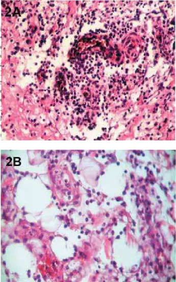
vessels and circulatory changes (dilatation and edema) around and within the cavity as a result of the material implantation in all four groups of adhesive systems (Figure 2AB). Groups 3 (Xeno III) and 4 (Transbond XT) showed the most 0 F observed due to the presence of sponge. In Group 2 (Concise), there was formation of granuloma and presence of multinuclear giant cells (Figure 2AB), which indicates the beginning of a repair process (Table 2).
At the end of the 15-day period, it was observed for all adhesive systems compared to that at day 7, except for Transbond XT (Figure 3A-B), which showed presence of neutrophils and abscess formation at the region where the material was implanted, thus indicating a very toxic effect on the tissue. Presence of granuloma and multinuclear
giant cells were observed in Groups 3 (Xeno III) and 5 (Transbond SEP) as well as in the Control Group (Figure 3A-B) (Table 2).
After 30 days, the Control Group and all four process characterized by discrete mononuclear
Control Concise Xeno III Transbond XT Transbond Plus SEP
Days Rats %" Rats %" Rats %" Rats %" Rats %"
7 days 2 4 2 4 2 4 2 4 2 4
15 days 2 4 2 4 2 4 2 4 2 4
30 days 2 4 2 4 2 4 2 4 2 4
Total 6 12 6 12 6 12 6 12 6 12
Table 1-&#'**+'/++
SEP: Self-Etching Primer
Figure 2- Photomicrographs of histological samples after 8+'</'!+ ' + =>>>% *? =>> N< ' *=>>>%*?=>>N
formation of granuloma with multinuclear giant samples (Figure 4). Presence of granulation tissue around the sponges and cell proliferation with 0 all adhesives were shown to be biocompatible on long-term basis (Table 2).
DISCUSSION
by in vivo models is questioned and criticized in literature30, depends of use of a biological system
that reproduces as close as possible the metabolic behavior of the target organ for the toxic effect of xenobiotics and of choice of appropriate parameters to evaluate toxic effects. Evaluating the biocompatibility of orthodontic adhesives by means of subcutaneous implants in rats is of great value as the tissue response in rats is similar to that expected when the same material is applied to the gingival tissue surrounding the area to receive orthodontic accessories.
Several studies have assessed the biocompatibility of dental materials5,7,8,15,20.
However, methodological divergence exists. In the present study, polyvinyl sponges saturated with the respective adhesives were inserted into rats subcutaneous tissue and then light cured17 in an
attempt to simulate actual clinical procedures. Costa, et al.6 (1999) have used polyvinyl
sponges saturated with adhesives that had not been photoactivated after surgical implantation, allowing the adhesive and their monomers to be in close contact with the subcutaneous connective tissue. Therefore, not only a cytotoxic effect of the dental adhesive was observed but also a persistent
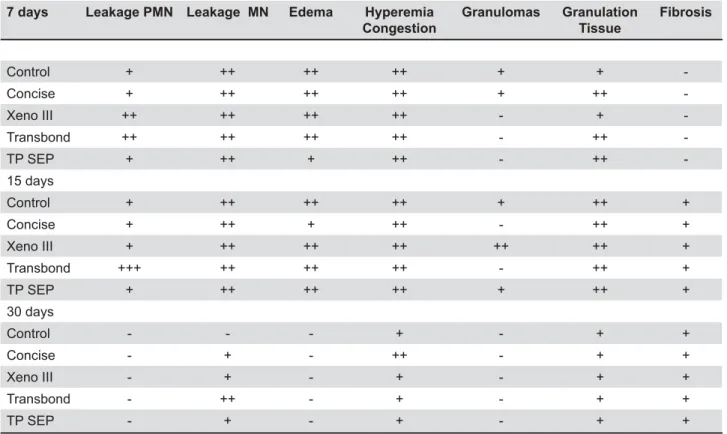
7 days Leakage PMN Leakage MN & ' Congestion
*%" Granulation Tissue
Fibrosis
Control + ++ ++ ++ + +
-Concise + ++ ++ ++ + ++
-Xeno III ++ ++ ++ ++ - +
-Transbond ++ ++ ++ ++ - ++
-TP SEP + ++ + ++ - ++
-15 days
Control + ++ ++ ++ + ++ +
Concise + ++ + ++ - ++ +
Xeno III + ++ ++ ++ ++ ++ +
Transbond +++ ++ ++ ++ - ++ +
TP SEP + ++ ++ ++ + ++ +
30 days
Control - - - + - + +
Concise - + - ++ - + +
Xeno III - + - + - + +
Transbond - ++ - + - + +
TP SEP - + - + - + +
Table 2- Mean values for the biopsy results regarding the samples studied during the 3 periods of time (7, 15, and 30 days after implantation)
SEP: Self-Etching Primer
compounds. According to the authors, these materials do not seem to be suitable for direct application to connective tissue.
Studies15,21,29 have demonstrated the cytotoxicity
of the compounds of adhesive systems, which can be explained by the different compositions, mechanisms, and application procedures as well as by methodological variations1. However, it is clear
that the choice for a given adhesive system should be based on its biocompatibility.
The most often studied method for in vivo
17.
; $ in different experimental groups makes it possible to establish the best biocompatibility by placing the material in contact with vascularized tissues and observing the different reactions. It is also important to use an innocuous substance in the control group in order to facilitate data interpretation17.
more intense reaction to both surgical procedures and implanted foreign body, and because such a % are not taken into account. After 7 days, a more $ to the adhesive rather than the surgical procedure.
relies on the control of the host defense system, which organizes itself to limit the aggressive action from the compounds existing in the adhesives $ 0 F intensity between the experimental groups, mainly regarding the higher level of cytotoxic from Xeno III and Transbond XT after 7 days.
Methacrylate monomers such as TEGDMA, Bis-GMA, UDMA, and HEMA, which are largely used in the composition of dentinal adhesives, can cause cell lesion10,12. TEGDMA, Bis-GMA, UDMA are
hydrophobic monomers that are often associated with HEMA. Diffusion of these monomers can be facilitated because HEMA increases the hydrophilic characteristic of the material. Under such conditions, the hydrophobic monomers can reach the cells and damage them10,12,23.
With respect to Xeno III, the presence of HEMA in association with ethanol seems to cause more cell damage. The ethanol in relation with the oral mucosa showed increased mucosal permeability13,24
and penetration of potential carcinogens across the mucosal permeability barrier13. It has been
reported that topical application of ethanol on the oral mucosa affects epithelial cell homeostasis2 and
alters mucosal structure18.
In the present study, a mild reaction was observed in the control group whose sponges were saturated with distilled water, whereas a moderate to severe reaction was found in all experimental groups. After 15 and 30 days, the the biocompatibility of the materials to be rated
in ascending order. In general, small necrotic areas with edema surrounded by cell proliferation,
blood vessels were observed. The presence of multinuclear giant cells suggests the formation of granulomas due to the presence of sponge and/ or adhesive material. Therefore, these events are described as a favorable tissue response regarding the biocompatibility of the material.
At the end of the 30-day period, it was possible to observe that all adhesive systems showed good biocompatibility, although Transbond XT was found to be more aggressive compared to other groups as formation of abscess occurred at the implant 0D Transbond XT is the least biocompatible adhesive.
According to in the literature, HEMA is an important toxic component released by most adhesive systems since several in vitro studies have ?; culture of cells3,25. Methacrylate monomers, such as
HEMA, are incorporated in the lipid bilayers of cell membranes which are solubilized by the unreacted monomers10. This mechanism of action of uncured
leached monomers on the cell membrane may be regarded as responsible for the high cytotoxicity of Transbond (Transbond XT, 3M Unitek) observed in the present investigation.
Traditionally, persulfate molecules have been used as initiators in redox water-based polymerization systems to decrease the amount of residual monomers after setting14. The high
cytotoxicity of adhesive systems is probably caused by leachable resin components, such as TEGDMA, Bis-GMA and HEMA, which has frequently been added to their chemical composition9.
However, it may be speculated that some minor adhesive components released into the connective tissue, such as HEMA, which presents low molecular weight, might be removed by local lymphatic drainage. This hypothesis should explain time and the connective tissue healing occurred for all experimental materials at 30 days following the implantation.
Sohoel, et al.22 (1994), after testing adhesives
in pigs, have suggested that orthodontic adhesives can be potentially allergenic for human being, particularly the “no-mix” ones, and lead to adverse reactions in both patients and practitioners. Such cytotoxicity can last two years after polymerisation27.
Thompson, et al.28 (1982) have concluded that
materials for long periods of time, particularly the subgingival and interproximal areas.
CONCLUSIONS
F' * and healing phenomena to characterize, and rate the experimental groups by comparing them to a control group. This allows us to state that Xeno III, Transbond SEP, and Concise adhesives had the best biocompatibility, since formation of chronic and multinuclear giant cells around the samples were observed. However, one cannot interpret the
in vivo animal
models as a human response. The hypothesis was rejected and one can state that, among the adhesives studied, Transbond XT was found to have the worst biocompatibility.
REFERENCES
1- al-Dawood A, Wennberg A. Biocompatibility of dentin bonding agents. Endod Dent Traumatol. 1993;9(1):1-7.
2- Axford SE, Ogden GR, Stewart AM, Saleh HA, Ross PE, Hopwood D. Fluid phase endocytosis within buccal mucosal cells of alcohol misusers. Oral Oncol. 1999;35(1):86-92.
3- Bouillaguet S, Wataha JC, Hanks CT, Ciucchi B, Holz J. In vitro cytotoxicity and dentin permeability of HEMA. J Endod. 1996;22(5):244-8.
4- Costa CA, Giro EM, Nascimento AB, Teixeira HM, Hebling J. Short-term evaluation of the pulpo-dentin complex response % % applied in deep cavities. Dent Mater. 2003;19(8):739-46. 5- Costa CA, Hebling J, Hanks CT. Current status of pulp capping with dentin adhesive systems: a review. Dent Mater. 2000;16(3):188-97.
6- Costa CA, Teixeira HM, Nascimento AB, Hebling J. Biocompatibility of an adhesive system and 2-hydroxyethylmethacrylate. ASDC J Dent Child. 1999;66(5):337-42.
7- Cox CF, Kim KM, Stevenson RG 3rd, Hafez AA. Histological evaluation of a self-priming etchant adhesive system. Compend Contin Educ Dent. 2003;24(8):17-20.
8- Souza Costa CA, Nascimento AB, Teixeira HM. Response of human pulps following acid conditioning and application of a bonding agent in deep cavities. Dent Mater. 2002;18(7):543-51. 9- Souza Costa CA, Hebling J, Garcia-Godoy F, Hanks CT. In vitro % 0 D 0
2003;24(21):3853-8.
10- Fujisawa S, Kadoma Y, Komoda Y. 1H and 13C NMR studies of the interaction of eugenol, phenol, and triethyleneglycol dimethacrylate with phospholipid liposomes as a model system for odontoblast membranes. J Dent Res. 1988;67(11):1438-41. 11- Geurtsen W, Spahl W, Muller K, Leyhausen G. Aqueous extracts from dentin adhesives contain cytotoxic chemicals. J Biomed Mater Res. 1999;48(16):772-7.
12- Hanks CT, Wataha JC, Sun Z. In vitro models of biocompatibility:
a review. Dent Mater. 1996;12(3):186-93.
13- Howie NM, Trigkas TK, Cruchley AT, Wertz PW, Squier CA, Williams DM. Short-term exposure to alcohol increases the permeability of human oral mucosa. Oral Dis. 2001;7(6):349-54. 14- Kakaboura A, Eliades G, Palaghias G. An FTIR study on the % 0 Dent Mater. 1996;12(3):173-8.
15- Kostoryz EL, Eick JD, Glaros AG, Judy BM, Welshons WV, Burmaster S, et al. Biocompatibility of hydroxylated metabolites of BISGMA and BFDGE. J Dent Res. 2003;82(5):367-71.
16- Lefeuvre M, Amjaad W, Goldberg M, Stanislawski L. TEGDMA induces mitochondrial damage and oxidative stress in human 0D 0+##!+'+!*!"#%0
17- Machado NP, Moysés MR, Pereira AAC, Pereira LJ, Ribeiro JCR, Dias SC. Study of dentinal adhesives compatibility using histological analysis. Braz J Oral Sci. 2007;6(20):1289-94. 18- Maier H, Tisch M. Epidemiology of laryngeal cancer: results of the Heidelberg case-control study. Acta Otolaryngol Suppl. 1997;527:160-4.
19- Moyses MR, Lopes WL, Pereira AAC, Ribeiro JCR, Dias SC, Pereira LJ. Biocompatibility of the Prime & Bond 2.1, Prime & Bond NT and Scothbond MP Primer adhesive systems. Braz J Oral Sci. 2006;5(18):1079-84.
20- Mussel RL, Sa Silva E, Costa AM, Mandarim-De-Lacerda CA. Mast cells in tissue response to dentistry materials: an adhesive resin, a calcium hydroxide and a glass ionomer cement. J Cell Mol Med. 2003;7(2):171-8.
21- Nagem-Filho H, Monteiro CR, Nagem HD, Lage-Marques JL. Effect of dental adhesives on the exudative phase of the 0 Odontol Bras. 2003;17(2):109-12.
22- Sohoel H, Gjerdet NR, Hensten-Pettersen A, Ruyter IE. Allergenic potential of two orthodontic bonding materials. Scand J Dent Res. 1994;102(2):126-9.
23- Souza PP, Aranha AM, Hebling J, Giro EM, Costa CA. In vitro
cytotoxicity and in vivo biocompatibility of contemporary % 0 ~ 0 +##++'*"% 44.
24- Squier CA, Cox P, Hall BK. Enhanced penetration of nitrosonornicotine across oral mucosa in the presence of ethanol. J Oral Pathol. 1986;15(5):276-9.
25- Stanislawski L, Daniau X, Lauti A, Goldberg M. Factors % glass ionomer cements. J Biomed Mater Res. 1999;48(3):277-88. 26- Stanislawski L, Lefeuvre M, Bourd K, Soheili-Majd E, Goldberg ;0 &~;% associated with early and drastic glutathione depletion with subsequent production of oxygen reactive species. J Biomed Mater Res A. 2003;66(3):476-82.
27- Tell RT, Sydiskis RJ, Isaacs RD, Davidson WM. Long-term cytotoxicity of orthodontic direct-bonding adhesives. Am J Orthod Dentofacial Orthop. 1988;93(5):419-22.
28- Thompson LR, Miller EG, Bowles WH. Leaching of unpolymerized materials from orthodontic bonding resin. J Dent Res. 1982;61(8):989-92.
29- Vajrabhaya LO, Pasasuk A, Harnirattisai C. Cytotoxicity evaluation of single component dentin bonding agents. Oper Dent. 2003;28(4):440-4.