Cop
yright
© ABE&M t
odos os dir
eit
os r
eser
vados
.
Parathyroid carcinoma and
hungry bone syndrome
Carcinoma de paratiroide e fome óssea
Monique Nakayama Ohe1, Rodrigo Oliveira Santos2, Flavio Hojaij2, Murilo Catafesta Neves2, Ilda Sizue Kunii1, Denise Orlandi1, Luisa Valle1,
Carla Martins1, Carolina Janovsky1, Rimarcs Ferreira3, Roseane Delcelo3, Ana Maria Domingos3, Marcio Abrahão2, Onivaldo Cervantes2,
Marise Lazaretti-Castro1, Jose Gilberto Henriques Vieira1
SUMMARY
We hereby report two patients with parathyroid carcinoma presenting extremely high calcium and PTH levels, severe bone disease, and palpable neck mass at diagnosis. They both underwent parathyroidectomy, and one of them evolved to lung metastasis. Important hypocalcemia was observed after surgery in both: after parathyroidectomy in one patient, and only after surgical removal of the metastasis in the other. Both required intravenous calcium replacement, thus revealing hungry bone syndrome (HBS). HBS usually relects rapid mineralization after correc-tion of hyperparathyroidism. The more severe the bone disease before surgery, the more prone the patient is to HBS after surgery. Despite being an unfavorable outcome, HBS state suggests that surgical removal of hypersecretory parathyroid tissue was accomplished. In this study, HBS was observed in both patients, who presented severe bone disease prior to surgery. HBS would be expected post-operatively in successful parathyroid carcinoma removal. Arq Bras Endocrinol Metab. 2013;57(1):79-86
SUMÁRIO
O presente artigo descreve o relato de dois pacientes com carcinoma de paratiroide que apre-sentavam valores intensamente elevados de cálcio sérico e de PTH, associado a doença óssea e presença de nódulo cervical palpável ao diagnóstico. Ambos foram submetidos à paratiroi-dectomia, sendo que um evoluiu com metástases pulmonares. Hipocalcemia importante foi observada após a paratiroidectomia em um paciente e somente após remoção cirúrgica das metástases pulmonares em outro. Ambos necessitaram de reposição endovenosa de cálcio, revelando, assim, o estado de fome óssea (FO). A presença da FO usualmente relete rápida mineralização óssea após correção do hiperparatiroidismo; assim, quanto mais severa a do-ença óssea previa à cirurgia, maior será a FO observada no pós-operatório desses pacientes. Embora inicialmente considerada um evento indesejável, a FO representa a bem-sucedida re-moção cirúrgica do tecido paratiroideano hipersecretor. Fome óssea deve ser esperada no pós--operatório do tratamento cirúrgico bem-sucedido do carcinoma de paratiroide. Arq Bras Endocrinol Metab. 2013;57(1):79-86
1 Division of Endocrinology and
Metabolism, Escola Paulista de Medicina, Universidade Federal de São Paulo (Unifesp/ EPM), Sao Paulo, SP, Brazil
2 Division of Head and Neck Surgery,
Unifesp/EPM, Sao Paulo, SP, Brazil
3 Department of Pathology, Unifesp/
EPM, Sao Paulo, SP, Brazil
Correspondence to:
Monique Nakayama Ohe Av. Cons. Rodrigues Alves, 804, ap. 51
04014-002 – Sao Paulo, SP, Brazil [email protected]
Received on Mar/19/2012 Accepted on Sept/8/2012
INTRODUCTION
P
arathyroid carcinoma is a rare endocrine malignan-cy that was irst described in 1904 by De Quervain (1). Subsequent descriptions of functioning parathy-roid carcinomas were reported in the 1930s (2). Since then, only a few hundred cases have been reported in the literature. Parathyroid carcinoma is an uncommonCop
yright
© ABE&M t
odos os dir
eit
os r
eser
vados
.
The cause of parathyroid carcinoma is yet to be established, and at present, there is no data point-ing out causal relationships between parathyroid carcinoma and risk factors. Recently, it has been de-scribed that both familial and sporadic forms (4) of parathyroid carcinomas are associated with va rious mutations in the HRPT2 gene on chromosome 1q25-1q32, suggesting that HRPT2 acts as a tumor suppressor gene (5).
Optimal treatment is related to early diagnosis with clinical and laboratory suspicion of parathyroid carcinoma prior to surgery, once parathyroidectomy with complete resection of the primary site, en bloc,
including the surrounding tissue, is intended (6). The natural history of parathyroid carcinoma is de-scribed as slow, but progressive. Morbidity and mortali-ty usually result from unremitting hypercalcemia and its complications, rather than mass effect of tumor growth (7). Metastasis is common, and the most frequent com-promised sites are lungs (40%), cervical lymph nodes (30%), and liver (10%) (8).
We have previously reported our experience with seven patients with parathyroid carcinoma (6,9) and, in this study, we present two other cases, with em-phasis on hungry bone disease (HBS).
Hungry bone syndrome is described after successful parathyroidectomy; it is related to rapid bone remine-ralization causing hypocalcemia and requiring calcium and vitamin D replacement (10,11).
CASE REPORTS
Case 1
A 65-year-old female patient was seen at the emergency ward at Hospital São Paulo – a Federal University Hos-pital – in 2008, complaining of fever, dysuria, nausea, loss of appetite, and loss of weight, as well as general malaise. She was initially diagnosed with acute exa-cerbation of chronic renal failure related to recurrent nephrolithiasis. At clinical examination, a palpable neck mass was observed. Laboratory tests revealed important hypercalcemia, with ionized calcium (iCa) of 1.9 mmol/L (reference values: 1.20-1.40 mmol/L) and intact parathyroid hormone levels (iPTH) of 2,800 pg/mL (reference values 10-65 pg/mL), in addition to evidence of renal failure, with serum creatinine values of 3.4 mg/dL, creatinine clearan-ce of 16.7 mL/min (Cockcroft-Gault), and
normo-chromic normocytic anemia. Besides, in renal ultra-sound examination, renal cystic lesion was observed.
The patient was diagnosed with primary hyper-parathyroidism and, thus, referred to surgery. In face of palpable neck mass at physical examination and markedly high levels of calcium and iPTH, she under-went en bloc resection of the lower right parathyroid,
with ipsilateral thyroidectomy and isthmusectomy, followed by excision of paratracheal and central neck nodes, considering a possible diagnosis of parathy-roid carcinoma. Intraoperative PTH (IO-PTH) was performed (Elecsys 1010 System, Roche, Mannheim, Germany) revealing 2,480 pg/mL at the induction of anesthesia, which dropped to 394 pg/mL 10 minutes after the removal of the parathyroid gland (84.1% de-crease). Pathological examination showed a parathy-roid carcinoma (sized 4.0 x 3.3 cm) with vascular invasion and tumor extension to adjacent thyroid. Just after surgery, the patient presented important decrease in phosphorus and calcium levels.
In the outcome, high levels of iCa and iPTH were observed (iPTH = 287 pg/mL; iCa = 1.55 mmol/L) two months after surgery, which evolved to markedly abnormal laboratory indings with to-tal serum calcium (tCa) of 13.8 mg/dL (reference value = 8.5-10.5), iPTH of 2,127 pg/mL, and iCa of 1.98 mmol/L one year after the surgical proce-dure. At this time, a 99mTc scintigraphy revealed hy-perconcentration in the inferior left parathyroid.
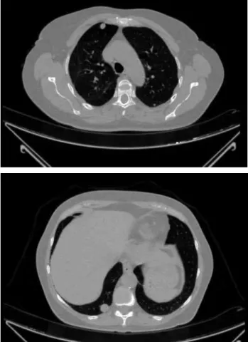
The patient underwent surgical re-exploration with removal of the left superior and inferior parathyroids along with left lobe thyroidectomy and neck nodal dis-section. Intraoperative PTH failed to drop: IO-PTH at the induction of anesthesia was 1,450 pg/mL, and dropped to 986 pg/mL; the patient remained hypercal-cemic in the outcome (iCa = 1.67 mmol/L). In a search for distant metastases, the patient underwent nary computerized tomography, and multiple pulmo-nary nodules were found (Figure 1). In July 2011, two years after the irst surgical procedure, the patient underwent surgical resection of lung metastases. Six nodules were removed. Histological analysis revealed a metastatic carcinoma in pulmonary nodule, showing cells with round and hyperchromatic nuclei (Figure 2).
Cop
yright
© ABE&M t
odos os dir
eit
os r
eser
vados
.
of 1.9 mg/dL (reference values: 2.5-5.0 mg/dL). We prescribed intravenous calcium and oral calcium car-bonate 500 mg, two pills, four times a day, associated with oral calcitriol 0.25 mg four times a day, plus cho-lecalciferol 3,000 UI a day in the irst two post-oper-ative days. We continued oral calcium, calcitriol and cholecalciferol replacement in lower doses for over two weeks, until the patient was discharged, which was done with her taking calcium carbonate 500 mg, three times a day, calcitriol 0.25 mg two times a day, and cholecalciferol 2,000 UI a day. Another important inding was related to bone mineral density recovery 6 months after lung metastasis removal (Table 1).
Table 1. Bone mineral density recovery after removal of lung metastasis
12/2008 12/2011
BMD (g/cm2) T-score BMD (g/cm2) T-score
L1-L4 0.814 -2.1 1.170 -0.1
Neck 0.779 -0.6 1.060 +0.2
Total femur 0.907 -0.3 1.144 +1.1
BMD: bone mineral density; L1-L4: lumbar spine; neck: femoral neck.
Figure 1. Pulmonary computed tomography: 1.0-cm nodule in the upper right anterior pulmonary lobe, and 1.7-cm nodule in the lower right pulmonary lobe.
Figure 2. (A) Metastatic carcinoma nodule in lung (HE, 100X). (B) Cells with round and hyperchromatic nuclei (HE, 200X).
A
B
Case 2
Cop
yright
© ABE&M t
odos os dir
eit
os r
eser
vados
.
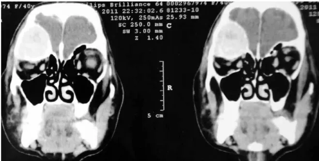
Figure 3. Computed tomography: supraorbital expansive lesion suggestive of brown tumor.
Figure 4. Computed tomography: expansive and heterogeneous lesion in the iliac bone, suggestive of brown tumor.
A computerized tomography revealed a supraorbital expansive lesion (Figure 3), and a 6.0 cm expansive and heterogeneous lesion in iliac bone (Figure 4), both sug-gestive of brown tumor; 99mTc scintigraphy revealed hyperconcentration around the lower left thyroid lobe that persisted in the late images, which was suggestive of parathyroid gland.
The patient was then referred to surgery consi-dering the hypothesis of parathyroid carcinoma in face of palpable neck mass at physical examina-tion and marked high levels of calcium and iPTH. Thus, she underwent en bloc resection of the lower
left parathyroid, with total thyroidectomy, followed by excision of bilateral paratracheal neck nodes (Fi-gure 5). The intraoperative aspect revealed an enlarged
parathyroid of 4.0 x 3.3 cm, with heterogeneous ap-pearance and attached to surrounding thyroid. The three remaining parathyroids presented normal ap-pearance. Intraoperative PTH was performed (Elec-sys 1010 System, Roche, Mannheim, Germany) and revealed 619 pg/mL at the induction of anesthesia, which dropped to 93 pg/mL 10 minutes after the re-moval of the parathyroid gland (85% percent decrease).
im-Cop
yright
© ABE&M t
odos os dir
eit
os r
eser
vados
.
Figure 5. En bloc resection: nodular thyroid, parathyroid (arrow), and lymph-nodes.
Cop
yright
© ABE&M t
odos os dir
eit
os r
eser
vados
.
portant bone lesions and high risk of hypocalcaemia, intravenous calcium replacement was prescribed. In the second post-operative day, after intravenous cal-cium replacement and taking calcal-cium carbonate 500 mg, two pills four times a day, associated with oral calcitriol 0.25 mg three times a day, plus cholecal-ciferol 3,000 UI a day, serum calcium was at lower normal limit of 8.5 mg/dL. She was discharged tak-ing calcium carbonate 500 mg, two pills three times a day, calcitriol 0.25 mg three times a day, plus cho-lecalciferol 3,000 UI a day. In the last six months of follow-up, she remained normocalcemic under oral calcium and cholecalciferol replacement.
DISCUSSION
Parathyroid carcinoma is the least common endocrine malignancy, with prevalence of 0.005% of all cancers (3). It is a rare malignant disease, likely to recur, and dificult to control. Several reports have highlighted the importance of en bloc resection, including thyroid
lobectomy with the isthmus, paratracheal, and central neck nodal dissection (8,12,13). This procedure, when performed as the initial therapeutic approach, provides patients with the best chance of cure. Recurrence after surgical excision of parathyroid carcinoma is common, with rates ranging from 33% to 78% (13). The repor-ted time from surgery to the irst recurrence (disease-free interval) varies greatly, from 1 month to 20 years, with the most commonly reported mean between 2 and 5 years (14). Metastasis to lungs, mediastinum, and lymph nodes are described and dificult to con-trol. When metastatic parathyroid tumors are found, surgical resection of metastatic tumors is the optimal treatment, once parathyroid carcinoma is refractory to radiation and cytotoxic reagents (8). Symptoms associated with metastatic lesions are typically due to hypercalcemia rather than tumor invasion (14). Studies reporting statistical estimates of disease-speciic survi-val in patients with parathyroid carcinoma have yielded 5-year survival rates ranging from 20% to 90%, and 10-year survival rates between 42% and 86%. Variations of the survival rates reported in these studies may relect the differences in histopathology deinitions, the pro-portions of unequivocal versus equivocal cases, and the
initial therapeutic interventions used.
In a recent article (15), the authors reviewed the outcomes of parathyroid cancer patients treated at the same institution over a 43-year period (between 1966
and 2009), and evaluated the factors associated with re-currence and mortality. Speciic factors that were inde-pendently associated with increased mortality included lymph node metastases, distant metastases, higher cal-cium level at recurrence, higher number of calcal-cium- calcium-lowering medications, and number of neck recurrences. Factors that were not associated with mortality included gender, race, age at diagnosis, calcium level at time of cancer diagnosis, tumor size, number of neck dissec-tions, time of irst recurrence, and decade in which treatment was initiated. Interestingly, the extent of ope-ration (parathyroidectomy alone vs. parathyroidectomy
with partial or total thyroidectomy) was not associated with mortality, either, nor was it associated with the number of recurrences or neck dissections necessary.
The authors usually resected en bloc only the soft
tis-sue that appears to be involved with the cancer (i.e. the
thyroid lobe, the esophagus, the trachea, the recurrent laryngeal nerve etc.) at the time of the initial operation,
and resect any lymph nodes of abnormal appearence. However, having the initial operation done by an ex-perienced parathyroid surgeon was associated with de-creased mortality. The authors concluded that if there is high pre-operative suspicion of parathyroid cancer because of severe hypercalcemia or palpable parathy-roid mass, one should consider referring the patient to a specialized tertiary center.
Parathyroid carcinoma is often misdiagnosed preop-eratively as primary hyperparathyroidism due to para-thyroid adenoma or hyperplasia. Clinical and laboratory indings may suggest carcinoma, but these indings are nonspeciic. Even so, there is no external independent standard reference for the diagnosis of parathyroid car-cinoma. Marked elevated serum calcium (often over 14 mg/dL), and intact parathyroid hormone levels (over ive times the upper normal limit) should raise a clinical suspicion of this rare entity. A palpable neck mass has been reported in approximately half of the patients with parathyroid carcinoma, but in less than 1% of patients with primary hyperparathyroidism (6,9,16). In this case study, we report two patients presenting marked high levels of serum calcium and intact PTH, with severe clinical symptoms and presence of palpable neck mass at diagnosis, thus raising the suspicion of parathyroid carcinoma. Taking this hypothesis into account, both patients underwent en bloc parathyroid resection in
Cop
yright
© ABE&M t
odos os dir
eit
os r
eser
vados
.
Regarding histopathology diagnosis, it is worth highlighting the current dificulties in pathological diag nosis concerning parathyroid carcinoma, once cel-lular pleomorphism and atypia are not reliable indica-tors of malignancy in endocrine tumors. Nevertheless, presence of trabecular growth pattern, capsular inva-sion, and vascular invasion are considered highly specif-ic indings concerning parathyroid carcinoma (17). In this case study, patients presented capsular or vascular invasion in the parathyroid lesion.
Lung metastasis was observed in the post-opera-tive outcome of one patient (case 1), two years after the irst parathyroidectomy. That patient underwent surgical removal of the lung metastasis and revealed marked hypocalcemia, thus requiring intravenous cal-cium infusion – the hungry bone syndrome was then installed.
The hungry bone syndrome is described after para-thyroidectomy, since correction of hyperparathyroid-ism is associated with rapid bone remineralization, causing severe and prolonged hypocalcemia that re-quires intensive intravenous calcium and oral vitamin D supplementation (10,11). HBS is considered a com-plication of parathyroid surgery observed in 13-30% of cases (18), and some studies advocate the use of bisphosphonates prior to surgery in order to prevent this post-operative outcome (19). The more severe the bone disease before surgery, the more prone the pa-tient is to the hungry bone syndrome after the surgi-cal procedure. Despite being an unfavorable outcome, the hungry bone syndrome state suggests that surgical removal of hypersecretive parathyroid tissue was ac-complished. In this study, HBS was observed in both patients: in case 1 after lung metastasis removal, and in case 2 after surgical resection of the parathyroid le-sion. Both patients presented severe bone disease prior to surgery. Therefore, it is worth highlighting that HBS would be expected post-operatively in successful para-thyroid carcinoma removal.
Hungry bone syndrome treatment requires cal-cium and vitamin D replacement. Calcal-cium carbo nate is usual ly recommended and intravenous calcium re-placement may be temporarily necessary in severe forms of HBS, as observed in both reported cases. Along with calcium, therapy with vitamin D is almost always required; 1,25(OH)2D3 (calcitriol), the active metabolite of vitamin D, maintains serum calcium, in part, by improving the eficiency of intestinal calcium absorption. Calcitriol is administered over a wide
dos-ing range – 0.25 to 2 mg/d – and can increase serum calcium concentration substantially within 3 days (20). A single daily dose is typically administered in modest doses (0.25 to 0.75 mg/d). When higher amounts are required, calcitriol is typically administered in divided doses. Vitamin D3 (cholecalciferol) is often used along with the active vitamin D metabolite (20). The longer half-life of cholecalciferol (2-3 weeks) helps to provide smoother control in view of the very short half-life of calcitriol, which is measured in hours.
In conclusion, parathyroid carcinoma is a rare and severe entity, with marked clinical and laboratory mani-festations at diagnosis. The optimal treatment is re-lated to early diagnosis with clinical and laboratorial suspicion for this entity prior to surgery, once com-plete resection of primary site is intended. Parathy-roid carcinoma is a disease likely to recur and dificult to control. Hungry bone syndrome observed relects the rapid mineralization after correction of hyperpara-thyroidism, and is related to bone disease severity prior to surgery. HBS may be expected as a post-operative outcome in successful parathyroid carcinoma.
Disclosure: no potential conlict of interest relevant to this article was reported.
REFERENCES
1. De Quervain F. Parastruma maligna aberrata [Malignant aberrant parathyroid] Deusche Zeitschr Chir. 1904;100:334-52.
2. Sainton P, Millot J. Malegne dún adenoma parathyroidiene eosi-nophile [Malignant eosinophilic parathyroid]. Au cours dune de Recklinghausen. Ann Anal Pathol. 1933;10:813.
3. Hundahl SA, Fleming ID, Fremgen AM, Menck HR. Two hundred eigth-six cases of parathyroid carcinoma treated in the US bet-ween 1985-1995: a National Cancer Data Base Report. The can College of Surgeons Commission on Cancer and the Ameri-can Cancer Society. Cancer. 1999;86:538-44.
4. Shattuck TM, Valimaki S, Obara T, Gaz RD, Clark OH, Shoback D, et al. Somatic and germ-line mutations of the HRPT2 gene in spora-dic parathyroid carcinoma. N Engl J Med. 2003;349:1722-9. 5. Carpten JD, Robbins CM, Villablanca A, Forsberg I, Presciuttini
S, Bailey-Wilson J, et al. HRPT2 encoding paraibromin, is mu-tated in hyperparathyroidism-jaw tumor syndrome. Nat Genet. 2002;32:676-80.
6. Vieira JGH, Ohe MN, Hauache OM, Oliveira UM, Delana JM, Gon-çalves A, et al. Carcinoma de paratiróide. Arq Bras Endocrinol Metab. 2005;49/5:811-5.
7. Obara T, Okamoto T, Ito Y, Yamashita T, Kawano M, Nishi T, et al. Surgical and medical management of patients with pul-monary metastasis from parathyroid carcinoma. Surgery. 1993;114:1040-9.
8. Shane E. Clinical review 122: parathyroid carcinoma. J Clin Endo-crinol Metab. 2001;86:485-93.
Cop
yright
© ABE&M t
odos os dir
eit
os r
eser
vados
.
clínicas e anátomo-patológicas de cinco casos. Arq Bras Endocri-nol Metab. 2001;45:148-56.
10. Headley CM. Hungry bone syndrome following parathyroidec-tomy. Anna J. 1998;25(3):283-9.
11. Rathi MS, Ajjan R, Orme SM. A case of parathyroid carcinoma with severe hungry bone syndrome and review of literature. Exp Clin Endocrinol Diabetes. 2008;116(8):487-90.
12. Fujimoto Y, Obata T, Ito Y, Kanazawa K, Aiyoshi Y, Nobori M. Sur-gical treatment of ten cases of parathyroid carcinoma: importan-ce of an initial en block resection. World J Surg. 1984;8:392-400. 13. Kebebew E, Arici C, Duh Q, Clark OH. Localization and reopera-tion results for persistent and recurrent parathyroid carcinoma. Arch Surg. 2001;136:878-85.
14. Silverberg SJ, Rubin MR, Faiman C, Peacock M, Shoback DM, Smallridge RC, et al. Cinacalcet hydrochloride reduces the serum calcium concentration in inoperable parathyroid carcinoma. J Clin Endocrinol Metab. 2007;92:3803-8.
15. Harari A, Waring A, Fernandez-Ravier G, Hwang J, Suh I, Mit-maker E, et al. Parathyroid carcinoma: a 43-year outcome and survival analysis. J Clin Endocrinol Metab. 2011;96:3679-86.
16. Cordeiro A, Montenegro F, Kulcsar M, Dellanegra LA, Tavares MR, Michaluart P Jr, et al. Parathyroid carcinoma. Am J Surg. 1998;175:52-5.
17. Obara T, Fujimoto Y, Hirayama A, Kanaji Y, Ito Y, Kodama T, et al. Flow cytometric DNA analysis of parathyroid tumors with special reference to its diagnostic and prognostic value in parathyroid carcinoma. Cancer. 1990;65(8):1789-93.
18. Ajmi S, Sfar R, Trimeche S, Ben Ali K, Nouira M. Scintigraphic indings in hungry bone syndrome following parathyroidectomy. Rev Esp Med Nucl. 2010;19(2):81-3.
19. França TC, Griz L, Pinho J, Diniz ET, Andrade LD, Lucena CS, et al. Bisphosphonates can reduce bone hunger after parathyroidec-tomy in patients with primary hyperparathyroidism and osteitis ibrosa cystica. Rev Bras Reumatol. 2011;51(2):131-7.