cop
yr
ight
© ABE&M todos os dir
eitos r
eser
v
ados
clinical case report
JOSÉ MIGUEL DORA
DÉBORA RODRIGUES SIQUEIRA
ERIKA L. SOUZA MEYER
MÁRCIA KHALED PUÑALES
ANA LUIZA MAIA
Thyroid Section, Endocrine Division, Hospital de Clínicas de Porto Alegre, Universidade Federal do Rio Grande do Sul, Porto Alegre, RS, Brazil.
Received in 1/9/2008 Accepted in 13/9/2008
ABSTRACT
Multiple endocrine neoplasia type 2A (MEN2A) is an autosomal dominant inherited condition that predisposes to the triad of medullary thyroid cancer (MTC), pheochromocytoma (Pheo), and primary hyperparathyroidism (PHT). Nearly 100% of MEN2A are associated with germ line mutation of the RET
proto-oncogene (RET), and DNA-based RET genotype analysis is now consid-ered essential for earlier diagnosis. The first manifestation of MEN2A is most often due to MTC, and less frequently to Pheo. Rarely, MEN2A is recognized during the search for PHT associated conditions. Most patients with primary hyperparathyroidism are asymptomatic, and the focus of the presentation may be the side effects of chronic hypercalcemia, osteoporosis, renal lithia-sis, peptic ulcer disease, and hypertension. Hypercalcemic pancreatitis is rare, being an uncommon first manifestation of PHT. Here, we report on a patient who presented recurrent pancreatitis as the first manifestation of MEN2A. In the present case, prompt sequential dosage of calcium, diagnosis of PHT, and genetic analysis would have resulted in pancreatitis prevention and early MEN2A management. (Arq Bras Endocrinol Metab 2008; 52/8:1332-1336)
Keywords: Pancreatitis; Multiple endocrine neoplasia type 2; Primary hyper-parathyroidism; Medullary thyroid carcinoma; Molecular diagnosis
RESUMO
Pancreatite como Primeira Manifestação de Neoplasia Endócrina Múltipla do Tipo 2.
Neoplasia endócrina múltipla do tipo 2 (NEM2A) é uma síndrome genética com herança autossômica dominante, que predispõe à tríade de carcinoma medular de tireóide (CMT), feocromocitoma (Feo) e hiperparatireoidismo primário (HPP). Aproximadamente 100% dos casos de NEM2A estão associa-dos a mutações germinativas do protooncogene RET (RET), e a análise mo-lecular do RET é atualmente considerada essencial para diagnóstico precoce. A primeira manifestação da NEM2A é geralmente em decorrência de CMT, e menos freqüentemente devido ao Feo. Raramente, a NEM2A é descoberta durante investigação para condições associadas ao HPP. A maioria dos pa-cientes com HPP é oligossintomática e a apresentação ocorre devido a sin-tomas relacionados à hipercalcemia, à osteoporose, à dispepsia, à hipertensão ou à litíase renal. A pancreatite hipercalcêmica é rara, sendo uma manifesta-ção incomum do HPP. Este artigo relata um caso de paciente que apresentou pancreatite recorrente como primeira manifestação de NEM2A. Neste caso, abordagem seqüencial com determinação do cálcio sérico, diagnóstico de HPP e análise genética poderiam ter resultado prevenção de pancreatite e mane-jo precoce da NEM2A. (Arq Bras Endocrinol Metab 2008; 52/8: 1332-1336)
cop
yr
ight
© ABE&M todos os dir
eitos r
eser
v
ados
INTRODUCTION
M
ultiple endocrine neoplasia type 2A (MEN2A) is a multi-glandular autosomal dominant inherited condition that affects tissues of neural crest origin. It is caused by germline mutations of the RET proto-onco-gene (RET) located on cromossome 10q11.2 (1). TheRET encodes a transmembrane tyrosine kinase recep-tor, expressed by cells of neural crest origin that regulates cell growth. The MEN2A gain-of-function mutations predispose to the triad of medullary thyroid cancer (MTC), pheochromocytoma (Pheo), and primary hyperparathyroidism (PHT) (1,2). The estimated pre-valence of MEN2A is around 1 in 40,000 individuals. With a penetrance of ~100%, MTC is the hallmark of MEN2A. In patients with MEN2A, C-cell hyperplasia develops early in life and can be viewed as the precursor lesion for MTC, which often arises multifocally and bi-laterally. Penetrance for Pheo and PHT is around ~40% and ~15%, respectively. Nearly 100% of MEN2A are associated with RET germ line mutation, and DNA-based RET genotype analysis is now considered essen-tial for earlier diagnosis and treatment (3). The first manifestation of MEN2A is most often due to MTC, with symptoms of Pheo less frequently being the first sign of the disease. Rarely, recognition of MEN2A oc-curs during the search for PHT associated conditions (3). Symptomatic manifestations of PHT are most commonly due to hypercalcemia, osteoporotic fractu-res or nephrolithiasis. Hypercalcemic pancreatitis is rare and is an uncommon first manifestation of PHT (4). In this article we describe a patient with recurrent episo-des of acute pancreatitis, a potentially lethal condition, as the first manifestation of MEN2A.
SUBJECTS AND METHODS
Case report
A 58-year-old male patient was referred to our institu-tion (Hospital de Clínicas de Porto Alegre, Universida-de FeUniversida-deral do Rio GranUniversida-de do Sul, Porto Alegre, RS, Brazil) for genetic evaluation due to a diagnosis of MTC and PHT. He had begun a clinical investigation for diffuse bone pain and hypercalcemia about two ye-ars before. Laboratorial evaluation disclosed serum pa-rathyroid hormone (PTH) levels of 1445 pg/ml (CLIA, normal range [NR] 7-53 pg/ml), serum cal-cium 11.7 mg/dl (NR 8.4-10.2 mg/dl), serum phos-phorus 1.9 mg/dl (NR 2.5-4.5 mg/dl), serum creatinine 1.0 mg/dl and urinary calcium of 414
mg/24h (NR 100-300 mg/24h). Imaging with 99m
Tc-Methylene diphosphonate (99mTc-MDP) demonstrated
alterations suggestive of metabolic bone disease. Bone density analysis showed osteoporosis of the lumbar spi-ne and femoral spi-neck (0.794 g/cm2 [T score of -3.3]
and 0.567 g/cm2 [T score of -3.6], respectively;
Gene-ral Electric GE Lunar DPX). Parathyroid 99m
Tc-2-Me-thoxy-isobutyl-isonitrile (99mTc-MIBI) scan showed
two images suggestive of parathyroid hyperfunction. Cervical ultrasonography (US) described two augmen-ted parathyroid glands measuring around 1.9 cm in diameter and two thyroid nodules measuring 1.6 cm and 1.8 cm in diameter. The two thyroid nodules were solid, irregular, heterogeneous, and had microcalcifica-tions. Serum calcitonin was 1140 pg/ml (CLIA, NR < 12.0 pg/ml). A cervical surgical intervention compri-sing dual parathyroidectomy and total thyroidectomy were performed. The anatomopathological exam sho-wed two hyperplastic parathyroid glands measuring 2.6 cm and 2.5 cm in diameter. The thyroid gland exami-nation disclosed C-cell hyperplasia and two encapsula-ted nodules measuring 2.4 cm and 0.8 cm in diameter, with cells displaying amphophilic cytoplasm on hema-toxylin and eosin stain, angiolymphatic and capsular invasion, accentuated mitotic activity and positive amyloid deposits, histological findings suggestive of MTC. Thyroid tissue immunohistochemistry (IMHC) confirmed the histological findings, displaying positivi-ty for calcitonin, synaptophysin and chromogranin A and negativity for thyroglobulin. The patient was then referred to our center.
METHODS
Genetic screening
Genomic DNA was prepared from peripheral blood leukocytes by standard procedures and RET mutations were screened by restriction enzyme digestion and/or direct sequencing as previously described (5). Briefly, Oligonucleotide primers for amplificationof different
pro-cop
yr
ight
© ABE&M todos os dir
eitos r
eser
v
ados
grammable thermal controller (MJ Research,Inc., Wal-tham, MA). Following PCR, the amplicon sizes were analyzedin 1.5% agarose gel and the products visualized by ethidiumbromide staining.Whenever necessary, the presenceof mutations is confirmed by direct sequencing of the PCRproduct using the Sanger method in an au-tomated sequencer, accordingto the manufacturer’s ins-tructions (Alf Express, PharmaciaBiotech AB, Uppsala, Sweden).Before undergoing genetic testing, our patient gave his written informed consent, asrequired by the institution’s Ethics Committee.
Laboratorial Investigation
Evaluation and follow up for patients with MEN2 at our center comprise a complete physical examination, basal plasma calcitonin (Immulite 2000, Diagnostic Products Corporation, Los Angeles, CA, USA; NR. Male < 12.0 pg/ml and female < 6.0 pg/ml]), plasma calcium and PTH levels (Immulite 2000 Intact PTH, Diagnostic Pro-ducts, Los Angeles, CA, USA). Further, urinary fractio-nated and total metanephrines, and catecholamines are measured by high-performance liquid chromatography to rule out pheochromocytoma, besides an extensive diag-nostic imaging investigation, which includes cervical US, cervical, thorax, and abdominal computed tomography (CT), and whole-body metaiodobenzylguanidine (131
I-MIBG) scintigraphy on selected patients.
RESULTS
Molecular analysis disclosed a C634Y RET proto-onco-gene mutation, thus confirming the clinical hypotheses of MEN2A. Laboratorial investigation showed persistent hyperparathyroidism (PTH 122 pg/ml and serum calci-um 10.9 mg/dl), a sercalci-um calcitonin of 1841 pg/ml, and cervical US demonstrating enlargement of lymph nodes. Thorax and abdominal CT were normal, and the comple-mentary evaluation for Pheo was negative.
Physical examination was unremarkable except for a fibrous scar on the neck due to thyroidectomy. The patient has undergone surgical intervention for resec-tion of lymph nodes in the central zone of the neck combined with lymph node dissection of both cervico-lateral compartments, and complement of total para-thyroidectomy, with implantation of slices of parathyroid tissue in the arm. Five out of 14 lymph nodes removed were positive for MTC and thymic metastasis was found. In the second year of follow-up at our institution,
urinary metanephrines increased and the abdominal CT showed a 1cm diameter nodule of in the left adrenal gland. The patient was submitted to a videolaparoscopic adrenalectomy and the anatomopathological exam con-firmed Pheo. Currently the patient is being followed, with persistent elevated calcitonin of 1841 pg/ml, and no detectable metastatic MTC on imaging studies.
Interestingly, a review of medical records revealed that until starting investigation for bone pain, the patient medical history was unremarkable, except for two prior episodes of acute pancreatitis two years and six months previously. On both occasions, evaluation for pancreati-tis etiology disclosed no anatomical anomaly or evidence of gallstones. Prior to the episodes, the patient was using no drugs, and had no history of alcohol ingestion, trau-ma, bug bites or infection. The patient had no evidence of autoimmunity, renal function was normal and serum triglycerides were within the normal range. Except for elevated serum calcium of 11.2 mg/dl, all the investiga-tion to determine pancreatitis etiology was unremark-able. Despite lack of evidence for biliary pancreatitis, the patient had undergone videolaparoscopic cholecystec-tomy afterthe first episode of pancreatitis. After the sec-ond acute pancreatitis episode the patient was discharged home from his hometown hospital with a presumptive diagnosis of hypercalcemic pancreatitis.
DISCUSSION AND CONCLUSIONS
cop
yr
ight
© ABE&M todos os dir
eitos r
eser
v
ados
and in the follow-up is warranted as a means of strati-fying risk though the application of the Ranson’s Criteria and for etiologic investigation.
Our patient underwent extensive investigation in search for the cause of pancreatitis in the two episodes reported. Patient history and laboratorial investigation do not support other etiologic diagnosis than hypercal-cemia. It is well known that, even when investigation discloses no evidence of biliary obstruction or gallstones, biliary pancreatitis is far more prevalent than hypercalce-mic pancreatitis (7). Indeed, based on this assumption, the patient was submitted to cholecystectomy after the first pancreatitis episode, but a recurrence of pancreatitis after cholecystectomy had occurred. Pancreatitis is asso-ciated with PHT, whereas hypercalcemia seems to be a major factor in the development of this condition. Some studies have described severe acute pancreatitis as a first manifestation of PHT (4,8,9). It is interesting however, that no association with MEN2A has been reported so far. This probably can be partially explained by the fact that PHT in MEN2 is usually mild, develops slowly and in most cases have no symptoms, with diagnostic been performed by screening.
The RET proto-oncogene is expressed in cells of neuronal and neuroepithelial origin and encodes a re-ceptor tyrosine kinase (10). Mutations on the highly conserved extracellular cysteine ligand-binding domain encoded by exons 10 and 11 induce constitutive tyro-sine kinase activity due to aberrant homodimerization (10,11). The transforming capacity of the c-RET ex-amined in transfected NIH-3T3 cells has been shown to be dependent on specific mutated codons with the C634R mutant showing a 3-to 5-fold higher trans-forming activity compared with any exon 10 Cys mu-tants (11). Although the three-dimensional structure of the RET extracellular domain is still unknown, it is likely that these cysteines likely form intramolecular di-sulfide bonds in the wild-type receptor, and the muta-tion results in an unpaired cysteine, which forms an activating intermolecular bridge (10,11).
Differences in dimerization induction intensities are a reasonable explanation for the phenotypes result-ing from mutations of the different cysteines. In fact, the international RET mutation consortium analysis studied 477 MEN2 families from 18 tertiary referral centers and demonstrated that specifically mutated
RET codons correlate with MEN2 variants (3,6,12,13). In agreement with the results of the consortium analy-sis, in our series, the most frequent phenotype was the
MEN2A syndrome with codon 634 mutation (5). The presence of RET 634 mutations predicts the develop-ment of MTC, Pheo and HPT, such as the case of the patient described here. However, there is a general consensus that MTC occurs before or concurrently with the development of Pheo in almost all cases of MEN2, and that the overwhelming cause of death in MEN2 is metastatic MTC. PHT is rarely the first man-ifestation of MEN2A (14). Our patient, not only pre-sented PHT as the first manifestation, but also presented recurrent episodes of acute pancreatitis be-fore being diagnosed with PHT. The diagnosis of PHT associated to MTC warranted prompt genetic screen-ing for RET proto-onocogene mutation, that con-firmed the clinical hypothesis of MEN2A. Although uncommon, the diagnosis of inherited forms of prima-ry hyperparathyroidism are clinically important since the management of the parathyroid tumors in familial parathyroid syndromes often differs from that of spo-radic primary hyperparathyroidism. Moreover, extra-parathyroidal manifestations of hereditary syndromes may need treatment, and awareness of familial cluster-ing should prompt systematic family screencluster-ing. Of note, the molecular diagnosis preformed in this case was crucial for the close surveillance of Pheo that has allowed early diagnosis and management of this poten-tial fatal disorder. HPT is also one of the major compo-nents of the multiple neoplasia type 1 (MEN1) (15).
cop
yr
ight
© ABE&M todos os dir
eitos r
eser
v
ados
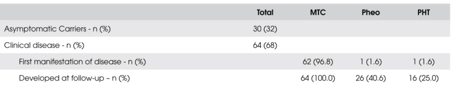
Table 1. Phenotype of the patients with Multiple Endocrine Neoplasia Type 2A (MEN2A) followed at our institution (n=94).
Total MTC Pheo PHT
Asymptomatic Carriers - n (%) 30 (32)
Clinical disease - n (%) 64 (68)
First manifestation of disease - n (%) 62 (96.8) 1 (1.6) 1 (1.6)
Developed at follow-up – n (%) 64 (100.0) 26 (40.6) 16 (25.0)
MTC = medullary thyroid carcinoma; Pheo = pheocromocytoma; PHT = primary hyperparathyroidism.
To our knowledge this is the first report of acute pancreatitis as the first manifestation of MEN2A. In this case, prompt sequential dosage of serum calcium and PTH, diagnosis of PHT and genetic analysis would have resulted in pancreatitis prevention and early MEN2A management.
Acknowledgements:We thank Conselho Nacional de Desenvol-vimento Científico e Tecnológico (CNPq), Fundação de Ampa-ro Pesquisa do Estado do Rio Grande do Sul (FAPERGS), and Fundo de Incentivo a Pesquisa (FIPE), Brazil. The authors de-clare that there is no conflict of interest that would impair the impartiality of this scientific study.
REFERENCES
1. Ponder BA. The phenotypes associated with RET mutations in
the multiple endocrine neoplasia type 2 syndromes. Cancer Res. 1999;59: 1736-42.
2. Mulligan LM, Kwok JB, Healey CS, Elsdon MJ, Eng C, Gardner
E, et al. Germ-line mutation of the RET proto-oncogene in
mul-tiple endocrine neoplasia type 2 A. Nature. 1993;363:458-60. 3. Eng C, Clayton D, Schuffenecker I, Lenoir G, Cote G, Gagel RF,
et al. The relationship between specific RET proto-oncogene
mutation and disease phenotype in multiple endocrine neo-plasia type 2. JAMA. 1996; 276:1575-9.
4. Sitges-Serra A, Alonso M, de Lecea C, Gores PF, Sutherland DE. Pancreatitis and hyperparathyroidism. Br J Surg. 1988;75:158-60.
5. Puñales MK, Graf H, Gross JL, Maia AL. RET Codon 634
Muta-tions in Multiple Endocrine Neoplasia Type 2: Variable Clinical Features and Clinical Outcome. J Clin Endocrinol Metab. 2003;88:2644-9.
6. Mulligan LM, Marsh DJ, Robinson BG, Schuffeenecker I,
Zede-nius J, Lips CJM, et al. RET mutation consortium.
Genotype-phenotype correlation in multiple endocrine neoplasia type 2:
report of international RET mutation consortium. J Intern Med.
1995;238:343-6.
7. Frossard J-L, Steer ML, Pastor CM. Acute pancreatitis. Lancet. 2008;371:143-52.
8. Carnaille B, Oudar C, Pattou F, Combemale F, Rocha J, Proye C. Pancreatitis and primary hyperparathyroidism: forty cases. Aus N Z J Surg. 1998;68:117-9.
9. Tsuboi K, Takamura M, Sato Y, Yokoyama H, Takeuchi M, Iaga-rashi M, et al. Severe acute pancreatitis as an initial manifesta-tion of primary hyperparathyroid adenoma in a pediatric patient. Pancreas. 2007;35:100-1.
10. Takahashi M, Cooper GM. Cloning and expression of the RET
protooncogene encoding a tyrosine-kinase with two potential transmembrane domain. Oncogene. 1998;3:571-6.
11. Santoro M, Carlomagno F, Romano A, Bottaro DP, Dathan NA,
Grieco M, et al. Activation of RET as a dominant transforming
gene by germline mutations of MEN 2A and MEN 2B. Science. 1995;267:381-3.
12. Ito S, Iwashita T, Murakami H, Iwata Y, Sobue-Ku, Takahashi
M. Biological properties of RET with cysteine mutations
cor-relate with multiple endocrine neoplasia type 2A, familial thy-roid carcinoma, and Hirschsprung’s disease phenotype. Cancer Res. 1997;14:2870-2.
13. Santoro M, Mellilo RM, Carlomagno F, Fusco A, Vecchio G.
Molecular mechanism of RET activation in human cancer. Ann
NY Acad Sci. 2002;117-21.
14. Schuffenecker I, Virally-Monod M, Brohet R, Goldgar D, Conte-Devolx B, Leclerc L, et al. Risk and Penetrance of Primary Hy-perparathyroidism in Multiple Endocrine Neoplasia Type 2A
Families with Mutations at Codon 634 of the RET
Proto-Onco-gene. J Clin Endocrinol Metab. 1998;83:487-91.
15. Hoff AO, Hauache OM. Neoplasia Endócrina Múltipla Tipo 1: diagnóstico clínico, laboratorial e molecular e tratamento das do-enças associadas. Arq Bras Endocrinol Metab. 2005;48:735-46.
Correspondence to:
Ana Luiza Maia
Serviço de Endocrinologia, Hospital de Clínicas de Porto Alegre