Re gulatio n o f antio xidant e nzym e
activitie s in m ale and fe m ale rat
m acro phage s by se x ste ro ids
1Departamento de Genética e Morfologia, Instituto de Ciências Biológicas,
Universidade de Brasília, Brasília, DF, Brasil
2Departamento de Fisiologia e Biofísica, Instituto de Ciências Biológicas,
Universidade de São Paulo, São Paulo, SP, Brasil R.B. Azevedo1,
Z.G.M. Lacava1,
C.K. Miyasaka2,
S.B. Chaves1 and R. Curi2
Abstract
Human and animal immune functions present sex dimorphism that seems to be mainly regulated by sex hormones. In the present study, the activities of the antioxidant enzymes total superoxide dismutase (SOD), catalase (CAT), and glutathione peroxidase (GSH-Px) were measured in intraperitoneal resident macrophages from adult male and female rats. In addition to comparing males and females, we also examined the regulation of these enzyme activities in macrophages by sex steroids. GSH-Px activity did not differ between male and female macrophages. However, both total SOD and CAT activities were markedly higher in females than in males (83 and 180%). Removal of the gonads in both males and females (comparison between castrated groups) increased the difference in SOD activity from 83 to 138% and reduced the difference in CAT activity from 180 to 86%. Castration and testosterone administration did not significantly modify the activi-ties of the antioxidant enzymes in male macrophages. Ovariectomy did not affect SOD or GSH-Px activity but markedly reduced (48%) CAT activity. This latter change was fully reversed by estrogen administration, whereas progesterone had a smaller effect. These results led us to conclude that differences in the SOD and CAT activities may partially explain some of the differences in immune function reported for males and females. Also, estrogen is a potent regulator of CAT in macrophages and therefore this enzyme activity in macrophages may vary considerably during the menstrual cycle.
Co rre spo nde nce
R.B. Azevedo
Departamento de Genética e Morfologia, ICB, UnB 70910-900 Brasília, DF Brasil
Research supported by FAPESP, CAPES, CNPq, Pronex and FAP-DF.
Received August 8, 2000 Accepted February 13, 2001
Ke y wo rds
·Antioxidant enzymes
·Macrophages
·Sex hormones
·Estrogen
·Progesterone
·Castration
Several aspects of both the humoral and cellular immune responses differ between males and females (1,2) and this dimorphism is mainly caused by sex steroid hormones (3). Specific receptors for gonadal steroids have been found in the lymphoid organs and macrophages (1). Macrophages play a cen-tral role in the inflammatory and immune responses (4). These cells are able to kill foreign organisms and expose antigens on
mem-branes (6). To protect themselves against the adverse effects of the ROS, these cells pre-sent a complex machinery of antioxidant compounds and enzymes, such as superox-ide dismutase (SOD), catalase (CAT), and glutathione peroxidase (GSH-Px) (7). The activities of these enzymes have been shown to be regulated by nutrients (6,7) and hor-mones (8).
The role of sex hormones in lipid peroxi-dation has been investigated in rat liver ho-mogenates. Male rats have a higher content of products of lipid peroxidation than fe-males (9). There is substantial evidence that estrogen presents antioxidant properties (10,11). The antioxidant effect of estrogen has been regarded as the main mechanism for this hormone to protect skeletal and car-diac muscles (12), uterus (13), and liver (9) from damage. In brain, progesterone instead of estrogen exerts a significant antioxidant effect (14). Contrary to female steroids, tes-tosterone has been shown to decrease the activities of SOD, CAT, and GSH-Px, lead-ing to lipid peroxidation (15).
On the basis of these considerations, we investigated the possible role of gonadal ster-oids in the protection against oxidative stress in macrophages. We compared the activities of the antioxidant enzymes (SOD, CAT, and GSH-Px) in female and male rat macro-phages and examined the effect of gonadec-tomy and the sex steroid hormone replace-ment in both male and female rats.
Male and female albino Wistar rats, 3 months old, were obtained from the Institute of Biomedical Sciences, São Paulo, SP, Bra-zil. The rats were kept at 23o
C on a light/dark cycle of 12/12 h (lights on from 7:00 am). All chemicals and enzymes were obtained from Boehringer-Mannheim GmbH (Mannheim, Germany), or Sigma Chemical Co. (St. Louis, MO, USA). The sex steroid hormones were obtained from Sigma. Male and female rats were gonadectomized under ether anesthe-sia. After one month, the rats were decapi-tated without anesthesia between 10:00 and
12:00 am. Some of the castrated animals were treated with steroid hormones as fol-lows: male castrated animals received 10 µg testosterone daily and ovariectomized rats received 200 ng estrogen or 1 µg progeste-rone, or both, daily. The hormones were injected subcutaneously for 4 days before obtaining the cells. The control group was treated with saline. The rats were killed 4 h after the last injection. The protocol of sex steroid hormone replacement was the same as used in a previous study by our group (2). Cells normally present in the intraperito-neal cavity of the rats were collected using 6 ml PBS, and allowed to adhere to glass flasks for 2 h at 37o
C, after which time the medium and nonadherent cells were removed, and the remaining cells were called resident mac-rophages (unstimulated). Cell viability was determined by Trypan blue exclusion (>95%). At least 92% of the cells were mac-rophages as determined by differential counts. The macrophages were homogenized with a polytron apparatus (PCU-2) at a pro-portion of 1:10 in 10 mM sodium phosphate buffer, pH 7.5. CAT activity was determined by monitoring hydrogen peroxide consump-tion at 230 nm and 30oC (16). GSH-Px activ-ity was measured as described by Wendel (17), by monitoring the decrease of NADPH concentration at 340 nm and 37oC. Total SOD activity (16) was measured on the basis of the rate of cytochrome c reduction by O2
-·
groups. ANOVA and Duncans test were used for comparison between treatments within the same group. Differences were considered to be significant at P<0.05.
GSH-Px activity did not differ between male and female macrophages (see Tables 1 and 2 for comparison). However, both total SOD and CAT activities were markedly higher in females than in males (83 and 180%, respectively). Removal of the gonads in both males and females (comparison be-tween castrated groups) raised the differ-ence in SOD activity to 138% and reduced the difference in CAT activity to 86%. Never-theless, the female rats always showed higher antioxidant enzyme activity. Even gonadec-tomized females presented higher CAT and SOD activities than the control male rats. One may then conclude that the capacity of female macrophages to protect themselves against oxidative stress is really higher than that observed in male cells. This might be an important mechanism for the high immu-noreactivity described for females (1,18).
Administration of testosterone has been shown to increase lipid peroxidation in rat testis (15). This effect is accompanied by a significant decrease in SOD, CAT, and GSH-Px activities. In this study, however, castra-tion and testosterone replacement did not significantly affect the activities of the anti-oxidant enzymes in male macrophages. Therefore, the pro-oxidant effect of testos-terone may be restricted to only a few tis-sues.
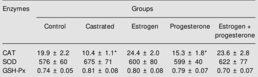
Ovariectomy did not affect SOD or GSH-Px activity but reduced CAT activity by 48% (Table 1). This change was fully reversed by estrogen administration whereas progeste-rone had a smaller effect (Table 1). On the other hand, the CAT activity of macrophages from rats treated with estrogen plus proges-terone did not differ from that of the groups which received only estrogen. Therefore, it is very unlikely that the two hormones pre-sent a synergistic effect on the regulation of CAT activity (Table 1). Since estrogen is a
potent regulator of CAT in macrophages, this enzyme activity may vary considerably during the menstrual cycle. Whether this fact has significant implications for macrophage function and thus for the immune and in-flammatory responses remains to be investi-gated.
Previous studies have shown that estro-gen affects several aspects of macrophage function. This hormone increases the ex-pression of surface markers and the produc-tion of interleukin-1 (19). Suppression of gonadal steroids by ovariectomy reduces
Table 2. Effect of castration and testosterone re-placement on catalase (CAT), total superoxide dis-mutase (SOD) and glutathione peroxidase (GSH-Px) specific activity of peritoneal macrophages from male rats.
Enzymes Groups
Control Castrated Testosterone
CAT 7.1 ± 0.6 5.6 ± 0.6 5.5 ± 0.48
SOD 314 ± 23 283 ± 33 295 ± 30
GSH-Px 0.82 ± 0.09 0.66 ± 0.08 0.62 ± 0.08
Specific activities are expressed as U/mg protein. Data are reported as means ± SEM for 5-8 rats. There w as no significant effect of either castration or hormone replacement. The CAT and SOD ac-tivities of male control and castrated rats w ere significantly low er than those of the correspond-ing female rats (Table 1) (P<0.05, Student t-test). For details of macrophage preparation and hor-mone administration, see text.
Table 1. Effect of castration and hormone replacement on catalase (CAT), total super-oxide dismutase (SOD) and glutathione peroxidase (GSH-Px) specific activity of perito-neal macrophages from female rats.
Enzymes Groups
Control Castrated Estrogen Progesterone Estrogen +
progesterone
CAT 19.9 ± 2.2 10.4 ± 1.1* 24.4 ± 2.0 15.3 ± 1.8* 23.6 ± 2.8
SOD 576 ± 60 675 ± 71 600 ± 80 599 ± 40 622 ± 77
GSH-Px 0.74 ± 0.05 0.81 ± 0.08 0.80 ± 0.08 0.79 ± 0.07 0.70 ± 0.07
hydrogen peroxide production and phago-cytic capacity by rat macrophages and the changes are abolished by estrogen treatment (2). Lacava and Luna (11) showed that ova-riectomy induces a significant increase in the frequency of structural chromosome ab-errations in peritoneal macrophages, which is also reversed by estrogen administration. These authors suggested the involvement of ROS in the chromosome damage caused by the lack of gonadal steroids.
Sex steroid hormones regulate the activi-ties of a number of enzymes of the glucose and glutamine metabolism in leukocytes. Azevedo et al. (2) showed that the activity of phosphate-dependent glutaminase is regu-lated by estrogen in rat peritoneal macro-phages. In the same study, the authors found a decrease in the phagocytic capacity and hydrogen peroxide production of macro-phages from castrated female rats. These changes were all reversed by estrogen treat-ment. In the present study evidence is pre-sented that in addition to the metabolic
en-zymes, estrogen also controls the activity of the antioxidant enzyme CAT. Changes in the oxidative defense system do impair macro-phage function (4,8). Then, the control of macrophage function by estrogen may in-volve several mechanisms including cell metabolism and oxidative stress.
The findings presented herein led us to conclude that differences in the enzymatic antioxidant capacity (mainly CAT and SOD) between male and female macrophages may partially explain the dimorphism widely re-ported for the immune function. Also, the role played by the female sex steroids in the high immunoreactivity observed in females may occur by modulation of the antioxidant defense system. Additional studies are needed to further address this important issue.
Ackno wle dgm e nts
The authors are indebted to J.R. Men-donça and G. de Souza for technical assis-tance.
Re fe re nce s
1. Grossman CJ (1985). Interactions be-tw een the gonadal steroids and immune system. Science, 227: 257-261. 2. Azevedo RB, Costa-Rosa LFBP, Lacava
ZGM & Curi R (1997). Gonadectomy im-pairs lymphocyte proliferation and macro-phage function in male and female rats. Correlation w ith key enzyme activities of glucose and glutamine metabolism. Cell Biochemistry and Function, 15: 293-298. 3. Ansar-Ahmed S, Penhale WJ & Talal N
(1985). Sex hormones in immune re-sponses and aut oim m une diseases.
American Journal of Pathology, 121: 531-551.
4. New sholme P, Costa-Rosa LFBP & Curi R (1996). The importance of macrophage fuel metabolism to its function. Cell Bio-chemistry and Function, 14: 1-10. 5. Curi R, New sholme P, Pithon-Curi TC,
Pires-de-M elo M , Garcia C, Homem-de-Bittencourt Jr PI & Guimarães ARP (1999). M etabolic fate of glutamine in
lympho-cytes, macrophages and neutrophils. Bra-zilian Journal of M edical and Biological Research, 32: 15-21.
6. M iyasaka CK, De-Souza JAA, Torres RP, M ancini-Filho J, Lajolo FM & Curi R (1998). Effect of the administration of fish oil by gavage on activities of antioxidant enzymes of rat lymphoid organs. General Pharmacology, 30: 759-762.
7. Harris ED (1992). Regulation of antioxi-dant enzymes. Journal of Nutrition, 122: 625-626.
8. Pereira B, Costa-Rosa LFBP, Bechara EJH, New sholme P & Curi R (1998). Changes in TBARS content and superox-ide dismutase, catalase and glutathione peroxidase activities in the lymphoid or-gans and skeletal muscles of adrenode-medullated rats. Brazilian Journal of M edi-cal and Biologiedi-cal Research, 31: 827-833. 9. Huh K, Shin US, Choi JW & Lee SI (1994). Effect of sex hormone on lipid peroxida-tion in liver. Archives of Pharmacal
Re-search, 17: 109-114.
10. Gomez-Zubeldia M A, Hernandez R, Vigue-ra J, Arbues JJ, Aparicio A & M illan JC (2000). Effect of bilateral ovariectomy and ovarian steroid hormones on the antioxi-dant systems and plasma malondialde-hyde levels in Wistar rats. Endocrine Re-search, 26: 97-107.
11. Lacava ZGM & Luna H (1994). The anti-clastogenic effect of tocopherol in perito-neal m acrophages of benznidazole-treated and ovariectomized mice. M uta-tion Research, 305: 145-150.
12. Persky AM , Green PS, Stubley L, How ell CO, Zaulyanov L, Brazeau GA & Simpkins JW (2000). Protective effect of estrogens against oxidative damage to heart and skeletal muscle in vivo and in vitro. Pro-ceedings of the Society for Experimental Biology and M edicine, 223: 59-66. 13. Diaz-Flores M , Baiza-Gutman LA, Pedron
estro-gens and progesterone. Life Sciences, 65: 2481-2488.
14. Pajovic SB, Saicic ZS, Spasic M B, Petrovic VM & M artinovic JV (1999). Effects of progesterone and estradiol benzoate on glutathione dependent antioxidant en-zyme activities in the brain of female rats.
General Physiology and Biophysics, 18: 35-44.
15. Chainy GB, Samantaray S & Samanta L (1997). Testosterone-induced changes in testicular antioxidant system. Andrologia, 29: 343-349.
16. Flohé L & Ötting F (1984). Superoxide dismutase assays. M ethods in Enzymol-ogy, 105: 93-104.
17. Wendel A (1981). Glutathione peroxidase.
M ethods in Enzymology, 77: 325-339.
18. Kimura M , Watanable H, Sato S & Abo T (1994). Female predominance of extra-thymic T cells in mice: statistical analysis.
Immunology Letters, 39: 259-267. 19. Flynn A (1986). Expression of Ia and the